Review
. 2017 Sep;24(9):T83-T96.
doi: 10.1530/ERC-17-0003. Epub 2017 Mar 1.

Cell death response to anti-mitotic drug treatment in cell culture, mouse tumor model and the clinic

Affiliations
Review

Cell death response to anti-mitotic drug treatment in cell culture, mouse tumor model and the clinic

Jue Shi et al. Endocr Relat Cancer. 2017 Sep.

Abstract

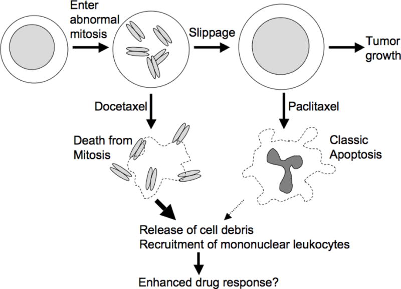
Anti-mitotic cancer drugs include classic microtubule-targeting drugs, such as taxanes and vinca alkaloids, and the newer spindle-targeting drugs, such as inhibitors of the motor protein; Kinesin-5 (aka KSP, Eg5, KIF11); and Aurora-A, Aurora-B and Polo-like kinases. Microtubule-targeting drugs are among the first line of chemotherapies for a wide spectrum of cancers, but patient responses vary greatly. We still lack understanding of how these drugs achieve a favorable therapeutic index, and why individual patient responses vary. Spindle-targeting drugs have so far shown disappointing results in the clinic, but it is possible that certain patients could benefit if we understand their mechanism of action better. Pre-clinical data from both cell culture and mouse tumor models showed that the cell death response is the most variable point of the drug action. Hence, in this review we focus on current mechanistic understanding of the cell death response to anti-mitotics. We first draw on extensive results from cell culture studies, and then cross-examine them with the more limited data from animal tumor models and the clinic. We end by discussing how cell type variation in cell death response might be harnessed to improve anti-mitotic chemotherapy by better patient stratification, new drug combinations and identification of novel targets for drug development.

Keywords: apoptosis; chemotherapy.


Conflict of interest statement

The authors declare no conflict of interest.

Figures

Figure 1
Figure 1
This diagram illustrates anti-mitotic drug responses at the phenotypic level observed by time-lapse microscopy [–22]. Similar responses are seen with microtubules-targeting drugs, Kinesin-5 inhibitors and Plk1 inhibitors. The timing of events, and the fraction of cells that undergo alternative outcomes, vary between cell lines and drug types. Duration of mitotic arrest varies from 10–20 hours, and fraction of cells that die from <5% to 100%. The 4N G1-arrest cells that slip out of mitotic arrest progress to distinct cellular state, likely depending on p53 activity (see text).
Figure 2
Figure 2
Cell death induced by anti-mitotic drugs during mitotic arrest is mediated by mitochondrial, intrinsic apoptosis triggered by depletion of an anti-apoptotic protein, Mcl-1. The other major negative regulator of mitotic death is Bcl-xL, whose expression level largely determines the sensitivity of different cancer cell lines to anti-mitotics-induced cell death in mitotic arrest.
Figure 3
Figure 3
Post-slippage cell death induced by anti-mitotic drugs is mediated by mitochondrial, intrinsic apoptosis triggered by DNA damage in the 4N G1-arrest cells. The degree of multinucleation in the post-slippage 4N cells determines the level of DNA damage and subsequent p53-mediated cell death activation. Taxanes engender significantly more mutlinucleation than the spindle-targeting Kinesin-5 inhibitor. Therefore, while different anti-mitotic drugs induce similar degree of cell death during mitotic arrest, the extent of post-slippage death that they trigger vary significantly.
Figure 4
Figure 4
Variation of apoptotic response to anti-mitotic drugs is much more significant than that of mitotic response between both different mouse tumors and culture human cancer cell lines. (A, B) Fifteen different mouse cancers (propagated by serial passage in mice) were treated with a single dose of paclitaxel. Tumor response was measured as growth delay (x-axis). Mitotic arrest and apoptosis were scored by quantitative histology at different times after drug injection, and peak responses were scored as % of cells in the tumor. (A) plots the average peak mitotic arrest response, and (B) the average peak apoptotic response, against average growth delay for different tumors. Note that most tumors showed a strong mitotic arrest response, but only tumors that respond well to drug show a strong apoptotic response. These figures are taken from Fig. 1 by Milross et al (1996) [29]. (C, D) Comparison of mitotic response (C) and apoptotic response (D) to three different anti-mitotic drugs, including paclitaxel (150nM, shown in black), nocodazole (500nM, shown in orange), and Kinesin-5 (Eg5) inhibitor (500nM, shown in green), for a panel of human cancer cell lines. The mitotic response corresponds to the percentage of mitotic cells after 24 hours of drug treatment, probed by phospho-Histone 3 antibody, and the apoptotic response corresponds to the percentage of apoptotic cells after 48 hours of drug treatment, probed by Parp1 cleavage. Cell lines are arranged in descending order of sensitivity to anti-mitotics-induced cell death. These figures are taken from Fig. 1 by Shi et al (2008) [18].
Figure 5
Figure 5
This cartoon summarizes the work of the Milas group on the effect of two taxanes on mouse model cancers [29, 65, 66]. Docetaxel was overall more effective than paclitaxel at the does used, though sensitivity of different tumor lines to the two drugs strongly correlated. In cancers that responded well, docetaxel promoted mainly death in mitosis, and paclitaxel mainly apoptosis after slippage. Death in mitosis following docetaxel treatment was correlated with extensive release of cell debris, including mitotic chromosomes, and massive recruitment of mononuclear leukocytes to the shrinking tumor. Subsequent work suggested that leukocyte recruitment enhances the therapeutic response [69]. Therefore, timing of cell death may be important in tumors for anti-mitotic response.

References

    1. Jordan MA, Wilson L. Microtubules as a target for anticancer drugs. Nat Rev Cancer. 2004;4:253–65. - PubMed
    1. Perez EA. Microtubule inhibitors: Differentiating tubulin-inhibiting agents based on mechanisms of action, clinical activity, and resistance. Mol Cancer Ther. 2009;8(8):2086–95. - PubMed
    1. Mukhtar E, Adhami VM, Mukhtar H. Targeting microtubules by natural agents for cancer therapy. Mol Cancer Ther. 2014;13(2):275–84. - PMC - PubMed
    1. Stanton RA, Gernert KM, Nettles JH, Aneja R. Drugs that target dynamic microtubules: a new molecular perspective. Med Res Rev. 2011;31(3):443–81. - PMC - PubMed
    1. Crown J, O’Leary M, Ooi WS. Docetaxel and paclitaxel in the treatment of breast cancer: a review of clinical experience. Oncologist. 2004;9(Suppl 2):24–32. - PubMed

Publication types

Substances