. 2017 May;206(1):199-213.
doi: 10.1534/genetics.116.193813. Epub 2017 Mar 1.

Adult Muscle Formation Requires Drosophila Moleskin for Proliferation of Wing Disc-Associated Muscle Precursors

Affiliations

Adult Muscle Formation Requires Drosophila Moleskin for Proliferation of Wing Disc-Associated Muscle Precursors

Kumar Vishal et al. Genetics. 2017 May.

Abstract

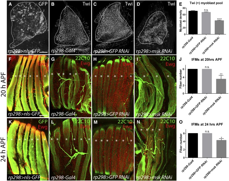
Adult muscle precursor (AMP) cells located in the notum of the larval wing disc undergo rapid amplification and eventual fusion to generate the Drosophila melanogaster indirect flight muscles (IFMs). Here we find that loss of Moleskin (Msk) function in these wing disc-associated myoblasts reduces the overall AMP pool size, resulting in the absence of IFM formation. This myoblast loss is due to a decrease in the AMP proliferative capacity and is independent of cell death. In contrast, disruption of Msk during pupal myoblast proliferation does not alter the AMP number, suggesting that Msk is specifically required for larval AMP proliferation. It has been previously shown that Wingless (Wg) signaling maintains expression of the Vestigial (Vg) transcription factor in proliferating myoblasts. However, other factors that influence Wg-mediated myoblast proliferation are largely unknown. Here we examine the interactions between Msk and the Wg pathway in regulation of the AMP pool size. We find that a myoblast-specific reduction of Msk results in the absence of Vg expression and a complete loss of the Wg pathway readout β-catenin/Armadillo (Arm). Moreover, msk RNA interference knockdown abolishes expression of the Wg target Ladybird (Lbe) in leg disc myoblasts. Collectively, our results provide strong evidence that Msk acts through the Wg signaling pathway to control myoblast pool size and muscle formation by regulating Arm stability or nuclear transport.

Keywords: Drosophila melanogaster; Moleskin; indirect flight muscles; proliferation.


Figures

Figure 1
Figure 1
Msk is required for the generation of the wing disc-associated myoblast pool. (A and B) The 1151-Gal4 driver is used to express nls-GFP in all larval wing disc-associated myoblasts at the L3 stage. (A) Low magnification of the wing disc (white dotted outline). The yellow boxed region shows the location of the larval myoblasts in the notum (B). (C, D, F, G, I, and J) Maximum projection confocal microscopy images of the AMP pool in (C, D, F, and I) control or (G and J) 1151 > msk RNAi L3 wing discs labeled with the myoblast markers (C, D, F, and G) Twi or (I and J) Mef2. Note that the myoblast pool (dotted line) is reduced upon disruption of (G and J) Msk compared to (C, D, and F) controls. (E, H, and K) Quantitation of myoblast density (per regions 1600 µm2) in control (1151-Gal4 or 1151 > GFP RNAi) and msk RNAi (1151 > msk RNAi) wing discs labeled with (E and H) Twi or (K) Mef2. (L–N) PH3 staining to monitor proliferating notum myoblasts. More Cut(+) myoblasts also stain for PH3 in (L) control compared to (M) msk RNAi discs. (N) Bar graph showing the fraction of PH3(+)/Cut(+) myoblasts. Mean ± SEM. n.s., not significant. **** P < 0.001, *** P < 0.005. Bar, 50 µm.
Figure 2
Figure 2
Abrogated Msk function during larval myoblast proliferation reduces DLM fiber number. (A–D and F–I) Maximum projection confocal micrographs of DLM fibers at (A–D) 20 hr APF or (F–I) 24 hr APF. (A and F) Pupal myoblasts labeled with Ewg (red) are being incorporated into the developing DLM fibers (*) marked by 1151-driven GFP (green) at (A) 20 hr APF or (F) 24 hr APF through reiterative myoblast fusion events. (B–D and G–I) Developing DLMs are stained with 22C10 (green) to mark muscle fibers and Ewg (red) to label myoblasts. (B and G) 1151-Gal4 or (C and H) 1151 > GFP RNAi control animals have six DLM fibers (*) at (B and C) 20 hr APF or (G and H) 24 hr APF. (D and I) Little fiber formation is seen in 1151 > msk RNAi animals. (E and J) Quantitation of fiber number shows there are significantly fewer DLM fibers upon knockdown with msk RNAi animals compared to controls. Mean ± SEM. n.s., not significant. **** P < 0.001. Bar, 50 µm.
Figure 3
Figure 3
Blocking Msk function in founder cells reduces both the myoblast pool size and fiber number. (A–D) Maximum projection confocal pictures of L3 wing disc-associated myoblasts. (A) rp298 expression is present in a subset of myoblasts as visualized by GFP expression. (B–D) The numbers of larval AMPs labeled with anti-Twi is similar in (B) rp298-Gal4 or (C) rp298 > GFP RNAi controls, but reduced in (D) rp298 > msk RNAi-expressing myoblasts. Dotted lines denote larval myoblast pool. (E) A bar graph showing a significant reduction in the density of myoblasts (per regions 1600 µm2) present in the notum of rp298 > msk RNAi wing discs compared to controls. (F–O) The consequences of msk RNAi knockdown in DLM fibers at (F–J) 20 hr APF or (K–O) 24 hr APF. Fibers are marked by 22C10 (green; *) and fused myonuclei are labeled with Ewg (red). Six fibers are present in (G, H, L, and M) controls, whereas (I and N) rp298 > msk RNAi individuals have less than six fibers. (J and O) Bar graphs showing significantly fewer fibers per hemi-segment at (J) 20 hr APF or (O) 24 hr APF. Mean ± SEM. n.s., not significant. **** P < 0.001, ** P < 0.01, *P < 0.05. Bar, 50 µm.
Figure 4
Figure 4
Knockdown of msk RNAi during pupal morphogenesis does not alter the number of myonuclei, but causes a minor delay in fiber formation. (A) Schematic showing the temperature-shift paradigm for msk RNAi induction during pupal development. (B–D and F–H) Maximum projection confocal micrographs of DLM fiber formation at 24 hr APF. Fibers are marked by 22C10 (red; *) and fused myonuclei are immunostained with Ewg (green). (B–D) Controls have the normal complement of six fibers. (F–H) A mild decrease in fiber number is observed upon msk RNAi knockdown. Note that complete fiber splitting is delayed (G, white ←). (E) Bar graph quantitates the small decrease in fiber number upon a reduction in Msk. (I) Quantitation reveals no difference in the number of myonuclei between (D) control and (H) experimental samples. Mean ± SEM. n.s., not significant. *** P < 0.005. Bar, 50 µm.
Figure 5
Figure 5
Msk regulates the expression of Wg-responsive genes in the wing imaginal disc and leg disc. (A–F) Myoblasts in L3 wing discs immunolabeled with Cut (red) and Vg (green) in (A–C) controls, compared to those with (D–F) disrupted Msk function (1151 > msk RNAi). While both Vg and Cut exhibit broad myoblast expression, (A and B; *) Vg accumulates at higher levels in the dorsal myoblasts while (A and C; ←) Cut protein is seen at increased levels in ventral myoblasts. (D and F) Cut staining is still present, while (D and E) Vg expression is absent upon induction of msk RNAi. (G–L) Effect of blocking Msk function on Lbe expression in leg disc-associated myoblasts. (G–I) In control animals, myoblasts are double labeled with Ebd (green) and Lbe (red, *). (J–L) Disruption of Msk function results in a significantly fewer Ebd(+) myoblasts accompanied by loss of Lbe expression. All images are Z-stack projections. Bar, 50 µm.
Figure 6
Figure 6
Moleskin acts upstream of Wg transcriptional complexes. (A and B) Maximum projection confocal images of wing disc-associated myoblasts marked by Ebd antibody in control and msk RNAi samples. (C) The myoblast density (per regions 1600 µm2) is significantly less in (B) msk RNAi samples compared to (A) controls. (D–G) Effect of overexpressing Msk in a DN-TCF mutant background. Myoblasts are marked by Twi in the notum region of L3 wing discs in Z-stack projections. (D) Overexpression of Msk alone does not alter the myoblast pool number. (E) Expression of DN-TCF results in reduced density of the myoblast pool. (F) Overexpression of Msk in a DN-TCF background does not rescue the reduction in myoblast number. (G) Quantification of the myoblast pool density in the indicated genotypes. (H–K) Effect of overexpressing armS10 in an msk RNAi mutant background in maximum intensity projections. (H) The Twi-labeled myoblast pool in armS10 wing discs is similar to controls. (I and J) A diminished myoblast pool is present in both (I) 1151 > msk RNAi and (J) 1151 > GFP; msk RNAi wing discs. (K) Overexpressing armS10 partially rescues the myoblast pool size. (L and M) Quantitation comparing the (L) myoblast density (per regions 1600 µm2) or (M) myoblast pool size per single confocal plane in the indicated genotypes. Mean ± SEM. n.s., not significant. **** P < 0.001, *** P < 0.005, **P < 0.01. Bar, 50 µm.
Figure 7
Figure 7
Disrupting Msk function results in loss of Arm protein. (A–G) Immunofluorescent double-labeling of Arm (green) and myoblasts (red) marked by Twi antibody in L3 wing discs. (A–C) Controls show an accumulation of Arm in a fraction of myoblasts depicted as maximum intensity projections. (D) Single plane orthogonal views through the notum wing disc showing colocalization of Twi(+) and Arm(+) myoblasts. (D′) Increased magnification of the XZ scan in D. The yellow ← points from the epithelium toward the myoblast layers in the notum. (E–G) There is no Arm accumulation in the remaining myoblasts in 1151 > msk RNAi wing discs shown as Z-stack projections. (H) Bar graph shows a significant reduction in the fraction of Arm(+) myoblasts upon a reduction of Msk. (I–K) Maximum projection confocal images of L3 wing discs double labeled with Wg (green) and Twi (red). (L) A single plane orthogonal section of the wing disc showing Wg-expressing epidermal cells and the overlying myoblasts. (L′) Increased magnification of the XZ scan in L. The yellow ← points away from the source of Wg toward the myoblasts. (M–O) Similar to controls, the myoblast pool (red) in msk RNAi animals is evenly distributed relative to the Wg(+) (green) cells. Also, there is no difference in Wg staining between the control and the experimental samples. (P) Orthogonal section of notum wing discs show that the myoblasts are juxtaposed next to a source of Wg, but maintain their location at the distal edge of the myoblast layer. (P′) Increased magnification of the XZ scan in P. The yellow ← points away from the source of Wg toward the myoblasts. Note that two different samples are shown in (M) and (P). In all orthogonal views, the upper panel corresponds to an XZ view of the red line and the green line is the location of the XZ view of the green line. Mean ± SEM. **** P < 0.001. Bar, 50 µm.
Figure 8
Figure 8
Msk acts through Sgg to regulate the wing disc myoblast pool size. (A–C and E–G) Maximum confocal projections of L3 notum myoblasts immunostained with Twi. (A and B) Qualitatively, less myoblasts are present upon overexpression of (B) sgg (WT) compared to (A) 1151 controls. (C) Overexpressing Msk in a sgg (WT) background partially rescues the myoblast number. (D) Quantification of myoblast density (per regions 1600 µm2) in (A–C). (E and F) Targeting a weak version of (F) activated sgg (214F) causes a reduction in the myoblast pool compared to (E) 1151 controls. (G) A significant increase in the myoblast pool size is seen in 1151 > msk FL; sgg (Y214F) samples. (H) A bar graph showing partial restoration of the myoblast pool (per regions 1600 µm2) upon overexpression of msk FL in a sgg (Y214F) background. Mean ± SEM. n.s., not significant. **** P < 0.001, *** P < 0.005. Bar, 50 µm.

Similar articles

Cited by

References

    1. Anant S., Roy S., VijayRaghavan K., 1998. Twist and Notch negatively regulate adult muscle differentiation in Drosophila. Development 125: 1361–1369. - PubMed
    1. Bate M., 1990. The embryonic development of larval muscles in Drosophila. Development 110: 791–804. - PubMed
    1. Bate M., Rushton E., Currie D. A., 1991. Cells with persistent twist expression are the embryonic precursors of adult muscles in Drosophila. Development 113: 79–89. - PubMed
    1. Bejsovec A., 2006. Flying at the head of the pack: Wnt biology in Drosophila. Oncogene 25: 7442–7449. - PubMed
    1. Benchabane H., Xin N., Tian A., Hafler B. P., Nguyen K., et al. , 2011. Jerky/Earthbound facilitates cell-specific Wnt/Wingless signalling by modulating β-catenin-TCF activity. EMBO J. 30: 1444–1458. - PMC - PubMed

MeSH terms

LinkOut - more resources