. 2017 May;66(5):1237-1246.
doi: 10.2337/db16-1057. Epub 2017 Mar 1.

Mast Cells Promote Seasonal White Adipose Beiging in Humans

Affiliations

Mast Cells Promote Seasonal White Adipose Beiging in Humans

Brian S Finlin et al. Diabetes. 2017 May.

Abstract

Human subcutaneous (SC) white adipose tissue (WAT) increases the expression of beige adipocyte genes in the winter. Studies in rodents suggest that a number of immune mediators are important in the beiging response. We studied the seasonal beiging response in SC WAT from lean humans. We measured the gene expression of various immune cell markers and performed multivariate analysis of the gene expression data to identify genes that predict UCP1. Interleukin (IL)-4 and, unexpectedly, the mast cell marker CPA3 predicted UCP1 gene expression. Therefore, we investigated the effects of mast cells on UCP1 induction by adipocytes. TIB64 mast cells responded to cold by releasing histamine and IL-4, and this medium stimulated UCP1 expression and lipolysis by 3T3-L1 adipocytes. Pharmacological block of mast cell degranulation potently inhibited histamine release by mast cells and inhibited adipocyte UCP1 mRNA induction by conditioned medium (CM). Consistently, the histamine receptor antagonist chlorpheniramine potently inhibited adipocyte UCP1 mRNA induction by mast cell CM. Together, these data show that mast cells sense colder temperatures, release factors that promote UCP1 expression, and are an important immune cell type in the beiging response of WAT.


Figures

Figure 1
Figure 1
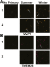
Seasonal changes in UCP1 and TMEM26 immunohistochemistry in thigh SC WAT. A: Histochemical analysis of UCP1 expression in thigh SC WAT of two lean subjects in the summer and winter. The left panel shows no primary antibody control. The arrows in the middle panels point to UCP1 staining in unilocular adipocytes in the summer. The arrows in the right panels point to UCP1 staining in cells between adipocytes in the winter. B: TMEM26 staining in the same subjects. The arrow points to TMEM26 staining in cells between adipocytes in the winter. Scale bars = 50 μm.
Figure 2
Figure 2
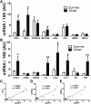
Seasonal changes in gene expression of beige adipocyte markers, immune cell markers, and cytokines. A and B: Gene expression in thigh SC adipose tissue was measured in the summer (n = 7) and winter (n = 9) by real-time RT-PCR, and the relative expression of each gene is represented in arbitrary units (AU). Data are mean ± SEM. *P < 0.05, **P < 0.01; #P = 0.06 (unpaired Student t test). CE: Gene expression of UCP1 and the indicated genes. Pearson correlation coefficients (r) and two-tailed P values are indicated (n = 16).
Figure 3
Figure 3
Mast cells respond to cold. TIB64 mast cells were incubated at 37°C (control) or at 30°C for 4 h and allowed to recover at 37°C for the indicated time. A: Histamine levels were measured in the media. B: IL-4 protein levels were measured in the media. C: TNFα protein levels were measured in the media. DI: RBM3, tryptase, CPA3, IL-4, TNFα, and UCP1 mRNA expression was measured in the mast cells. Data are mean ± SEM (n = 3). *P < 0.05, **P < 0.01, ***P < 0.001 (ANOVA with Tukey post hoc test).
Figure 4
Figure 4
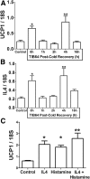
Mast cell CM from cold-shocked mast cells induces UCP1 expression in adipocytes. Mast cell CM from Fig. 3 was applied to differentiated 3T3-L1 adipocytes for 4 h at 37°C. A and B: Adipocyte gene expression was measured for UCP1 and IL-4. C: Differentiated 3T3-L1 adipocytes were treated with recombinant mouse IL-4 (1 nmol/L), histamine (10 nmol/L), or a combination of both for 3 h. Data are mean ± SEM (n = 3). *P < 0.05, **P < 0.01 (ANOVA with Dunnett post hoc test).
Figure 5
Figure 5
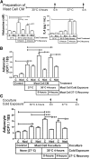
Identification of factors in mast cell CM that induce adipocyte UCP1 expression. A: TIB64 mast cells were incubated at 37°C (control) or 30°C (cold) for 4 h in the absence (control) or presence of nedocromil, and the media were harvested (0 h). Another set of 30°C-incubated mast cells was allowed to recover at 37°C for 4 h, and the media were harvested (time course shown above the graph). Shown are the histamine and IL-4 protein levels measured in the media. B: Mast cell CM from A was warmed to 37°C and applied to differentiated 3T3-L1 adipocytes for 4 h at 37°C, and UCP1 mRNA was measured. C: Differentiated 3T3-L1 adipocytes in six-well dishes were cocultured with mast cells in inserts as indicated or were cultured alone (control). Nedocromil was added as indicated. The coculture system was incubated at 30°C or 37°C, and the adipocytes were harvested after 4 h of coculture at 37°C or 0 or 4 h after incubating the coculture at 30°C for 4 h (time course shown above the graph). UCP1 mRNA expression was measured in the adipocytes. Data are mean ± SEM (n = 3). *P < 0.05, **P < 0.01, ***P < 0.001 (ANOVA with a Tukey post hoc test). C, control; Ned, nedocromil.
Figure 6
Figure 6
Histamine in mast cell CM from cold-treated mast cells induces UCP1 expression in adipocytes through a PKA-dependent mechanism. A: Differentiated 3T3-L1 adipocytes were treated with vehicle (control), 5 μmol/L chlorpheniramine, 50 μmol/L H89, 10 μmol/L propranolol, or 100 nmol/L SB202190 in adipocyte differentiation media for 30 min as indicated. The adipocytes were then incubated with 50% mast cell CM (from cold-treated mast cells allowed to recover for 4 h at 37°C [cold CM]) and 50% adipocyte differentiation media containing the same final concentration of inhibitors or vehicle control for 4 h. The cells were harvested, and UCP1 mRNA expression was measured. Data are mean ± SEM (n = 3). **P < 0.01, ***P < 0.001 compared with cold CM treatment (ANOVA with Dunnett post hoc test). B: Differentiated 3T3-L1 or ADHASC adipocytes were treated with vehicle (control) or histamine (10 nmol/L) for 4 h as indicated, and UCP1 immunohistochemistry was performed as described in research design and methods. Representative images of UCP1 staining (orange) and DAPI (blue) identify nuclei. Scale bar = 50 μm. Quantification of UCP1 staining are shown below the images. Data are mean ± SEM (n = 3). *P < 0.05, **P < 0.01 (unpaired Student t test). C: Differentiated 3T3-L1 adipocytes were incubated for 4 h at 37°C with media (control) or CM from mast cells incubated for 8 h at 37°C (CM) or 4 h at 30°C and then 4 h at 37°C (CM cold); 5 μmol/L norepinephrine (NE) was used as a positive control. The media were then replaced with potassium ringers containing 2% fatty acid–free BSA, and the cells were incubated overnight at 37°C. These media were harvested, and free fatty acid concentrations were determined. Data are mean ± SEM (n = 5; NE: n = 3). *P < 0.05, ***P < 0.001 (ANOVA with a Tukey post hoc test).

Similar articles

Cited by

References

    1. Cannon B, Nedergaard J. Brown adipose tissue: function and physiological significance. Physiol Rev 2004;84:277–359 - PubMed
    1. Rothwell NJ, Stock MJ. A role for brown adipose tissue in diet-induced thermogenesis. Nature 1979;281:31–35 - PubMed
    1. Lowell BB, S-Susulic V, Hamann A, et al. . Development of obesity in transgenic mice after genetic ablation of brown adipose tissue. Nature 1993;366:740–742 - PubMed
    1. Virtanen KA, Lidell ME, Orava J, et al. . Functional brown adipose tissue in healthy adults. N Engl J Med 2009;360:1518–1525 - PubMed
    1. Cypess AM, Lehman S, Williams G, et al. . Identification and importance of brown adipose tissue in adult humans. N Engl J Med 2009;360:1509–1517 - PMC - PubMed

Publication types

MeSH terms