. 2017 Mar 1;10(3):259-270.
doi: 10.1242/dmm.027409.

Systemic autoimmunity induced by the TLR7/8 agonist Resiquimod causes myocarditis and dilated cardiomyopathy in a new mouse model of autoimmune heart disease

Affiliations

Systemic autoimmunity induced by the TLR7/8 agonist Resiquimod causes myocarditis and dilated cardiomyopathy in a new mouse model of autoimmune heart disease

Muneer G Hasham et al. Dis Model Mech. .

Abstract

Systemic autoimmune diseases such as systemic lupus erythematosus (SLE) and rheumatoid arthritis (RA) show significant heart involvement and cardiovascular morbidity, which can be due to systemically increased levels of inflammation or direct autoreactivity targeting cardiac tissue. Despite high clinical relevance, cardiac damage secondary to systemic autoimmunity lacks inducible rodent models. Here, we characterise immune-mediated cardiac tissue damage in a new model of SLE induced by topical application of the Toll-like receptor 7/8 (TLR7/8) agonist Resiquimod. We observe a cardiac phenotype reminiscent of autoimmune-mediated dilated cardiomyopathy, and identify auto-antibodies as major contributors to cardiac tissue damage. Resiquimod-induced heart disease is a highly relevant mouse model for mechanistic and therapeutic studies aiming to protect the heart during autoimmunity.

Keywords: Autoimmunity; Dilated cardiomyopathy; Heart disease; Model; Myocarditis; Resiquimod; Toll-like receptor 7/8.


Conflict of interest statement

Competing interests

The authors declare no competing or financial interests.

Figures

Fig. 1.
Fig. 1.
Resiquimod treatment induces morphological changes and functional impairment of the heart. (A) Examples of m-mode traces of a heart from a Resiquimod-treated mouse compared with traces of a control mouse heart. (B) Quantification of LV end-systolic and end-diastolic volumes (ESV and EDV), LV mass, fractional shortening and ejection fraction in Resiquimod-treated mice measured at baseline, and at 2 week intervals until week 8. (C) Diastolic function of Resiquimod-treated mice over time assessed by Doppler echocardiography measurements of early (A) and late (E) ventricular filling velocities across the mitral valve. For B and C n=8/time point (week 8: n=3), data pooled from two independent repeats. (D) Cardiac MR images demonstrating how parameters of cardiac structure and function were assessed. 10-frame movies including end diastolic and end systolic images of 7 cross-sections (white lines) from base to apex were acquired and segmented to obtain the outer and inner border of the LV to determine end diastolic and end systolic mass (EDM, ESM), and EDV and ESV, as well as ejection fraction. (E) Quantification of LV ESV and EDV, LV mass and ejection fraction in Resiquimod-treated mice measured at baseline and 2 and 4 weeks after the start of treatment. n=5/group, two independent repeats. Values represent mean±s.e.m.; *P<0.05, **P<0.005, one-tailed, paired Student's t-test.
Fig. 2.
Fig. 2.
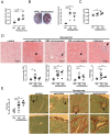
Resiquimod treatment induces cardiac tissue damage as seen in autoimmune disease. (A) Splenomegaly as a measure of systemic immune activation 2 and 4 weeks after start of treatment. (B) Macroscopic observation of morphological changes, severe hyperaemia and haemorrhagic lesions of hearts ex vivo 2 weeks and corresponding quantification 2 and 4 weeks after start of treatment. Macroscopic haemorrhagic lesions were scored on a scale according to: no lesions (=); lesions cover <10% of heart surface (1); lesions cover 10-30% of heart surface (2); lesions cover 30-50% of heart surface (3); lesions cover >50% of heart surface (4). (C) Heart/body mass ratio 2 and 4 weeks after start of treatment. (D) H&E-stained paraffin-embedded heart sections (top) showing eosinophilic cardiomyocytes, islands of hyperaemia and extravasation of red blood cells (RBC), cardiomyocyte (CM) vacuolisation and mononuclear cell infiltration as indicated by black arrows 400× magnification. Original micrograph is cropped to show an example of the respective damage parameter. Corresponding semi-quantitative scores are shown below. (E) PicoSirius Red-stained frozen heart sections showing epicardial, interstitial and perivascular fibrosis. Top row: 200× magnification; bottom row, 3.3× magnification of area of interest in panel above. Individual cardiac damage parameters were scored on a scale from 0 to 3 (none, mild, moderate, severe) in five fields of view in four heart cross-sections at papillary muscle level per mouse. n=4-10/group (each symbol represents one individual mouse), one representative experiment shown of >3 independent repeats. Values represent mean±s.e.m. *P<0.05, **P<0.005 and ***P<0.001, two-tailed, unpaired Student's t-test.
Fig. 3.
Fig. 3.
The Resiquimod-induced cellular immune response in the heart, mediastinal lymph nodes and spleen. (A) Representative blots obtained by flow cytometry showing differences in immune cell populations in the hearts of control and Resiquimod-treated mice (left) and corresponding total counts of CD45+, CD3+, CD19+, CD11b+ immune cells/g heart tissue in Resiquimod-treated versus untreated mice (right). (B) Frequency of CD3+, CD19+, CD11b+ immune cells among total CD45+ immune cells in heart tissue, mediastinal lymph nodes and spleens in Resiquimod-treated versus untreated mice. n=4/group, data pooled from two independent experiments. Values represent mean±s.e.m. *P<0.05 and **P<0.005, two-tailed, unpaired Student's t-test.
Fig. 4.
Fig. 4.
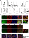
Resiquimod induces anti-cardiac auto-antibodies. (A) Levels of anti-dsDNA at week 4 after Resiquimod treatment. (B) ELISA to detect relative levels of antibody isotypes as indicated (light and heavy chains) in serum of untreated and Resiquimod-treated mice. Data pooled from two independent experiments. (C) Levels of anti-cardiac auto-antibodies of IgM and IgG isotype. (D) Levels of anti-cardiac auto-antibodies with IgG1, IgG2a and IgG2b heavy chains. (E-G) Immunofluorescence staining using anti-mouse IgG-Alexa Fluor 488 (E), anti-mouse IgM-FITC (F), anti-mouse IgG1/IgG2a/IgG2b/IgG3 and a secondary anti-rat IgG-Alexa Fluor 488 (G) to detect in vivo deposited antibodies and immuno-complexes as indicated by white arrows. Green: IgG/IgM/IgG1/IgG2a/IgG2b/IgG3 staining; red: wheat germ agglutinin staining membranes; blue: DAPI staining of nuclei. 400× original magnification. Areas of interest (a,b in E and a in F): additional 2× magnification. *P<0.05, **P<0.005, two-tailed, paired/unpaired Student's t-test.
Fig. 5.
Fig. 5.
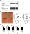
Transfer and mutant studies indicate that adaptive immune cells induce cardiac damage. (A) Donor mice were treated with Resiquimod as described above and spleens and sera were harvested one week after cessation of treatment. 2×10⁷ splenocytes/recipient mouse were pre-stimulated in vitro with 5 μg/ml concanavalin A for 2 days and injected via the tail vein. 200 μl serum/mouse was injected three times per mouse 1 week apart. Control mice were either treated with Resiquimod as per standard 2 week treatment protocol or left untreated. Hearts of recipient and control mice were harvested 3 weeks after start of treatment. (B) H&E- or PicoSirius Red-stained paraffin-embedded heart sections (representative examples) were scored for degree of mononuclear cell infiltration and fibrosis on a scale from 0-3 (none, mild, moderate, severe) in five fields of view in four heart cross-sections at papillary muscle levels per mouse. (C) Systolic and diastolic function of ‘Group A’ splenocyte recipient mice 4 weeks after splenocyte transfer. Quantification of LV end-systolic and end-diastolic volumes, LV mass, fractional shortening and ejection fraction measured at baseline, and after 4 weeks. n=3/time point. Diastolic function assessed by Doppler echocardiography measurements of early (A) and late (E) ventricular filling velocities. n=3/time point. Values represent mean±s.e.m. *P<0.05, **P<0.005 and ***P<0.001, two-tailed, paired/unpaired Student's t-test.
Fig. 6.
Fig. 6.
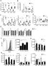
Genetic variation defines the responses to Resiquimod treatment and the severity of cardiac damage. (A) Splenomegaly as a measure of systemic immune activation 2 weeks after start of treatment in CFN, C57Bl/6J (B6), FVB/NJ (FVB) and NOD/ShiLtJ (NOD). (B) Levels of anti-dsDNA at week 2 after Resiquimod treatment. (C) Semi-quantitative scores of degree of mononuclear cell infiltration and fibrosis based on H&E- and PicoSirius Red-stained paraffin-embedded heart sections. Individual damage parameters were scored on a scale from 0-3 (none, mild, moderate, severe) in five fields of view in four heart cross-sections in the myocardium at papillary muscle level per mouse. (D) Heart/body mass ratio 2 weeks after start of treatment. (E) Quantification of LV end-systolic volumes, LV mass, fractional shortening and ejection fraction in Resiquimod-treated mice measured at baseline (CTRL) and 6 weeks after start of treatment (Resiquimod). n=5. (F) Expression level of TLR7 in splenocytes as measured by mean fluorescence intensity (MFI) using flow cytometry; n=5. (G) Total cell count in culture at day 4 of splenocytes stimulated with 1 mg/ml Resiquimod. (H) Total T-cell, B-cell and myeloid cell counts in culture at day 4 in splenocytes stimulated with 1 mg/ml Resiquimod obtained by manual counting and flow cytometry staining against CD45+, CD3+, CD19+, CD11b+ immune cells; n=3. Values represent mean±s.e.m. *P<0.05, **P<0.005, ***P<0.001 and ****P<0.0001, two-tailed, unpaired Student's t-test. Asterisks directly above individual columns indicate a significant difference between the respective parental strain and CFN mice.

Similar articles

Cited by

References

    1. Abou-Raya A. and Abou-Raya S. (2006). Inflammation: a pivotal link between autoimmune diseases and atherosclerosis. Autoimmun Rev. 5, 331-337. 10.1016/j.autrev.2005.12.006 - DOI - PubMed
    1. Abusamieh M. and Ash J. (2004). Atherosclerosis and systemic lupus erythematosus. Cardiol. Rev. 12, 267-275. 10.1097/01.crd.0000124843.85660.ed - DOI - PubMed
    1. Askanas V., Engel W. K. and Mirabella M. (1994). Idiopathic inflammatory myopathies: inclusion-body myositis, polymyositis, and dermatomyositis. Curr. Opin. Neurol. 7, 448-456. 10.1097/00019052-199410000-00013 - DOI - PubMed
    1. Baldeviano G. C., Barin J. G., Talor M. V., Srinivasan S., Bedja D., Zheng D. and Cihakova D. (2010). Interleukin-17A is dispensable for myocarditis but essential for the progression to dilated cardiomyopathy. Circ. Res. 106, 1646-1655. 10.1161/CIRCRESAHA.109.213157 - DOI - PubMed
    1. Beranek J. T. (2001). Quick disposal of dead cardiomyocytes: an ultimate proof of apoptosis. J. Heart Lung. Transplant. 20, 923-924. 10.1016/S1053-2498(01)00270-4 - DOI - PubMed

Publication types

MeSH terms