. 2017 Apr;49(4):606-612.
doi: 10.1038/ng.3804. Epub 2017 Feb 27.

Biallelic mutations in human DCC cause developmental split-brain syndrome

Affiliations

Biallelic mutations in human DCC cause developmental split-brain syndrome

Saumya S Jamuar et al. Nat Genet. 2017 Apr.

Abstract

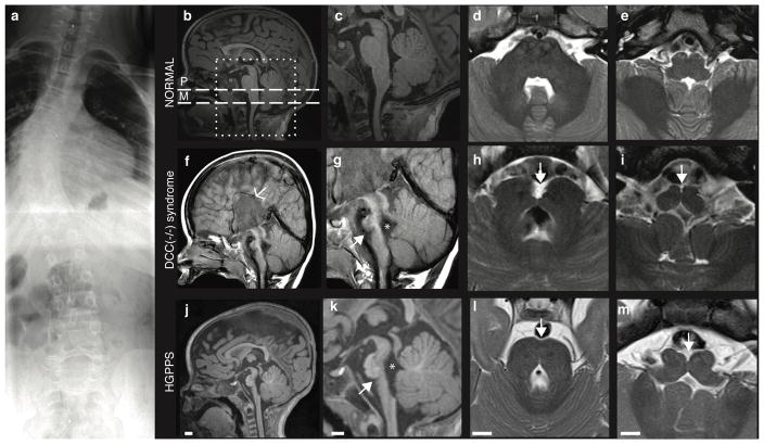
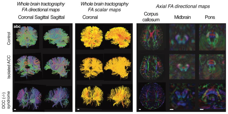
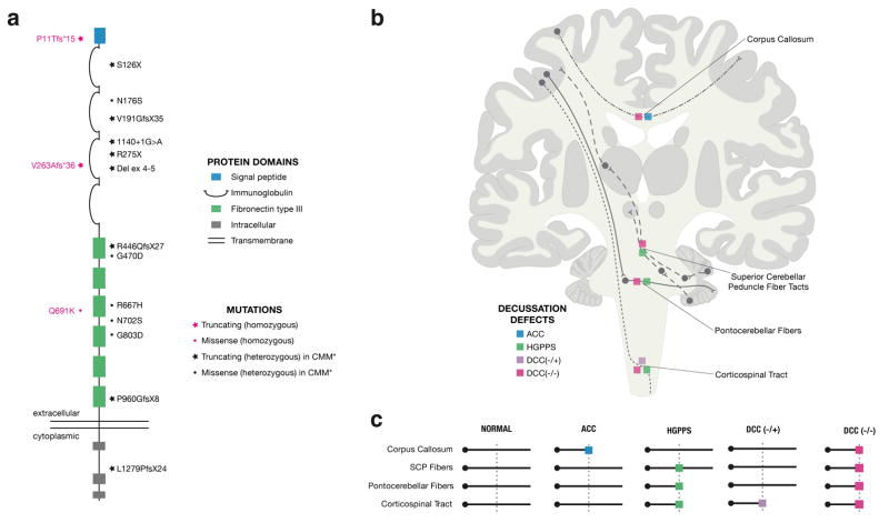
Motor, sensory, and integrative activities of the brain are coordinated by a series of midline-bridging neuronal commissures whose development is tightly regulated. Here we report a new human syndrome in which these commissures are widely disrupted, thus causing clinical manifestations of horizontal gaze palsy, scoliosis, and intellectual disability. Affected individuals were found to possess biallelic loss-of-function mutations in the gene encoding the axon-guidance receptor 'deleted in colorectal carcinoma' (DCC), which has been implicated in congenital mirror movements when it is mutated in the heterozygous state but whose biallelic loss-of-function human phenotype has not been reported. Structural MRI and diffusion tractography demonstrated broad disorganization of white-matter tracts throughout the human central nervous system (CNS), including loss of all commissural tracts at multiple levels of the neuraxis. Combined with data from animal models, these findings show that DCC is a master regulator of midline crossing and development of white-matter projections throughout the human CNS.


Conflict of interest statement

COMPETING FINANCIAL INTEREST

The authors disclose that T.W.Y. is co-founder of Claritas Genomics, a gene diagnostic and genomic medicine company, and S.S.J. is co-founder of Global Gene Corporation Pte Ltd, a gene diagnostic company; however, T.W.Y. and S.S.J. declare no competing financial interests related to the publication of this work. The remaining authors do not have any financial conflict of interest that might be construed to influence the results or interpretation of this work.

Figures

Figure 1
Figure 1. Radiographic features of the DCC (−/−) syndrome
(a) Spine film from individual II:7 from Family 1 shows thoracolumbar scoliosis. (b–m) MRI features of affected individual II:2 from Family 2 compared to age matched control and individual with horizontal gaze palsy and progressive scoliosis (HGPPS). (b,f,j) Sagittal images showing agenesis of corpus callosum (arrow) and absent anterior commissure in the affected individual but intact corpus callosum in control and HGPPS individuals. (c,g,k) Zoomed in version (box from Figure 1b) of the sagittal image shows the brainstem abnormalities. The 4th ventricle is enlarged (*). The pons is hypoplastic (arrow) in both affected and HGPPS individuals, but especially in DCC(−/−) individuals, with associated elongation of the midbrain and medulla. All are dysmorphic, in particular the pons and midbrain with midline cleft, compared to control. (d,h,l) Axial images through pons (dashed line “P” from Figure 1b) show hypoplasia of the pons and middle cerebellar peduncle and the cleft in the brainstem in the affected and HGPPS individuals (arrow). In addition, the anterior surface of the pons appears irregular (e,i,m) Axial images through medulla (dashed line “M” from Figure 1b) show hypoplasia and “butterfly” appearance of the medulla in the affected and HGPPS individuals (arrow). Scale bars= 1 cm.
Figure 2
Figure 2. Mapping and identification of mutations in DCC
(a–c) Pedigrees of Families 1 (a), 2 (b), and 3 (c). Asterisks indicate family members whose DNA was available (d) Sequencing of the junction PCR products in Family 1 confirmed a 7682 bp intragenic deletion (in red; chr18:49,867,184–49,874,866, hg19 coordinates) that is predicted to result in deletion of 21 amino acids (in grey bar) from the DCC protein, including part of the signal peptide, the Ig-like C2-type 1 domain and the exon 1- intron 1 splice junction. The predicted amino acid sequence also leads to a premature stop codon 15 amino acids downstream from the first changed amino acid. gDNA= genomic DNA, AA= amino acid. M (in blue) indicates translation start site for the predominant isoform of DCC (NM_005215.3). (e) Sanger traces from DCC sequencing in Family 2 revealed homozygous out-of-frame 7 bp deletion in exon 4 (c.788_794delTTTCTGG) in the proband, and heterozygous deletion in the parents. (f) Sanger trace from DCC sequencing in Family 3 revealed a homozygous missense variant NM_005215.3: c.2071C>A; p.Gln691Lys. (g) The residue glutamine (Gln) at position 691 is a highly conserved residue in all species down to C. elegans, with the exception of frazzled in D. melanogaster, which is known to be significantly divergent from other orthologs. (h) Schematic of DCC exon-intron structure with location of the mutations in the three families reported.
Figure 3
Figure 3. Axon guidance defects in patients with DCC (−/−) syndrome
(a,b) Whole brain diffusion tensor tractography (DTT) of control individual, individual with isolated agenesis of the corpus callosum, and individual with DCC (−/−) syndrome. (a) Directional maps tracts are pseudocolored to visualize the directions of anisotropic diffusion. Green tracts are going in an anterior posterior direction, red tracts are commissural tracts (left to right), and blue tracts visualize inferior to superior tract directions. Both individuals with ACC and DCC(−/−) demonstrate absence of normal interhemispheric commissural fibers. In addition, the DCC(−/−) brain exhibits a paucity of reconstructable tracts. (b) Color scalar map is a fractional anisotropy (FA) map, where the scale is a fractional direction scale with units from 0–1. “0” (yellow colored fibers) means the diffusion direction is isotropic, and fibers that are yellow show isotropic diffusion. “1” (red colored fibers) indicate anisotropic diffusion and visualizes fibers where the diffusion is anisotropic. Fibers in the DCC(−/−) brain exhibit lower anisotropy and the organization of fibers in general appears disorganized as compared to the control and isolated ACC individuals. (c) Axial FA directional maps of human brain at the levels of the corpus callosum, midbrain, and pons. Both ACC and DCC(−/−) brains lack normal crossing fibers (red). In addition, in control and ACC individuals, midbrain color FA maps demonstrate a distinctive “red dot” sign denoting midline decussation of fibers of the superior cerebellar peduncle which are missing from the DCC(−/−) brain. Similarly, in the pons, control and ACC individuals exhibit distinct crossing fibers representing midline crossing pontocerebellar fibers; these fibers, too, are missing in the DCC(−/−) individual. Scale bars= 1 cm.
Figure 4
Figure 4. Expression patterns of DCC and Netrin-1 in developing brain
In situ hybridization patterns in E11.5, E13.5, E15.5 and E18.5 mice showing robust early expression in specific, yet largely complementary cell populations in the developing telencephalon, midbrain and hindbrain. DCC is particularly highly expressed at early stages in developing neurons of the cortical plate and brainstem nuclei; DCC expression subsequently tapers off with age. Similarly, Ntn expression is highest in midline structures of the telencephalic expansion and the ventral floor of the developing third and fourth ventricle; its expression also becomes much broader as development proceeds. Tel= telencephalon, Mb= midbrain, Hb= hindbrain, Sp= subpallium, III= third ventricle, IV= fourth ventricle, EGL= external granule layer of cerebellum.
Figure 5
Figure 5. Genotype-phenotype correlations in DCC and other related disorders of midline axon guidance
(a) Structure of the DCC protein and location of the DCC mutations identified in our report (in magenta) and in congenital mirror movements (in black). DCC encodes a transmembrane receptor with four immunoglobulin and six fibronectin type III domains, and three conserved cytoplasmic domains. In Family 1, homozygous loss of exon 1 results in a deletion of the signal peptide, as well as the first immunoglobulin-like C2 domain, and is predicted to lead to a nonfunctional protein. In Family 2, homozygous frameshift in exon 4 is predicted to lead to a nonfunctional protein. In Family 3, the missense mutation (Gln691Lys) affects the third fibronectin type III domain. Individuals with congenital mirror movements have truncating heterozygous mutations in DCC; however, these individuals have normal brain MRI and normal intellect. (b,c) Major tract abnormalities in the DCC (−/−) syndrome and other human disorders of axon guidance: Anatomical schematic of the ACC (blue box) vs. HGPPS (green box) vs. DCC (−/+) (purple box) vs. DCC (−/−) (magenta box) mutant phenotypes (C) and summary schematic (D) depicting disruptions in different commissural tracts in each disorder. In ACC, only supratentorial tracts are affected, while HGPPS affects only infratentorial tracts. In DCC (−/+), corticospinal tracts are functionally disrupted. In DCC (−/−), midline crossing along the entire length of the neuraxis is disrupted. ACC= agenesis of corpus callosum, HGPPS= horizontal gaze palsy with progressive scoliosis.

References

    1. Edwards TJ, Sherr EH, Barkovich AJ, Richards LJ. Clinical, genetic and imaging findings identify new causes for corpus callosum development syndromes. Brain. 2014;137:1579–613. - PMC - PubMed
    1. Izzi L, Charron F. Midline axon guidance and human genetic disorders. Clinical genetics. 2011;80:226–34. - PubMed
    1. Nugent AA, Kolpak AL, Engle EC. Human disorders of axon guidance. Current opinion in neurobiology. 2012;22:837–43. - PMC - PubMed
    1. Van Battum EY, Brignani S, Pasterkamp RJ. Axon guidance proteins in neurological disorders. The Lancet Neurology. 2015;14:532–46. - PubMed
    1. Jen JC, et al. Mutations in a human ROBO gene disrupt hindbrain axon pathway crossing and morphogenesis. Science. 2004;304:1509–13. - PMC - PubMed

Substances