

FDG Whole-Body PET/MRI in Oncology: a Systematic Review
- PMID: 28250855
- PMCID: PMC5313459
- DOI: 10.1007/s13139-016-0411-3
FDG Whole-Body PET/MRI in Oncology: a Systematic Review
Abstract
The recent advance in hybrid imaging techniques enables offering simultaneous positron emission tomography (PET)/magnetic resonance imaging (MRI) in various clinical fields. 18F-fluorodeoxyglucose (FDG) PET has been widely used for diagnosis and evaluation of oncologic patients. The growing evidence from research and clinical experiences demonstrated that PET/MRI with FDG can provide comparable or superior diagnostic performance more than conventional radiological imaging such as computed tomography (CT), MRI or PET/CT in various cancers. Combined analysis using structural information and functional/molecular information of tumors can draw additional diagnostic information based on PET/MRI. Further studies including determination of the diagnostic efficacy, optimizing the examination protocol, and analysis of the hybrid imaging results is necessary for extending the FDG PET/MRI application in clinical oncology.
Keywords: FDG; Oncology; PET/MRI; Positron emission tomography/magnetic resonance imaging.
Conflict of interest statement
Conflict of Interest
Hyun Woo Kwon, Ann-Katharina Becker, Jin Mo Goo, and Gi Jeong Cheon declare that they have no conflict of interest.
Ethical Statement
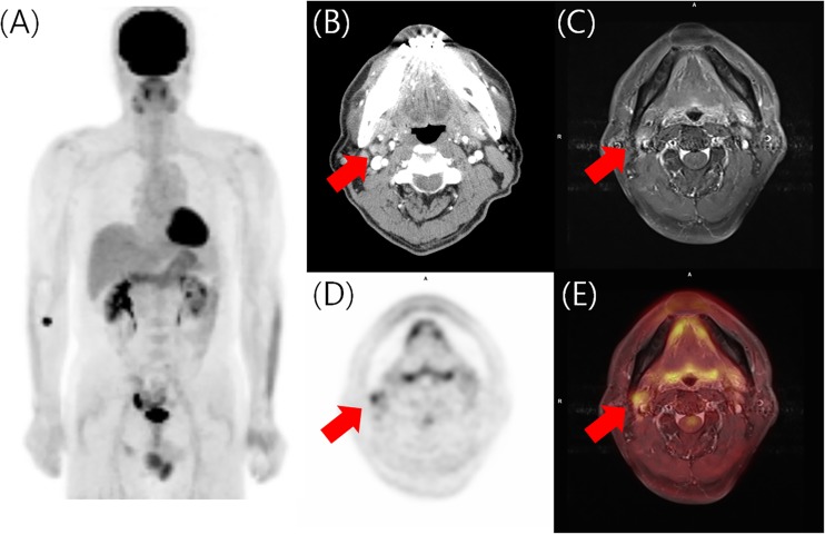
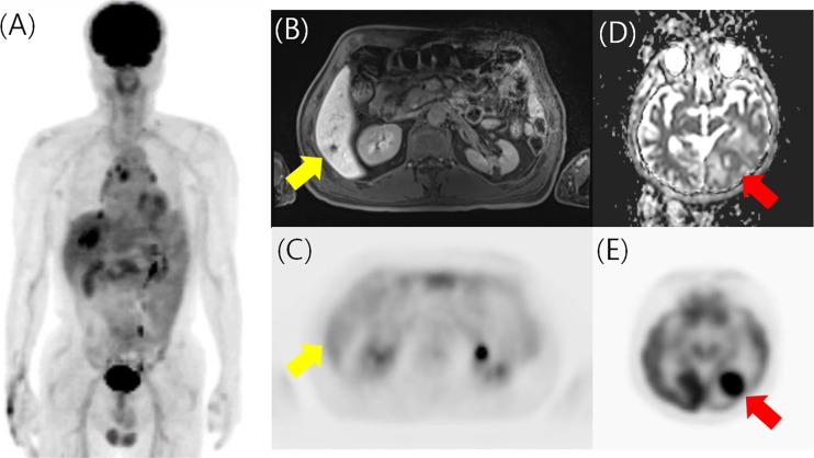
This paper dose not contains any patient information and only includes a representative case originated from previous research approved by the Institutional Review Board of our hospital (1306-055-495). The manuscript has not been published before, or is not under consideration for publication anywhere else. This manuscript has been approved by all co-authors.
Figures
References
-
- Wiesmuller M, Quick HH, Navalpakkam B, Lell MM, Uder M, Ritt P, et al. Comparison of lesion detection and quantitation of tracer uptake between PET from a simultaneously acquiring whole-body PET/MR hybrid scanner and PET from PET/CT. Eur J Nucl Med Mol Imaging. 2013;40:12–21. doi: 10.1007/s00259-012-2249-y. - DOI - PubMed
-
- Pace L, Nicolai E, Luongo A, Aiello M, Catalano OA, Soricelli A, et al. Comparison of whole-body PET/CT and PET/MRI in breast cancer patients: lesion detection and quantitation of 18F-deoxyglucose uptake in lesions and in normal organ tissues. Eur J Radiol. 2014;83:289–96. doi: 10.1016/j.ejrad.2013.11.002. - DOI - PubMed
Publication types
LinkOut - more resources
Full Text Sources
Other Literature Sources