Myocardial Bridge and Acute Plaque Rupture
- PMID: 28251167
- PMCID: PMC5317013
- DOI: 10.1177/2324709616680227
Myocardial Bridge and Acute Plaque Rupture
Abstract
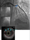
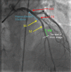
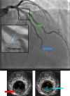
A myocardial bridge (MB) is a common anatomic variant, most frequently located in the left anterior descending coronary artery, where a portion of the coronary artery is covered by myocardium. Importantly, MBs are known to result in a proximal atherosclerotic lesion. It has recently been postulated that these lesions predispose patients to acute coronary events, even in cases of otherwise low-risk patients. One such mechanism may involve acute plaque rupture. In this article, we report 2 cases of patients with MBs who presented with acute coronary syndromes despite having low cardiovascular risk. Their presentation was life-risking and both were treated urgently and studied with coronary angiographies and intravascular ultrasound. This latter modality confirmed a rupture of an atherosclerotic plaque proximal to the MB as a likely cause of the acute events. These cases, of unexplained acute coronary syndrome in low-risk patients, raise the question of alternative processes leading to the event and the role MB play as an underlying cause of ruptured plaques. In some cases, an active investigation for this entity may be warranted, due to the prognostic implications of the different therapeutic modalities, should an MB be discovered.
Keywords: acute coronary syndrome; intravascular ultrasound; myocardial bridge.
Conflict of interest statement
Declaration of Conflicting Interests: The author(s) declared no potential conflicts of interest with respect to the research, authorship, and/or publication of this article.
Figures


References
-
- Ishii T, Asuwa N, Masuda S, Ishikawa Y. The effect of a myocardial bridge on coronary atherosclerosis and ischaemia. J Pathol. 1998;185:4-9. - PubMed
-
- Angelini P, Trivellato M, Donis J, Leachman RD. Myocardial bridges: a review. Prog Cardiovasc Dis. 1983;26:75-88. - PubMed
-
- Ishikawa Y, Akasaka Y, Ito K, et al. Significance of anatomical properties of myocardial bridge on atherosclerosis evolution in the left anterior descending coronary artery. Atherosclerosis. 2006;186:380-389. - PubMed
-
- Ishikawa Y, Akasaka Y, Suzuki K, et al. Anatomic properties of myocardial bridge predisposing to myocardial infarction. Circulation. 2009;120:376-383. - PubMed
-
- Ishikawa Y, Akasaka Y, Akishima-Fukasawa Y, et al. Histopathologic profiles of coronary atherosclerosis by myocardial bridge underlying myocardial infarction. Atherosclerosis. 2013;226:118-123. - PubMed
LinkOut - more resources
Full Text Sources
Other Literature Sources
Miscellaneous

