. 2016 Oct 10;2(10):1710-1721.
doi: 10.1021/acsbiomaterials.6b00246. Epub 2016 Jun 17.

Bioprinting the Cancer Microenvironment

Affiliations

Bioprinting the Cancer Microenvironment

Yu Shrike Zhang et al. ACS Biomater Sci Eng. .

Abstract

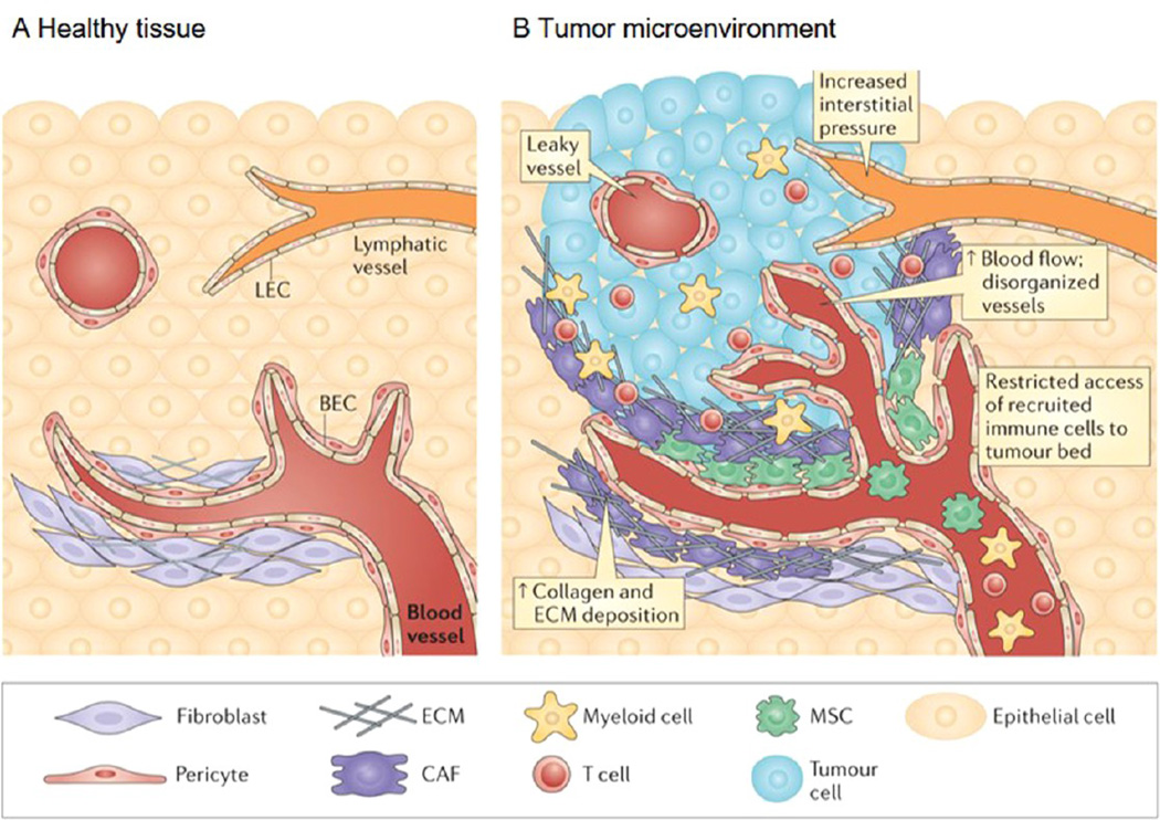
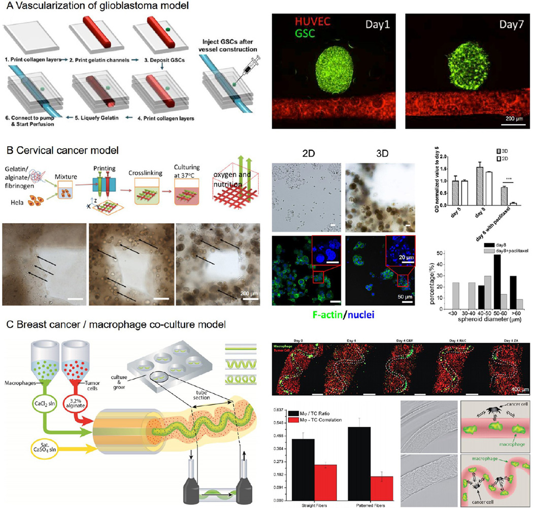
Cancer is intrinsically complex, comprising both heterogeneous cellular compositions and microenvironmental cues. During the various stages of cancer initiation, development, and metastasis, cell-cell interactions (involving vascular and immune cells besides cancerous cells) as well as cell-extracellular matrix (ECM) interactions (e.g., alteration in stiffness and composition of the surrounding matrix) play major roles. Conventional cancer models both two- and three-dimensional (2D and 3D) present numerous limitations as they lack good vascularization and cannot mimic the complexity of tumors, thereby restricting their use as biomimetic models for applications such as drug screening and fundamental cancer biology studies. Bioprinting as an emerging biofabrication platform enables the creation of high-resolution 3D structures and has been extensively used in the past decade to model multiple organs and diseases. More recently, this versatile technique has further found its application in studying cancer genesis, growth, metastasis, and drug responses through creation of accurate models that recreate the complexity of the cancer microenvironment. In this review we will focus first on cancer biology and limitations with current cancer models. We then detail the current bioprinting strategies including the selection of bioinks for capturing the properties of the tumor matrices, after which we discuss bioprinting of vascular structures that are critical toward construction of complex 3D cancer organoids. We finally conclude with current literature on bioprinted cancer models and propose future perspectives.

Keywords: bioprinting; cancer biology; cancer model; drug screening; vascularization.


Figures

Figure 1
Figure 1
Healthy tissue and the tumor microenvironment. (A) Stromal cells present in interstitial spaces surrounding the parenchyma of various organs promote tissue integrity by providing growth factors and structural support. Blood endothelial cells (BECs) and pericytes maintain the integrity of blood vessels and ensure the supply of oxygen and other nutrients to the tissue. Lymphatic vessels composed of lymphatic endothelial cells (LECs) drain interstitial fluid. Fibroblasts are constantly remodeling the ECM to cope with mechanical stress within connective tissue. (B) Neoplastic transformation is often accompanied by the formation of a tumor bed and profound alterations in the surrounding connective tissue and stroma, a process that culminates in the establishment of a pathological tumor microenvironment. An imbalance between pro- and antiangiogenic factors results in the formation of aberrant vasculature, characterized by numerous leaky blood vessels. Increased interstitial pressure and inadequate drainage by the lymphatic vessels is also observed. The increased hydrostatic load, along with tumor-secreted molecules (not shown), induces the recruitment of circulating mesenchymal stem cells (MSCs), the activation of cancer-associated fibroblasts (CAFs) and a marked accumulation of ECM. Finally, various chemokines and cytokines in the tumor microenvironment attract activated T cells and myeloid cells to the tumor lesion, but tortuous blood vessels and dense ECM often hinder their access to the tumor nest. Although the makeup of the cellular and extracellular milieu can differ between tumor types and stages of growth, it is becoming clear that changes in the cellular architecture of the tumor microenvironment can influence tumor growth, metastasis, and drug resistance. Adapted with permission from ref 10. Copyright 2015 Nature Publishing Group.
Figure 2
Figure 2. Conventional cancer models
(A) Schematic illustration of cancer models based on (i) 2D monolayer (co)cultures, (ii) spheroid 3D (co)cultures derived from microwells, hanging drops, and spinner flask methods, (iii) hydrogel-embedded 3D (co)cultures, and (iv) porous scaffold-enabled 3D (co)cultures. Adapted with permission from ref 28. Copyright 2014 Wiley–VCH. (B) Multicellular tumor spheroids composed of A375 human melanoma cells were characterized by hematoxylin and eosin (H&E), Mason’s trichrome, and Ki67 staining. Adapted with permission from ref 31. Copyright 2014 Nature Publishing Group. (C) (i) Matrigel-embedded 3D OSCC-3 human oral cancer model stained for Ki67 (red) and β-catenin (green), or laminin V (red) and caspase-3 (green); and (ii) PLG porous scaffold-based 3D OSCC-3 human oral cancer model stained by H&E and hydroxyprobe. (D) Comparisons of tumor growth, angiogenic factor secretion, and vascularization in 3D Matrigel-embedded model, PLG scaffold-based model, and in vivo. Adapted with permission from ref 38. Copyright 2007 Nature Publishing Group.
Figure 3
Figure 3. Common bioprinting modalities
(A) Thermal inkjet printers electrically heat the printhead to produce air-pressure pulses that force droplets from the nozzle, whereas piezoelectric printers utilize pulses formed by piezoelectric actuation for droplet ejection. (B) Microextrusion bioprinters use pneumatic or mechanical (piston or screw) dispensing systems to extrude continuous bioinks. (C) Laser-assisted bioprinters use laser focused on an absorbing substrate to generate localized pressures that propel cell-containing bioink onto a collector substrate. Adapted with permission from ref 51. Copyright 2014 Nature Publishing Group. (D) Schematic of the stereolithography-based 3D bioprinter. A frame is used to support the custom precision translation stages and projector system; light from a UV laser then illuminates a digital micromirror device (DMD) projector; a lens is adopted to project the image of the DMD pattern onto the bioink for layer-by-layer cross-linking, which along with vertical movement of the stage generates 3D bioprinted structures. Adapted with permission from ref 65. Copyright 2015 Nature Publishing Group.
Figure 4
Figure 4. Examples of bioprinted vascular structures
(A) Sacrificial bioprinting by templating against an open, interconnected, self-supporting carbohydrate glass lattice, where endothelialization of channel walls and formation of intervessel junctions could be achieved. The carbohydrate template was dissolved in culture medium. Adapted with permission from ref 95. Copyright 2012 Nature Publishing Group. (B) Sacrificial bioprinting by templating against pluronic. A heterogeneous engineered tissue construct was produced, in which blue, red, and green filaments corresponded to bioprinted 10T1/2 fibroblast (blue), human neonatal dermal fibroblast cells (green), and HUVECs (red). The pluronic template was liquefied at <4 °C and removed by vacuum suction. Adapted with permission from ref 85. Copyright 2014 Wiley–VCH. (C) Sacrificial bioprinting by templating against gelatin, where endothelial sprouting into the surrounding matrix was shown to occur. The gelatin template was liquefied at <37 °C and removed spontaneously. Adapted with permission from ref 97. Copyright 2014 Springer. (D) Sacrificial bioprinting by templating against agarose, where the channels could be subsequently endothelialized. The solidified agarose template was removed by pulling or vacuum suction. Adapted with permission from ref 96. Copyright 2014 Royal Society of Chemistry. (E) Schematic showing a microscale capillary tip sweeping out a complex pattern as the bioink was injected into the granular shear-thinning gel medium. Freeform vascular patterns could be generated using this embedded bioprinting approach. Top image dapted with permission from ref 98. Copyright 2015 American Association for the Advancement of Science. Bottom image adapted with permission from ref 82. Copyright 2015 Wiley–VCH.
Figure 5
Figure 5. Cancer bioprinting
(A) Construction of a glioblastoma vascular niche by injecting a spheroid of glioma stem cells (green) besides a sacrificially bioprinted perfusable vascular channel (red). Adapted with permission from ref 102. Copyright 2015 Institute of Electrical and Electronics Engineers. (B) Fabrication of a cervical cancer model by direct 3D bioprinting of Hela cell-laden bioink. The cells proliferated and clustered (black arrows) within the bioprinted microfibrous structure. The bioprinted 3D cervical cancer model showed drug-induced toxicity at higher resistance than the 2D cultures. Adapted with permission from ref 103. Copyright 2014 IOPscience. (C) Bioprinting of a breast cancer/ macrophage coculture model, where the relative geometry of the two cell populations was shown to affect the tumor-immune cell interactions. Adapted with permission from ref 106. Copyright 2015 Wiley–VCH.
Figure 6
Figure 6. Multimaterial bioprinting
(A) Extrusion-based multimaterial bioprinting system capable of depositing multiple bioinks along with polymeric scaffold for fabrication of large-scale vascularized functional tissue constructs. Adapted with permission from ref 110. Copyright 2016 Nature Publishing Group. (B) Multimaterial DMD stereolithography bioprinting system capable of high-resolution microscale patterning of the complex tissue microarchitecture to achieve functionality. Adapted with permission from ref 61. Copyright 2016 the National Academy of Sciences of the United States.

Similar articles

Cited by

References

    1. Stewart BW, Wild CP, editors. World Cancer Report 2014. International Agency for Research on Cancer. Lyon, France: World Health Organization; 2014.
    1. Harris AL. Hypoxia—a key regulatory factor in tumour growth. Nature Reviews Cancer. 2002;2:38–47. - PubMed
    1. Denko NC. Hypoxia, HIF1 and glucose metabolism in the solid tumour. Nat. Rev. Cancer. 2008;8:705–713. - PubMed
    1. Brown JM, Wilson W. R Exploiting tumour hypoxia in cancer treatment. Nat. Rev. Cancer. 2004;4:437–447. - PubMed
    1. Hanahan D, Weinberg RA. Hallmarks of cancer: the next generation. Cell. 2011;144:646–674. - PubMed