. 2017 Mar 14;114(11):E2106-E2115.
doi: 10.1073/pnas.1612444114. Epub 2017 Mar 1.

Structure of human IFIT1 with capped RNA reveals adaptable mRNA binding and mechanisms for sensing N1 and N2 ribose 2'-O methylations

Affiliations

Structure of human IFIT1 with capped RNA reveals adaptable mRNA binding and mechanisms for sensing N1 and N2 ribose 2'-O methylations

Yazan M Abbas et al. Proc Natl Acad Sci U S A. .

Abstract

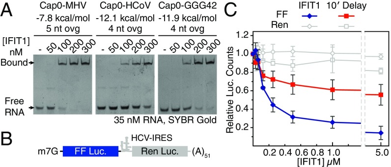
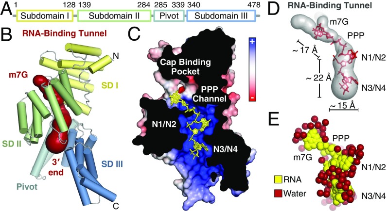
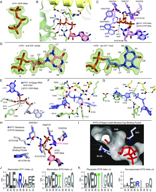
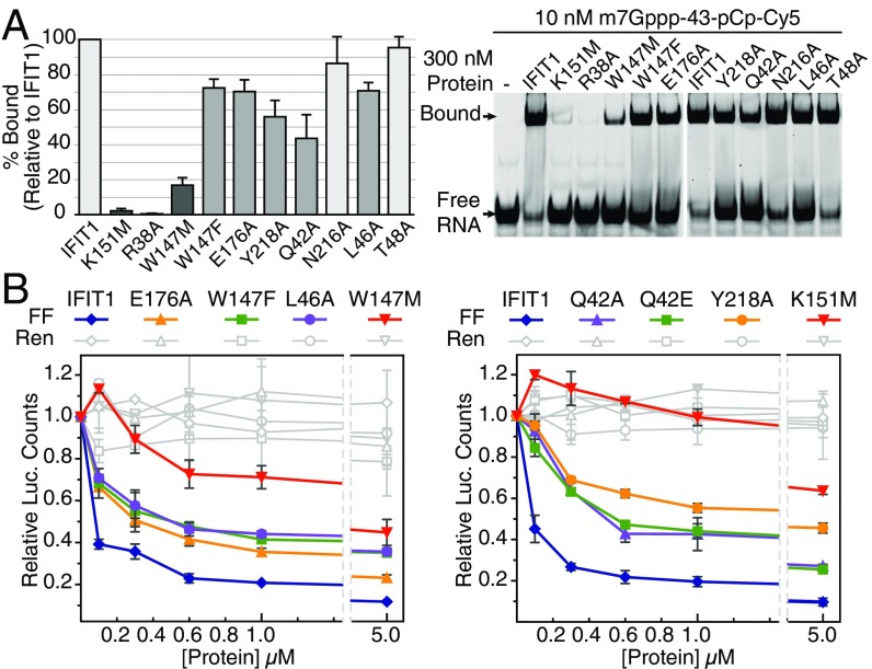
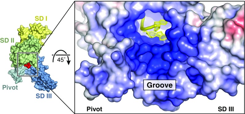
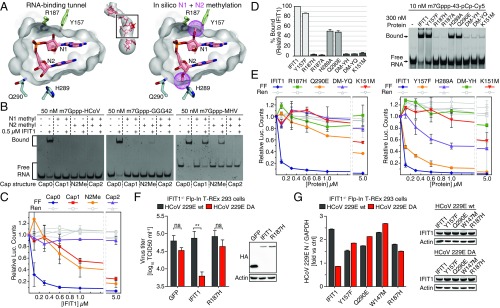
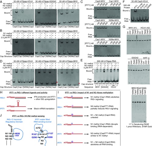
IFIT1 (IFN-induced protein with tetratricopeptide repeats-1) is an effector of the host innate immune antiviral response that prevents propagation of virus infection by selectively inhibiting translation of viral mRNA. It relies on its ability to compete with the translation initiation factor eIF4F to specifically recognize foreign capped mRNAs, while remaining inactive against host mRNAs marked by ribose 2'-O methylation at the first cap-proximal nucleotide (N1). We report here several crystal structures of RNA-bound human IFIT1, including a 1.6-Å complex with capped RNA. IFIT1 forms a water-filled, positively charged RNA-binding tunnel with a separate hydrophobic extension that unexpectedly engages the cap in multiple conformations (syn and anti) giving rise to a relatively plastic and nonspecific mode of binding, in stark contrast to eIF4E. Cap-proximal nucleotides encircled by the tunnel provide affinity to compete with eIF4F while allowing IFIT1 to select against N1 methylated mRNA. Gel-shift binding assays confirm that N1 methylation interferes with IFIT1 binding, but in an RNA-dependent manner, whereas translation assays reveal that N1 methylation alone is not sufficient to prevent mRNA recognition at high IFIT1 concentrations. Structural and functional analysis show that 2'-O methylation at N2, another abundant mRNA modification, is also detrimental for RNA binding, thus revealing a potentially synergistic role for it in self- versus nonself-mRNA discernment. Finally, structure-guided mutational analysis confirms the importance of RNA binding for IFIT1 restriction of a human coronavirus mutant lacking viral N1 methylation. Our structural and biochemical analysis sheds new light on the molecular basis for IFIT1 translational inhibition of capped viral RNA.

Keywords: 2′-O methylation; IFIT1 crystal structure; innate immunity; mRNA cap recognition; self vs. nonself.


Conflict of interest statement

The authors declare no conflict of interest.

Figures

Fig. S1.
Fig. S1.
RNA binding and inhibition of in vitro translation. (A) Schematic of mRNA cap structure. Note that italics are used to distinguish m7G atoms from the cap-proximal nucleotides N1 and N2. (B) Predicted secondary structure and thermodynamic stability for each RNA determined by the Mfold web server. MHV is predicted to have a 5-nt overhang whereas HCoV is predicted to have only a 4-nt overhang. Also, MHV is predicted to have a less stable hairpin than HCoV [compare their respective minimum free energy (ΔG), more negative indicates greater stability]. (C) EMSA between IFIT1 and OH- or PPP-RNA. PPP-ss44 is a previously characterized RNA, which binds IFIT1 in its PPP- form, likely because it lacks any 5′- secondary structure. PPP-MHV also binds to IFIT1, but PPP-HCoV and PPP-GGG42 do not. (D) Translation assay with IFIT5. At 5 µM, the inhibition of FF translation is likely because of weak cap binding or nonspecific RNA binding. Data represent the mean of two independent measurements performed in duplicate ± SD.
Fig. 1.
Fig. 1.
RNA binding and inhibition of in vitro translation by human IFIT1. (A) EMSAs between human IFIT1 and capped-RNA visualized by SYBR Gold staining. Cap0-MHV, first 41 nt of MHV strain A59; Cap0-HCoV, first 42 nt of HCoV strain 229E; Cap0-GGG42, ACU to GGG modification of HCoV. The RNA secondary structure minimum free energy (kcal/mol) and 5′-overhang length (ovg) are indicated (see also Fig. S1B). (B) Schematic of bicistronic mRNA reporter. (C) Translation assay with IFIT1 titrated into Krebs extracts programmed with Cap0/m7Gppp reporter, and titration following a 10-min preincubation of the reporter with extracts. FF and Ren luciferase (luc.) activities at each concentration were normalized against buffer control, which was set to 1. Data represent the mean of two independent measurements performed in duplicate ± SD.
Fig. 2.
Fig. 2.
Overall structure of monomeric, RNA-bound human IFIT1. (A) Schematic of IFIT1 subdomains. (B) Cartoon representation of human IFIT1 colored by subdomain (SD) and surface representation of the tunnel (dark red) determined by CAVER (50). (C) Cross-section of IFIT1 colored by surface electrostatic potential from negative (−10 kTe−1; red) to positive (+10 kTe−1; blue) with capped-RNA (yellow sticks). (D) Dimensions of the IFIT1 tunnel (gray surface) and capped RNA (red sticks). (E) Waters surrounding the RNA inside the tunnel.
Fig. S2.
Fig. S2.
Overall structure of monomeric, RNA-bound human IFIT1. (A) Secondary structure, TPR motif, and subdomain organization of human IFIT1, with TPR motif sequence numbers above. Note that because of an insertion between α9 and α10, TPR4 was annotated based on sequence and structure rather than sequence alone. All other TPR motifs were predicted based on sequence using TPRpred (https://toolkit.tuebingen.mpg.de/tprpred). (B) Overall structure of monomeric human IFIT1 with subdomains colored according to A. The non-TPR helices α1 and α2 are bridged by loop L1, which houses a highly conserved CHFTW motif (residues 19–23) (see Fig. S5) that mediates contacts with α7. Subdomains I and II are linked by loop L2. The two non-TPR pivot helices (α15/α16) connect subdomains II and III. (C) TPR4 (α9/α10) has an unusually long intra-TPR loop (purple) which forms a lid over the 3′ exit of the RNA-binding tunnel. Additionally, it mediates contacts between subdomains II and III (indicated by purple/blue dashes). The sequence of this loop is not conserved, but the insertion is found in almost all IFIT1- and IFIT5-like proteins. The double-headed arrow indicates putative subdomain III motions between RNA-free and RNA-bound forms of IFIT1, based on the structural analysis of human IFIT5. (D) Superposition of RNA-bound IFIT1 over RNA-bound IFIT5 (PDB ID code 4HOR). The two structures share a high degree of global similarity. The C-terminal regions appear to diverge, but that is because of intersubdomain angle differences between IFIT1 and IFIT5. (E) Superposition of RNA bound IFIT1 over the structure of RNA free, N-terminal IFIT1 (PDB ID code 4HOU). In the partial structure of RNA-free, N-terminal IFIT1, the cap-binding loop (blue) adopts a different conformation than in full-length (yellow; see also F). However, in one of the two molecules of the asymmetric unit (of 4HOU), this loop has high B-factors (loop average 61 Å2, protein average 46 Å2), whereas in the other it is disordered, indicating that this loop is mobile in the absence of RNA. The TPR4 loop (green) is also disordered in the structure of N-terminal IFIT1 (4HOU), likely because of the lack of RNA and missing subdomain contacts. (F) The cap-binding pocket is formed at the interface of subdomains I and II, with one wall formed by helix α2 and the cap-binding loop (connecting α2 and α3), and the other by subdomain II.
Fig. 3.
Fig. 3.
IFIT1 mRNA cap-binding mechanism. (A) The IFIT1 PPP (blue) adopts an extended conformation compared with the “bent” IFIT5 PPP (pink). The γ-phosphate from PPP–RNA-bound IFIT1 points toward the nearby unoccupied cap-binding pocket. (B) Protein cross-section and close-up the cap-binding pocket. This view is rotated by ∼180° compared with Fig. 2C. (C) Simulated annealing 2FoFc omit map of the m7Gppp-moiety contoured at 1σ. Syn- and anti-configuration carbons are colored light blue and salmon, respectively. (D) Surface/stick representation of residues (colored by subdomain) abutting the m7G base moiety from above and below. The interplanar distance between m7G and Trp-147 is 3.4–3.7 Å. (E and F) Cartoon/stick representation of residues interacting with the m7Gppp-moiety. See also Movie S1. Shown are both conformations of the m7GpppA dinucleotide, which was modeled as a single residue during model building and refinement. (G) Cross-sections of the IFIT1 and IFIT5 RNA-binding tunnels (gray/black) with m7Gppp- or PPP-RNA (red sticks). The asterisk (*) shows where K48 and Q41 block the IFIT5 putative cap-binding pocket (Fig. S3 HK).
Fig. S3.
Fig. S3.
IFIT1 PPP- and m7Gppp- binding mechanism. (A) Simulated annealing 2FoFc omit map of the PPP moiety contoured at 1σ. (B) Cartoon/stick representation of residues making specific contacts with the triphosphate group from PPP-RNA–bound IFIT1. (C) Superposition of PPP-RNA bound IFIT1 residues (blue carbons with orange phosphates) over PPP-RNA–bound IFIT5 residues (pink carbons with yellow-orange phosphates). The metal from PPP-RNA–bound IFIT5 is represented with a magenta sphere. The PPP from IFIT1 is in a different conformation, and makes additional H-bonds with Y256, Y218, and R187 not present in IFIT5. R38/D34 from IFIT1 are replaced with T37/E33 in IFIT5. Depending on the region, IFIT5 residue positions are offset by −1 or −2 compared with IFIT1 residue positions. (D) Simulated annealing 2FoFc omit map of the m7Gppp moiety contoured at 1σ. Syn- and anti-configuration carbons are colored light blue and salmon, respectively. The hydrogen bond between the β-phosphate and the 3′-OH is indicated with the dashed line. Because of the constraints imposed on the base and bridging PPP binding, the ribose sugar pucker adapts by switching from a C2′-endo conformation in the anti-configuration, to C1′-exo in the syn-configuration. The final refined occupancies are ∼0.5 for each conformation. (E) Superposition of PPP-RNA bound IFIT1 (gray) over m7Gppp-RNA bound IFIT1 (green side-chain carbons and blue RNA carbons). The extended PPP conformation is common to both PPP- and m7Gppp-RNA binding, except that the γ-phosphate of m7Gppp- is repositioned at the entrance of the cap-binding pocket, away from Y256 and toward Y218, for more optimal m7G binding inside the pocket. (F) Water-mediated hydrogen bonds between IFIT1 and the m7Gppp moiety. Starting with the water at Q42 (*) and going clockwise, the isotropic B-factors of these waters are 27.6, 18.2, 17.6, 18.0, 19.4, 24.1, 15.3, 17.8 Å2, most of which are lower than the crystal isotropic B factor average (30.65 Å2). (G) Coordination between residues involved in m7Gppp-RNA binding. R187 is coordinated by Y218 and Y157; R38 by D34; K151 by R38 and Q42; and W147 by E176. (H) Superposition of m7Gppp-RNA–bound IFIT1 over IFIT5 PPP-RNA. In IFIT5, T37 facilitates recognition of a bent/compact PPP conformation that is further stabilized by a metal bound to the α- and γ-phosphates and coordinated by E33. This draws in K150 (which is H-bonded to T37), Q41 (which interacts with the PPP directly), and K48 (which is coordinated by Q41). (I) In IFIT5, K48 sits atop the cap-binding residue to block access to the cap-binding pocket as indicated by the attempt to model m7Gppp. In IFIT1, K49 from its cap-binding loop is facing the solvent. Also, in IFIT1, Q42 is not involved in binding the PPP directly. (J) WebLogo (weblogo.berkeley.edu/) sequence consensus of helix α2 from 86 IFIT1- and IFIT1B-like sequences, and 60 IFIT5-like sequences. (K) The mammalian IFIT5 sequences were split into placentals and nonplacentals (i.e., Tazmanian devil, opossum, platypus). Only nonplacental IFIT5-like sequences have an arginine at position 37 (human IFIT5 numbering).
Fig. 4.
Fig. 4.
IFIT1 can accommodate multiple forms and conformations of the cap. (A) m7G base interactions at its periphery in the anti- or syn-modes of binding. The water H-bonded to Q42 (*) is 3.3 Å away from N3 in the anti-mode. (B, Left) The Gppp-moiety adopts only anti, and approaches N216 to form a weak H-bond through O6; m7Gppp-RNA bound IFIT1 is superposed in gray. (Right) The water structure surrounding the G moiety changes compared with m7G, and satisfies almost all H-bond donor and acceptor groups. The arrows depict the movement of waters from the m7Gppp-bound form (gray circles) to the Gppp-bound form (red spheres). The water molecule H-bonded to Q42 and N3 becomes more ordered in the Gppp-RNA structure (same resolution and crystal form but B-factor decreases from 28 Å2 to 15.3 Å2). The water at N7 (§) appears only in the Gppp-bound form. The water at N1 appears to be in two conformations and was modeled as two water molecules.
Fig. S4.
Fig. S4.
IFIT1 can accommodate multiple forms and conformations of the cap and mutational analysis of cap recognition. (A) Simulated annealing 2FoFc omit map of the Gppp moiety contoured at 1σ. (B) Simulated annealing 2FoFc omit map of m7Gppp moiety bound to monomeric IFIT1 N216A, contoured at 1σ. (C) EMSA between IFIT1 and m7Gppp- or Gppp-RNA. For HCoV, the lack of N7-methyl appears to enhance RNA binding. Whether this is a common feature of all IFIT1-like proteins remains to be validated. (D) EMSA between Gppp-GGG42 and IFIT1, IFIT1 N216D, or IFIT1 N216A. (E) In vitro translation assay with extracts programed with Cap0-, Gppp-, or Appp-capped reporter. Data represent the mean of two independent measurements performed in duplicate ± SD. (F) EMSA between IFIT1 and Appp- or PPP-GGG42 (Left), and between IFIT1 and Gppp- or Appp-GGG42 (Right). (G) Simulated annealing 2FoFc omit map of the PEG molecules inside the cap-binding pocket in PPP-RNA bound IFIT1, contoured at 1σ. The m7G from m7Gppp-RNA bound IFIT1 is superposed in light blue. The PEG molecules interact nonspecifically with residues lining the cap-binding pocket. (H) At physiological pH, the m7G moiety exists in an equilibrium between a cationic Keto tautomer, and a zwitterionic enolate tautomer in which N1 (red atom) is deprotonated. (I) Schematic of residues lining the cap-binding pocket and PPP channel. Residues are colored by subdomain, and red lines indicate hydrogen bonds or salt bridges. (J) EMSA between IFIT1 mutants and PPP-ss44 or OH-ss44. Except for R38A and K151M (which target the bridging triphosphate), all mutants retain binding to PPP-ss44, confirming that the protein fold is not affected by the mutations. Dashed lines indicate lanes that were cropped out. (K) Y50 and F45 from the cap-binding loop are distal from the m7G moiety. F45 from subdomain I packs against E280 and T251 from subdomain II, whereas Y50 stacks against R93, which in turn is salt-bridged to E176. These interactions may be important for subdomain contacts, and/or preorganizing the cap-binding loop. (L) EMSA between pCp-Cy5 labeled m7Gppp-43 and IFIT1, IFIT1 N216D, or IFIT1 Q42E, similar to Fig. 5A. (M) Composite WebLogo sequence consensus from 86 IFIT1- and IFIT1B-like sequences showing residues involved in m7Gppp-RNA binding. See also Fig. S5.
Fig. 5.
Fig. 5.
Functional validation of cap recognition. (A) Mutational analysis of cap-binding residues investigated by fluorescent EMSA with 3′-end–labeled (pCp-Cy5) RNA. (Left) Quantification of percent bound (Upper band, Right) for each mutant normalized against IFIT1. Data represent the mean of three independent measurements ± SD. (B) In vitro translation assays with RNA binding mutants and Cap0 reporter. Data represent the mean of two measurements ± SD.
Fig. S5.
Fig. S5.
ClustalO sequence alignment of select IFIT sequences from humans, mice, and rabbits. Filled triangles indicate residues directly in contact with the RNA. Open triangles are positively charged residues lining the protein surface outside of the tunnel (in the groove). Secondary structure elements are indicated for human IFIT1 (colored by subdomain) and human IFIT5 (PDB ID code 4HOR). This figure was prepared using ESPript (espript.ibcp.fr/ESPript/ESPript/). National Center for Biotechnology Information reference numbers for mIfit1, mIfit1b, and mIfitc are NP_032357.2, NP_444447.1, and NP_001103987.1, respectively.
Fig. 6.
Fig. 6.
Comparison with the canonical cap-binding proteins eIF4E (PDB ID code 1EJ1), CBC (PDB ID code 1H2T), and VP39 (PDB ID code 1V39). In IFIT1, some of the nearby water molecules are shown as transparent red spheres, and T48 adopts two conformations.
Fig. S6.
Fig. S6.
RNA binding mechanism at N1-N4. (A) Interactions between IFIT1 and RNA at N1/N2 and (B) at N3/N4. The protein is oriented such that we are looking along the RNA-binding tunnel from the 3′-exit (similar orientation as Fig. S2C). Note that K336 and the backbone carbonyl of G190 are H-bonded to N2 and N4, respectively. Concerning Fig. 8 and the multiple roles of R187, Y157, H289, and Q290: R187 packs against the ribose of N1, coordinates Y218 and Y157 (Fig. S3G), and interacts with m7G and bridging PPP (Fig. 3 E and F); Y157 contacts the N1 ribose and N1 adenine through van der Waals; Q290 is H-bonded to the 3′ oxygen of N2 and the internucleotide phosphate between N2 and N3; H289 is H-bonded to the 2′-OH of N2, and contacts the ribose of N3 via van der Waals. V372 could potentially clash with methylation at the N6 position of the first adenosine (see Discussion). (C) Simulated annealing 2FoFc omit map of N1/N2 contoured at 1σ. Two views rotated by 180° are shown. The dinucleotide conformation here resembles that found in CpG dinucleotides of Z-form RNA and UUCG tetraloop sequences (39). The defining feature of this rare dinucleotide motif is the antiparallel arrangement of the two riboses (with their respective O4′ atoms pointing toward each other), and a lone pair–π stack between the O4′ atom of N1 and the base of N2. (D) Simulated annealing 2FoFc omit map of N3/N4 contoured at 1σ. N3/N4 adopt standard A-form helical geometry. (E) Hydrogen-bonds between the RNA bases and the waters (red spheres) inside the tunnel. N6 methylation of the first adenosine could disrupt water mediated interactions at the first nucleotide (see Discussion). (F) N1 and N2 adopt C2′-endo and C3′-endo conformations respectively. RNA nucleotides are typically in equilibrium between the two, but generally favor the C3′-endo conformation.
Fig. 7.
Fig. 7.
IFIT1 forms a positively charged, solvent-exposed RNA binding groove. Fig. S5 for residues in this region.
Fig. 8.
Fig. 8.
Sensing of N1 and N2 ribose 2′-O methylation by IFIT1. (A, Left) Cross-section of the IFIT1 tunnel van der Waals surface (gray) and residues predicted to clash with N1 and N2 methylations. (Right) In silico rigid body modeling of N1 and N2 methylations (purple dots). (B) SYBR Gold-stained EMSAs between 0.5 µM IFIT1 and differentially methylated m7Gppp-RNA. The dashed lines demarcate lanes with different cap structures, as indicated by the labels below the gel. Fig. S7 AE for additional gel shifts. (C) Translation assay with differentially methylated reporter mRNA. Data represent the mean of three independent measurements performed in duplicate ± SD. (D) Mutational analysis of 2′-O methyl sensing residues investigated by fluorescent EMSA similar to Fig. 5A. (E) In vitro translation assays with 2′-O methyl sensing mutants and Cap0 reporter. Data represent the mean of two measurements ± SD. (F) Flp-In T-REx 293 IFIT1 knockout cells were cotransfected with expression plasmids for CD13 and either GFP control, wild-type IFIT1, or IFIT1 R187H and infected with HCoV 229E wild-type or HCoV 239E DA. Virus titers in supernatants were determined by TCID50 18 h postinfection. Data represent the mean of three independent experiments ± SD. **P < 0.01 as analyzed by two-way ANOVA with Bonferroni posttest. The Western blots show expression of proteins at the time of infection. (G) Similar to F, except IFIT1 knockout cells were reconstituted with the indicated IFIT1 constructs or GFP, and virus growth determined by quantitative PCR. Data represent the mean fold-change ± SD of triplicate measurements of the viral N-gene signal, relative to GFP control (ctrl). One representative experiment of three is shown. Western blots show protein expression at the end of the experiment.
Fig. S7.
Fig. S7.
IFIT1 senses N1 and N2 ribose 2′-O methylation (A and B) EMSAs between 1 µM or 2.5 µM IFIT1 and differentially methylated m7Gppp-RNA (C) Comparison of IFIT1 binding to 35 nM Cap0-MHV, Cap1-MHV, or N2Me-MHV. Cap1 methylation reduces the apparent affinity to m7Gppp-MHV by ∼fourfold, as the apparent Kd for Cap1-MHV is in the 200–300 nM range, whereas the apparent Kd for Cap0-MHV is ∼75 nM (see Fig. 1A). (D) EMSAs between 5 µM IFIT1 and differentially methylated m7Gppp-RNA. Note that at these protein concentrations, a second band is more prominent at the top of the gel, which are likely nonspecific or higher-order interactions. For Cap2-GGG42 and Cap2-MHV, the red arrowhead points to the smeared band corresponding to unbound RNA. (E) Similar to D, except a higher concentration of RNA was used to improve the staining sensitivity. This gel-shift demonstrates a clear additive effect of N1+N2 for GGG42. (F) SYBR Gold-stained EMSA between IFIT1 mutants targeting 2′-O methyl-sensing residues and Cap0-HCoV RNA. (G) 1× TBE, 17% (vol/vol) 8M urea denaturing PAGE of RNA used in this study. (HI) Comparison between IFIT1 and RIG-I self vs. nonself discernment of capped RNAs (H) In addition to recognizing base-paired, blunt-ended RNAs with a 5′-PPP, RIG-I is activated by m7Gppp/Cap0 containing RNAs (8, 9). Recognition of viral, self, or synthetic Cap0-RNAs by RIG-I leads to IFN production, and IFIT1 and other ISG up-regulation. In contrast, IFIT1 targets single-stranded mRNAs, or structured mRNAs if they have an ∼4-nt overhang, leading to inhibition of mRNA translation. (I) N1 methylation (i.e., Cap1-RNA) has been shown to abolish RIG-I activity, whereas N2 methylation alone has a partial effect; the combination of N1+N2 (i.e., Cap2-RNA) has not been tested with RIG-I. In contrast, N1 and N2 methylation alone have a similar effect on IFIT1 RNA binding, and N1+N2 methylation has an additive and potentially synergistic effect in preventing RNA binding, which may be important for protecting inherently susceptible self-mRNAs from IFIT1 recognition. (J) RIG-I is a multidomain protein that binds RNA through a central helicase domain (not drawn) and a C-terminal Regulatory domain (RD). The RIG-I RD houses a basic cleft, which recognizes the blunt-end, the bridging PPP, and the 2′-OH of N1, but makes no contacts with the m7G moiety. From the RD, H830 hydrogen-bonds with the N1 2′-OH to prevent binding of Cap1-RNA, whereas C829 makes minimal contacts with N2 but is predicted to clash with methylation at this position. Mutation of H830A does not affect Cap0-RNA binding but impairs RIG-I ability to discern self (Cap1) from nonself (Cap0) RNA. IFIT1 cap recognition differs, using a narrow tunnel that makes contacts with the m7G, the bridging PPP, and the cap-proximal nucleotides. Several conserved residues mediate 2′-O methyl-sensing and general RNA binding, and their mutation is deleterious for IFIT1 activity. These differences between IFIT1 and RIG-I reflect their complementary roles in self vs. nonself-mRNA discernment.

References

    1. Pelletier J, Graff J, Ruggero D, Sonenberg N. Targeting the eIF4F translation initiation complex: A critical nexus for cancer development. Cancer Res. 2015;75(2):250–263. - PMC - PubMed
    1. Bélanger F, Stepinski J, Darzynkiewicz E, Pelletier J. Characterization of hMTr1, a human Cap1 2'-O-ribose methyltransferase. J Biol Chem. 2010;285(43):33037–33044. - PMC - PubMed
    1. Werner M, et al. 2′-O-ribose methylation of cap2 in human: Function and evolution in a horizontally mobile family. Nucleic Acids Res. 2011;39(11):4756–4768. - PMC - PubMed
    1. Daffis S, et al. 2′-O methylation of the viral mRNA cap evades host restriction by IFIT family members. Nature. 2010;468(7322):452–456. - PMC - PubMed
    1. Züst R, et al. Ribose 2′-O-methylation provides a molecular signature for the distinction of self and non-self mRNA dependent on the RNA sensor Mda5. Nat Immunol. 2011;12(2):137–143. - PMC - PubMed

Publication types

MeSH terms