

Observation and Clinical Pattern in Patients with White Dot Syndromes: The Role of Color Photography in Monitoring Ocular Changes in Long-Term Observation
- PMID: 28253223
- PMCID: PMC5345700
- DOI: 10.12659/msm.901744
Observation and Clinical Pattern in Patients with White Dot Syndromes: The Role of Color Photography in Monitoring Ocular Changes in Long-Term Observation
Abstract
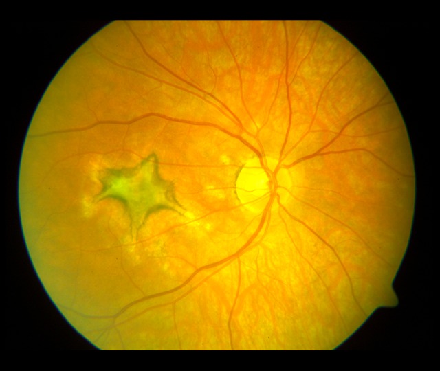
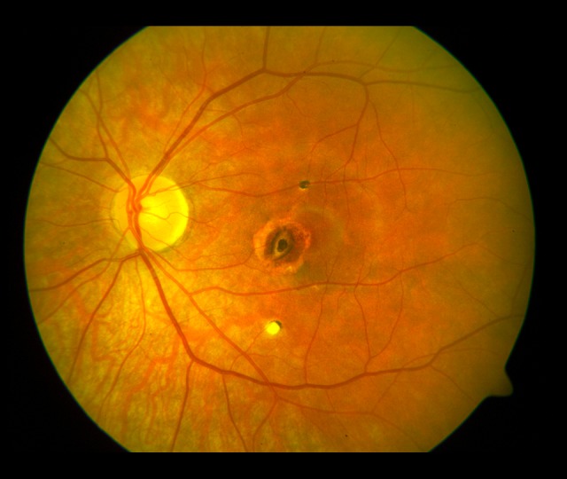
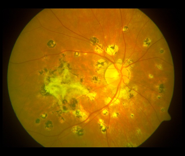
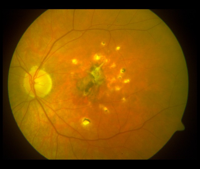
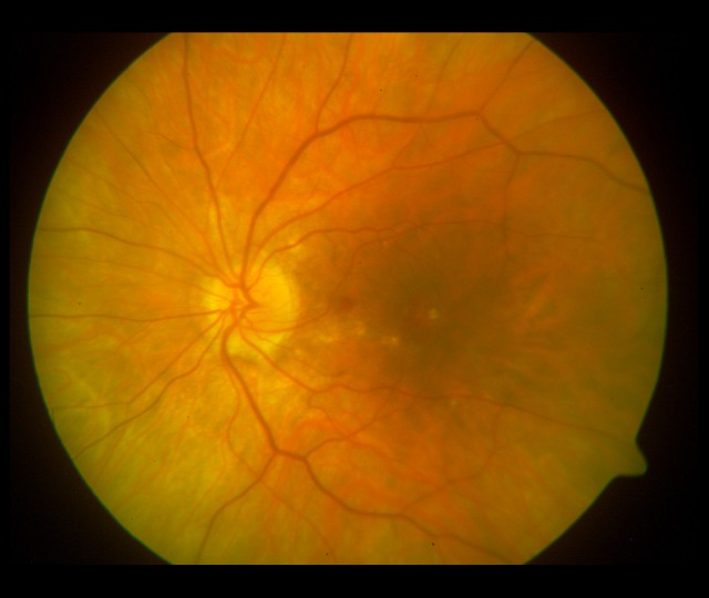
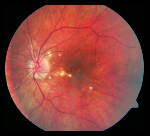
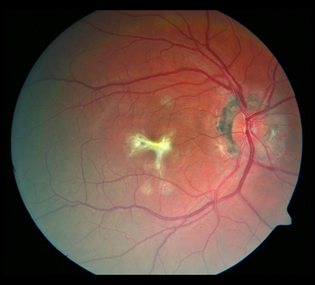
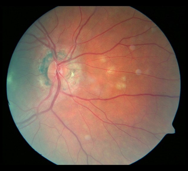
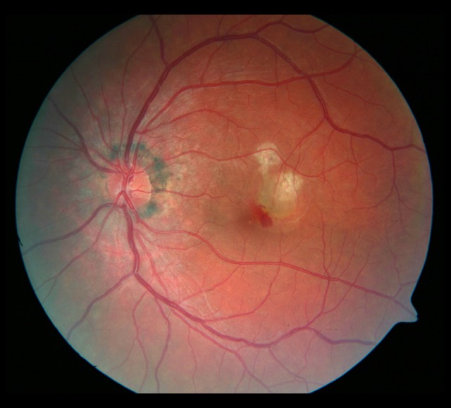
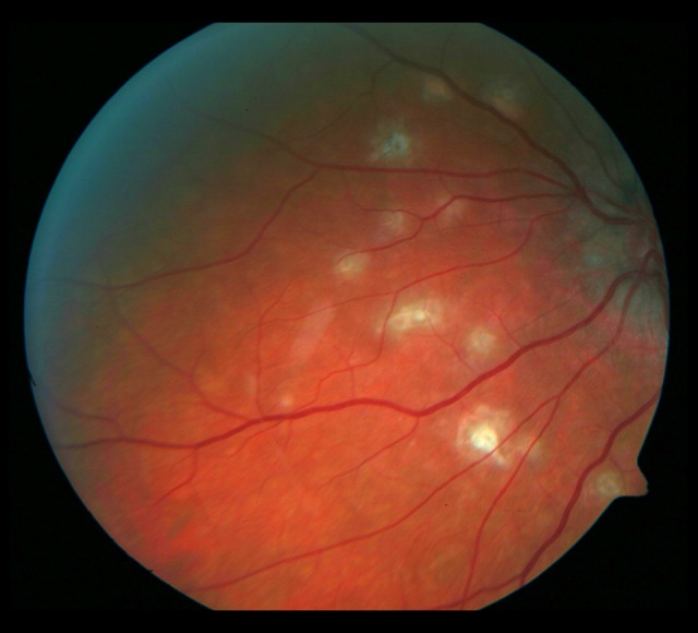
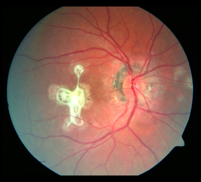
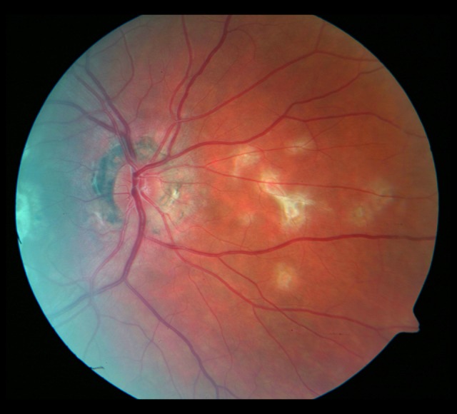
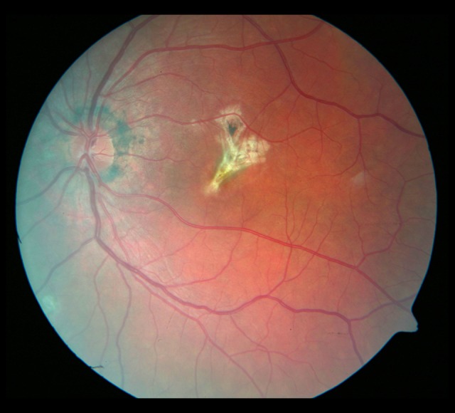
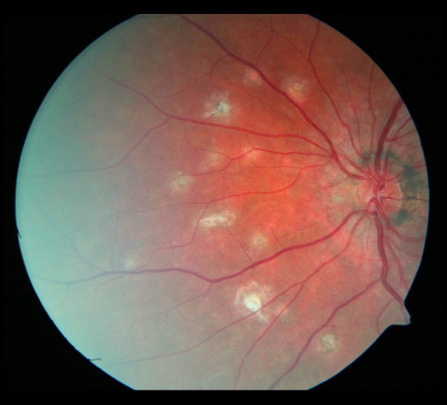
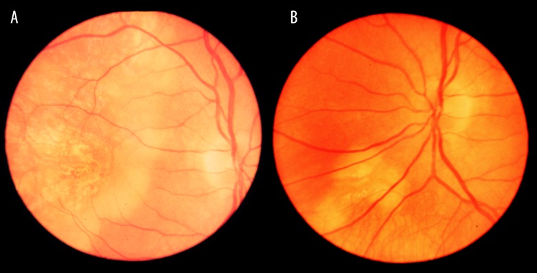
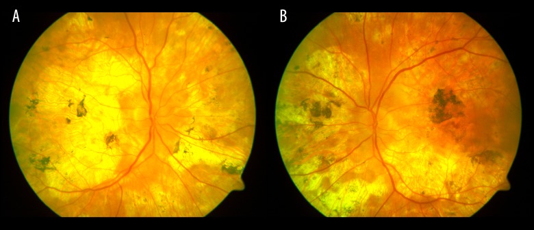
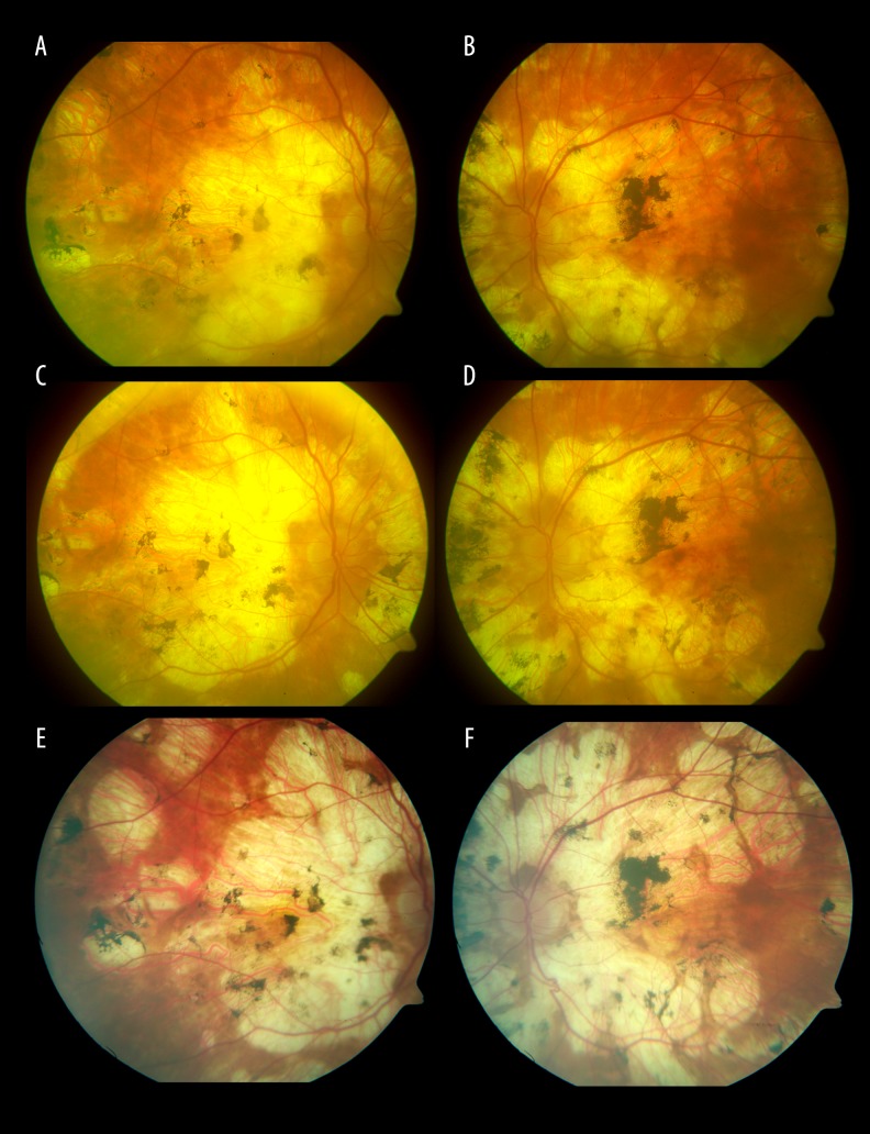
BACKGROUND The aim of this study was to assess the clinical course and distinctive features of different white dot syndromes (WDS) in patients attending the Ophthalmology Department, Medical University of Warsaw in the years 1995-2015. MATERIAL AND METHODS Sixty-two (62) patients (43 females and 19 males), aged 18 to 77 years, referred with a WDS were included in this prospective study, with observation period ranging from 5 months to 16 years. All patients underwent a complete ophthalmological examination and multimodal imaging studies. RESULTS In this cohort of 62 patients, the following WDS entities were identified: multifocal choroiditis with panuveitis (MFCPU), multifocal choroiditis (MFC), punctate inner choroidopathy (PIC), birdshot, acute posterior multifocal placoid pigment epitheliopathy (APMPPE), subretinal fibrosis and uveitis, multiple evanescent white dot syndrome (MEWDS), serpiginous choroiditis, and single cases of acute annular outer retinopathy (AAOR). CONCLUSIONS The study was performed at a Polish referral center and may to some extent reflect the varied geographical distribution of white dot syndromes, as none of the subjects was found to suffer from acute zonal occult outer retinopathy (AZOOR), acute macular neuroretinopathy (AMN), or diffuse unilateral subacute neuroretinitis (DUSN). Long-term follow-up is warranted by the evolution of lesions in the eye fundus, while management depends on correct diagnosis of WDS. When the posterior pole is involved in some cases of the WDS an immunosuppressive treatment, the use of the PDT or anti-VEGF injections were necessary.
Conflict of interest statement
All the authors declare no conflicts of interest.
Figures
Similar articles
-
[White dot syndromes : Principles, diagnostics, and treatment].Ophthalmologe. 2019 Dec;116(12):1235-1256. doi: 10.1007/s00347-019-01012-5. Ophthalmologe. 2019. PMID: 31748943 German.
-
[White dot syndrome].Ophthalmologe. 2008 Jan;105(1):91-108; quiz 109. doi: 10.1007/s00347-007-1687-6. Ophthalmologe. 2008. PMID: 18210124 Review. German.
-
White Dot Syndromes.2023 Mar 13. In: StatPearls [Internet]. Treasure Island (FL): StatPearls Publishing; 2025 Jan–. 2023 Mar 13. In: StatPearls [Internet]. Treasure Island (FL): StatPearls Publishing; 2025 Jan–. PMID: 32491777 Free Books & Documents.
-
The white dot syndromes.Compr Ophthalmol Update. 2007 Jul-Aug;8(4):179-200; discussion 203-4. Compr Ophthalmol Update. 2007. PMID: 17999832 Review.
-
An update of multimodal imaging in white dot syndrome.Oman J Ophthalmol. 2024 Oct 24;17(3):325-333. doi: 10.4103/ojo.ojo_116_24. eCollection 2024 Sep-Dec. Oman J Ophthalmol. 2024. PMID: 39651513 Free PMC article. Review.
Cited by
-
Multimodal Imaging of a Case of Monitoring of Acute Posterior Multifocal Placoid Pigment Epitheliopathy (APMPPE): Long-Term Follow-Up.Case Rep Ophthalmol Med. 2025 Feb 14;2025:9924678. doi: 10.1155/crop/9924678. eCollection 2025. Case Rep Ophthalmol Med. 2025. PMID: 39990599 Free PMC article.
-
BTS clinical statement for the diagnosis and management of ocular tuberculosis.BMJ Open Respir Res. 2022 Mar;9(1):e001225. doi: 10.1136/bmjresp-2022-001225. BMJ Open Respir Res. 2022. PMID: 35379660 Free PMC article. Review.
-
Imageology features of different types of multifocal choroiditis.BMC Ophthalmol. 2019 Feb 1;19(1):39. doi: 10.1186/s12886-019-1045-x. BMC Ophthalmol. 2019. PMID: 30709392 Free PMC article.
References
-
- Jampol LM, Becker KG. White spot syndromes of the retina: A hypothesis based on the common genetic hypothesis of autoimmune/inflammatory disease. Am J Ophthalmol. 2003;135(3):376–79. - PubMed
-
- Quillen DA, Davis JB, Gottlieb JL, et al. The white dot syndromes. Am Journal Ophthalmol. 2004;137(3):538–50. - PubMed
MeSH terms
LinkOut - more resources
Full Text Sources
Medical
