. 2017 Mar 2;12(3):e0173028.
doi: 10.1371/journal.pone.0173028. eCollection 2017.

Mangiferin protects against adverse skeletal muscle changes and enhances muscle oxidative capacity in obese rats

Affiliations

Mangiferin protects against adverse skeletal muscle changes and enhances muscle oxidative capacity in obese rats

Luz M Acevedo et al. PLoS One. .

Abstract

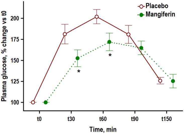
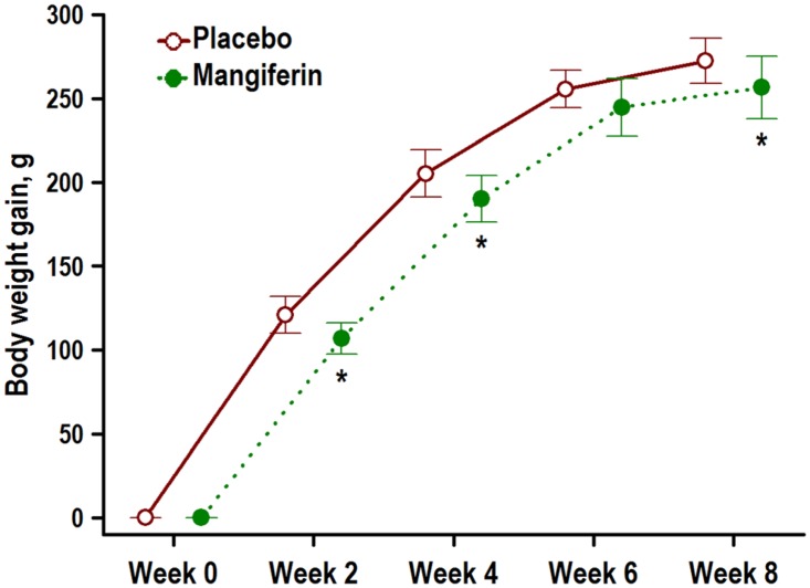
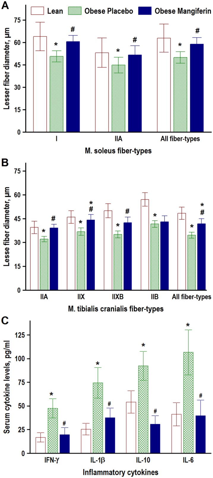
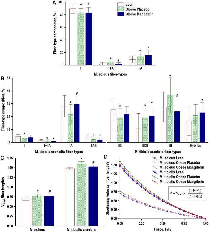
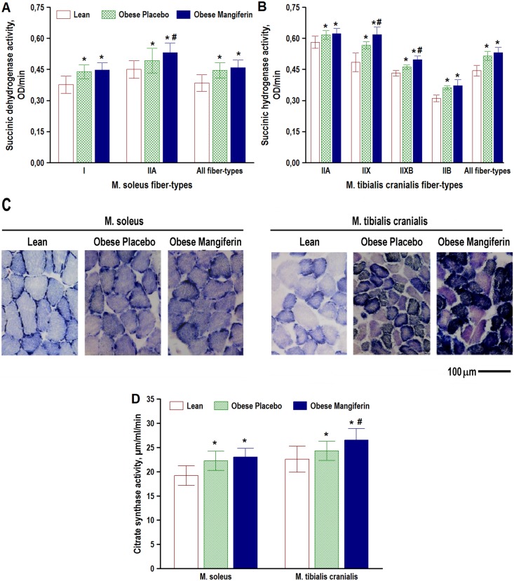
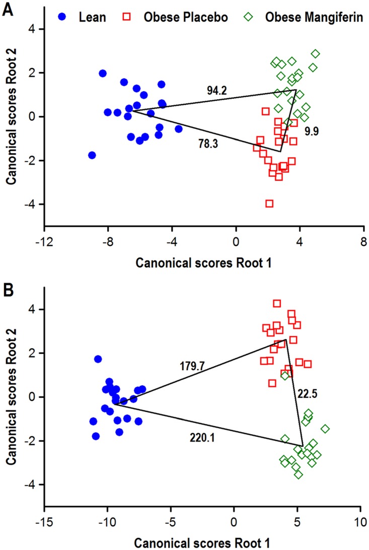
Obesity-related skeletal muscle changes include muscle atrophy, slow-to-fast fiber-type transformation, and impaired mitochondrial oxidative capacity. These changes relate with increased risk of insulin resistance. Mangiferin, the major component of the plant Mangifera indica, is a well-known anti-inflammatory, anti-diabetic, and antihyperlipidemic agent. This study tested the hypothesis that mangiferin treatment counteracts obesity-induced fiber atrophy and slow-to-fast fiber transition, and favors an oxidative phenotype in skeletal muscle of obese rats. Obese Zucker rats were fed gelatin pellets with (15 mg/kg BW/day) or without (placebo group) mangiferin for 8 weeks. Lean Zucker rats received the same gelatin pellets without mangiferin and served as non-obese and non-diabetic controls. Lesser diameter, fiber composition, and histochemical succinic dehydrogenase activity (an oxidative marker) of myosin-based fiber-types were assessed in soleus and tibialis cranialis muscles. A multivariate discriminant analysis encompassing all fiber-type features indicated that obese rats treated with mangiferin displayed skeletal muscle phenotypes significantly different compared with both lean and obese control rats. Mangiferin significantly decreased inflammatory cytokines, preserved skeletal muscle mass, fiber cross-sectional size, and fiber-type composition, and enhanced muscle fiber oxidative capacity. These data demonstrate that mangiferin attenuated adverse skeletal muscle changes in obese rats.


Conflict of interest statement

Competing Interests: The authors have declared that no competing interests exist.

Figures

Fig 1
Fig 1. Mangiferin attenuates hyperglycemic response in obese rats after glucose oral administration.
Zucker rats with the obese (fa/fa) phenotype (2 months old) were treated with placebo or mangiferin (15 mg/kg BW/day) for 8 weeks (n = 10 rats/group), and plasma glucose was monitored every 30 minutes after oral glucose administration (2 g/kg BW). Graph shows means ± SD plasma glucose change vs basal levels over time. * P < 0.05 indicates significant differences between Placebo and Mangiferin groups obtained by unpaired Student’s t test.
Fig 2
Fig 2. Mangiferin mitigates overweight in obese rats.
Zucker rats with the obese (fa/fa) phenotype (2 months old) were treated with placebo or with mangiferin (15 mg/kg BW/days) for 8 weeks, and body weight was monitored every two weeks. Graph shows means ± SD body weight gain over time, n = 10 rats/group. * P < 0.05 indicates significant differences between Placebo and Mangiferin groups obtained by unpaired Student’s t test.
Fig 3
Fig 3. Effect of mangiferin treatment on muscle fiber-size and serum levels of inflammatory cytokines in obese rats.
Mean lesser fiber diameters of soleus (A) and tibialis cranialis (B) muscle fiber-types, and plasma levels of interferon-γ (IFN- γ) and interleukin (IL)-1β, IL-6 and IL-10 (C) were determined in lean (FA/Fa or FA/fa) Zucker rats and obese (fa/fa) Zucker rats that were treated with either placebo (Obese Placebo) or 15 mg/kg BW/day of Mangiferin for 8 weeks (Obese Mangiferin). For clarity, muscle fiber-types with a percentage below 5% (see Fig 4) are excluded. Values are means ± SD, n = 20 rats/group in A and B, n = 10 rats/group in C. * P < 0.05 vs Lean, # P<0.05 vs Obese Placebo (one-way ANOVA with Fisher LSD post-hoc test).
Fig 4
Fig 4. Effect of mangiferin treatment on muscle fiber-type composition, and predicted muscle maximal shortening velocity (Vmax) and the muscle force-velocity relationship in obese rats.
The percentages of myosin-based fiber-types (A, B), and predicted Vmax (C) and force-velocity curves (D), dereived according to the Hill-type mathematical model (inset) [42], were determined in Zucker rats with the lean (Fa/Fa or Fa/fa) phenotype (Lean), and Zucker rats with the obese (fa/fa) phenotype that were treated with either placebo (Obese Placebo) or 15 mg/kg BW/day of Mangiferin for 8 weeks (Obese Mangiferin). Bars show means ± SD, n = 20 rats/group. * P < 0.05 vs Lean, # P < 0.05 vs Obese Placebo (one-way ANOVA with Fisher LSD post-hoc test). Data in D are expressed as means of 20 rats/group. V, shortening velocity; Vmax, maximal shortening velocity; P, isometric tension; P0, maximal isometric tension.
Fig 5
Fig 5. Effect of mangiferin treatment on muscle oxidative capacity in obese rats.
The histochemical succinic dehydrogenase activity of individual muscle fiber-types (A, B and C), and the biochemical citrate synthase activity of muscle homogenates (D) were compared in Zucker rats with the lean (Fa/Fa or Fa/fa) phenotype (Lean, and Zucker rats with the obese (fa/fa) phenotype that were treated with either placebo (Obese Placebo) or 15 mg/kg BW/day of mangiferin for 8 weeks (Obese Mangiferin). For clarity, muscle fiber-types with a percentage below 5% (see Fig 4) are excluded in A and B graphs. Values are means ± SD, n = 20 rats/group. * P < 0.05 vs Lean, # P < 0.05 vs Obese Placebo (one-way ANOVA with Fisher LSD post-hoc test). Note in C the darker staining intensity in muscle fibers of both muscles in the two groups of obese rats compared with the lean rat.
Fig 6
Fig 6. Phenotypic classification of individual hind limb muscles of obese Zucker rats treated with mangiferin in relation to lean and obese controls by means of multivariate discriminant analysis.
Individual soleus (A) and tibialis cranialis (B) muscles of all experimental rats were classified into different groups as a function of a cohort of phenotypic muscle characteristics by means of discriminant canonical analysis. The discriminant model for the soleus muscle included the following muscle variables: wet weight, muscle-somatic index, predicted maximal shortening velocity, and percentage, lesser diameter and SDH histochemical activity of type I fibres. The same parameters were included in the statistical model for the tibialis cranialis muscles, but fiber-types IIA, IIX, IIXB and IIB were considered instead of type I fibers. The squares of the Mahalanobis distances between pairs of groups indicative of the overall phenotypic divergences are indicated; P values for the statistical significance of these comparisons between pairs of groups were always significant (P < 0.001).

References

    1. Couturier A, Ringseis R, Mooren FC, Kruger K, Most E, Eder K (2013) Carnitine supplementation to obese Zucker rats prevents obesity-induced type I to type I muscle fiber transition and favors an oxidative phenotype of skeletal muscle. Nutr Metab (Lond) 10: 48. - PMC - PubMed
    1. Denies MS, Johnson J, Maliphol AB, Bruno M, Kim A, Rizvi A, et al. (2014) Diet-induced obesity alters skeletal muscle fiber types of male but not female mice. Physiol Rep 2: e00204 10.1002/phy2.204 - DOI - PMC - PubMed
    1. Perry BD, Caldow MK, Brennan-Speranza TC, Sbaraglia M, Jerums G, Garnham A, et al. (2016) Muscle atrophy in patients with Type 2 Diabetes Mellitus: roles of inflammatory pathways, physical activity and exercise. Exerc Immunol Rev 22: 94–109. - PMC - PubMed
    1. Pompeani N, Rybalka E, Latchman H, Murphy RM, Croft K, Hayes A (2014) Skeletal muscle atrophy in sedentary Zucker obese rats is not caused by calpain-mediated muscle damage or lipid peroxidation induced by oxidative stress. J Negat Results Biomed 13: 19 10.1186/s12952-014-0019-z - DOI - PMC - PubMed
    1. Ringseis R, Rosenbaum S, Gessner DK, Herges L, Kubens JF, Mooren FC, et al. (2013) Supplementing obese Zucker rats with niacin induces the transition of glycolytic to oxidative skeletal muscle fibers. J Nutr 143: 125–131. 10.3945/jn.112.164038 - DOI - PubMed