Review
. 2017 Apr 3;216(4):875-887.
doi: 10.1083/jcb.201610111. Epub 2017 Mar 2.

Telomere-driven diseases and telomere-targeting therapies

Affiliations
Review

Telomere-driven diseases and telomere-targeting therapies

Paula Martínez et al. J Cell Biol. .

Abstract

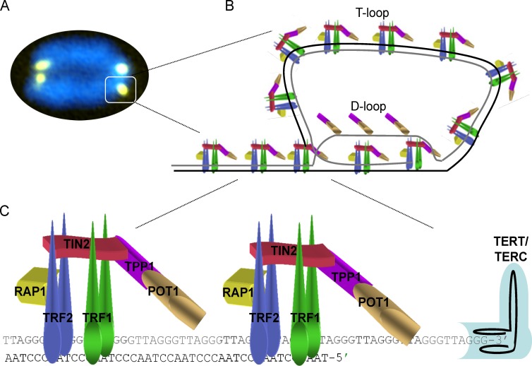
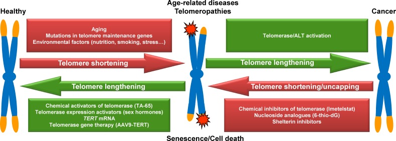
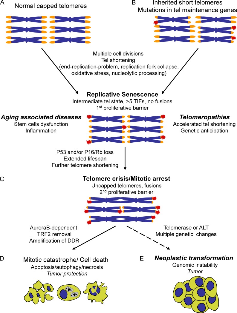
Telomeres, the protective ends of linear chromosomes, shorten throughout an individual's lifetime. Telomere shortening is proposed to be a primary molecular cause of aging. Short telomeres block the proliferative capacity of stem cells, affecting their potential to regenerate tissues, and trigger the development of age-associated diseases. Mutations in telomere maintenance genes are associated with pathologies referred to as telomere syndromes, including Hoyeraal-Hreidarsson syndrome, dyskeratosis congenita, pulmonary fibrosis, aplastic anemia, and liver fibrosis. Telomere shortening induces chromosomal instability that, in the absence of functional tumor suppressor genes, can contribute to tumorigenesis. In addition, mutations in telomere length maintenance genes and in shelterin components, the protein complex that protects telomeres, have been found to be associated with different types of cancer. These observations have encouraged the development of therapeutic strategies to treat and prevent telomere-associated diseases, namely aging-related diseases, including cancer. Here we review the molecular mechanisms underlying telomere-driven diseases and highlight recent advances in the preclinical development of telomere-targeted therapies using mouse models.


Figures

Figure 1.
Figure 1.
The shelterin complex and the structure of telomeres. (A) Representative image of a metaphasic chromosome stained with DAPI (blue) and the telomeric DNA–specific peptide nucleic acid probe (yellow). (B) Schematic model of the shelterin complex bound to a telomere in a T-loop configuration. Telomeres contain a double-stranded region of TTAGGG repeats and a 150–200-nucleotide-long single-stranded DNA overhang of a G-rich strand. The G-strand overhang (gray) invades the telomeric double-stranded DNA region to form a protective T-loop, with a displacement D-loop at the invasion site. (C) Schematic representation of telomere-bound proteins, the shelterin complex, and telomerase. The shelterin complex is composed of the telomeric repeat binding factor 1 (TRF1, also known as TERF1), TRF2 (also known as TERF2), repressor-activator protein 1 (RAP1, also known as TERF2IP1), POT1-TIN2 organizing protein (TPP1, also known as ACD), TIN2 (also known as TIFN2), and protection of telomeres protein 1 (POT1). TRF1, TRF2, and POT1 bind directly to telomeric DNA repeats, with TRF1 and TRF2 binding to telomeric double-stranded DNA and POT1 to the 3′ single-stranded G-overhang. TIN2 binds TRF1 and TRF2 through independent domains and recruits the TPP1–POT1 complex, constituting the bridge among the different shelterin components. Telomerase is a two-partner enzyme, the catalytic subunit (TERT) and the RNA template (TERC), that recognizes the 3′-OH at the end of the G-strand overhang and elongates the telomere.
Figure 2.
Figure 2.
Natural factors and therapeutic interventions affecting telomere-mediated diseases. Telomere shortening naturally occurs as a consequence of cell division throughout life, whose pace can be influenced by genetic and environmental factors. Shortened unprotected telomeres elicit a DDR that induces cellular senescence, impacting the regenerative capacity of tissues and giving rise to a whole range of age-associated diseases as well as the so-called telomeropathies, in which tissue degeneration occurs prematurely as a consequence of inherited defects in telomere maintenance. Several therapeutic interventions are being assessed to counteract telomere shortening: among others, chemical activators of telomerase (TA-65), activators of the telomerase reverse transcription (TERT) transcription (sex hormones), intracellular administration of TERT mRNA, and telomerase gene therapy (AAV9-TERT). Spontaneous mutations that activate telomerase expression or ALT result in telomere lengthening that, if occurring in genetically unstable checkpoint deficient cells, allows them to divide unlimitedly and eventually become cancer cells. Several therapeutic strategies based on chemical induction of telomere dysfunction have been assessed as anticancer therapies. Among others, we highlight the use of a chemical inhibitor of telomerase (imetelstat), a nucleoside analogue (6-thio-dG), and the use of molecules that inhibit shelterin components.
Figure 3.
Figure 3.
Impact of telomere shortening on aging-associated diseases, telomeropathies, and cancer. (A) Young, healthy cells contain long, fully protected telomeres that progressively shorten with increased cell divisions because of the end replication problem, replication fork collapse, nucleolytic processing, and oxidative stress. This progressive telomere shortening eventually leads to some critically short deprotected telomeres that have been termed intermediate-state telomeres. In this intermediate state of deprotection, telomeres retain enough shelterin to inhibit fusions but induce a DDR characterized by formation of the so-called telomere-induced focus (TIF). Replicative senescence is triggered when at least five telomeres become dysfunctional (more than five telomere-induced focuses), a critical damage threshold to elicit a DDR characterized by p53 activation. Telomere attrition in stem cell compartments impairs their tissue and self-renewal capacity and is considered to be one of the primary molecular causes of aging and the onset of aging-associated diseases. (B) Telomeropathies or telomere syndromes develop when telomere attrition occurs prematurely as a consequence of germline mutations in genes coding for factors involved in telomere maintenance and repair. Successive telomere shortening across generations exhibits genetic anticipation, whereby diseases show a progressively earlier age of onset and an aggravation of symptoms. (C) Senescence can be bypassed by acquisition of loss-of-function mutations in p53 and p16/Rb tumor suppressor genes that permit further proliferation during a period of time that has been named the extended life span period, during which cells experience further telomere shortening, eventually entering the uncapped state, when they do not retain any protective properties and fuse. (D) Fused telomeres lead to a mitotic arrest checkpoint during which telomere dysfunction is amplified by Aurora B–dependent TRF2 removal, causing cell death through apoptosis, necrosis, or autophagy in crisis, a second proliferative barrier that protects against tumor development. (E) Reactivation of either telomerase activity or ALT in some very rare crisis cells allows these premalignant cells to escape crisis and divide unlimitedly (immortalization). Fused chromosomes in postcrisis cells lead to large-scale genomic rearrangements that promote acquisition of oncogenic mutations and malignant traits required for a fully malignant phenotype, cancer.

References

    1. Agrawal A., Dang S., and Gabrani R.. 2012. Recent patents on anti-telomerase cancer therapy. Recent Patents Anticancer. Drug Discov. 7:102–117. 10.2174/157489212798357958 - DOI - PubMed
    1. Akbay E.A., Peña C.G., Ruder D., Michel J.A., Nakada Y., Pathak S., Multani A.S., Chang S., and Castrillon D.H.. 2013. Cooperation between p53 and the telomere-protecting shelterin component Pot1a in endometrial carcinogenesis. Oncogene. 32:2211–2219. 10.1038/onc.2012.232 - DOI - PMC - PubMed
    1. Alder J.K., Chen J.J., Lancaster L., Danoff S., Su S.C., Cogan J.D., Vulto I., Xie M., Qi X., Tuder R.M., et al. . 2008. Short telomeres are a risk factor for idiopathic pulmonary fibrosis. Proc. Natl. Acad. Sci. USA. 105:13051–13056. 10.1073/pnas.0804280105 - DOI - PMC - PubMed
    1. Alder J.K., Guo N., Kembou F., Parry E.M., Anderson C.J., Gorgy A.I., Walsh M.F., Sussan T., Biswal S., Mitzner W., et al. . 2011. Telomere length is a determinant of emphysema susceptibility. Am. J. Respir. Crit. Care Med. 184:904–912. 10.1164/rccm.201103-0520OC - DOI - PMC - PubMed
    1. Alder J.K., Barkauskas C.E., Limjunyawong N., Stanley S.E., Kembou F., Tuder R.M., Hogan B.L., Mitzner W., and Armanios M.. 2015a Telomere dysfunction causes alveolar stem cell failure. Proc. Natl. Acad. Sci. USA. 112:5099–5104. 10.1073/pnas.1504780112 - DOI - PMC - PubMed

Publication types

Supplementary concepts