Review
. 2017 Apr;69(2):93-139.
doi: 10.1124/pr.116.013078.

International Union of Basic and Clinical Pharmacology. CI. Structures and Small Molecule Modulators of Mammalian Adenylyl Cyclases

Affiliations
Review

International Union of Basic and Clinical Pharmacology. CI. Structures and Small Molecule Modulators of Mammalian Adenylyl Cyclases

Carmen W Dessauer et al. Pharmacol Rev. 2017 Apr.

Abstract

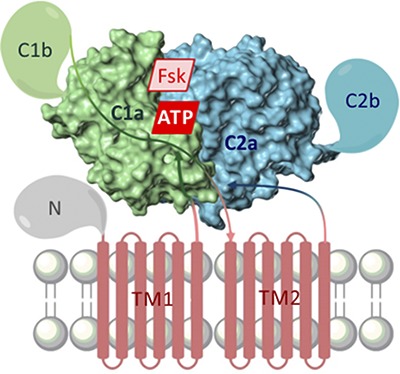
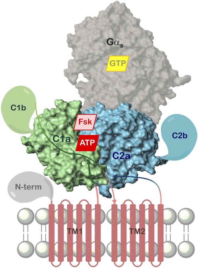
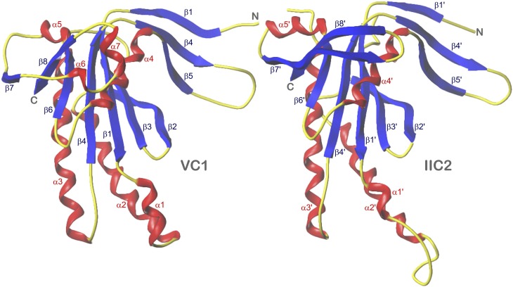
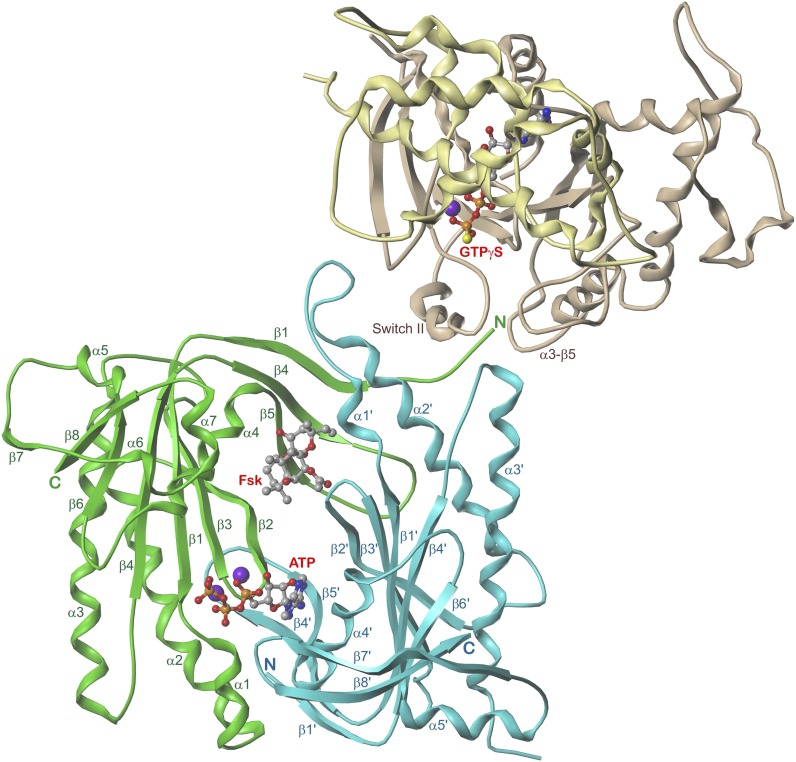
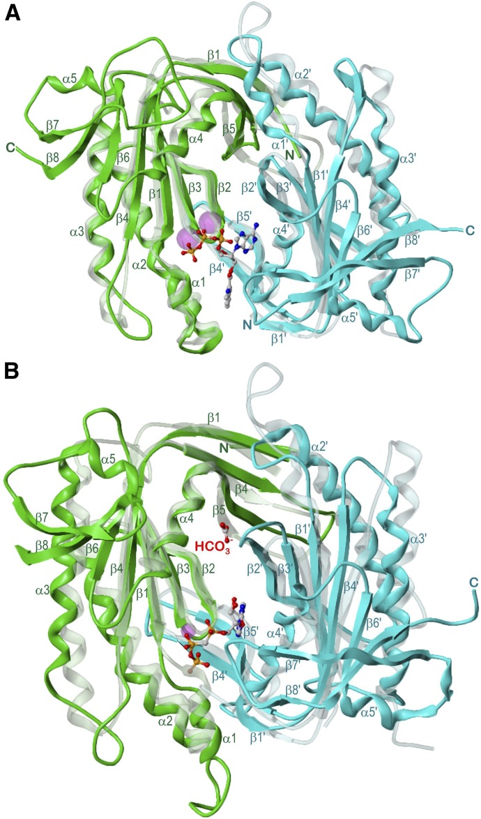
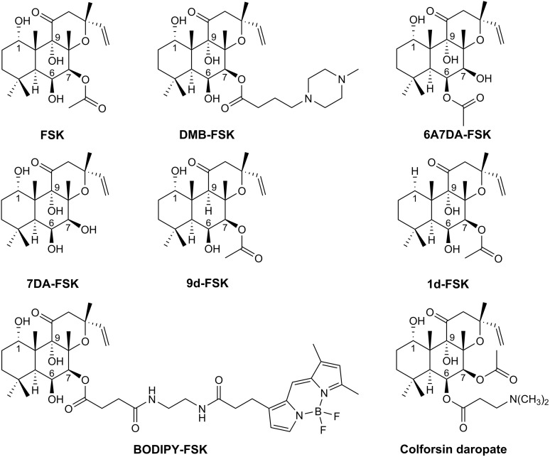
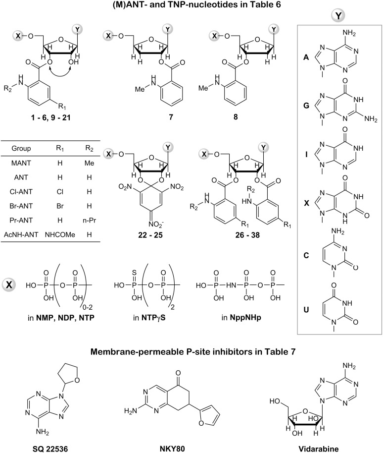
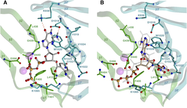
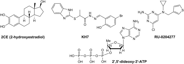
Adenylyl cyclases (ACs) generate the second messenger cAMP from ATP. Mammalian cells express nine transmembrane AC (mAC) isoforms (AC1-9) and a soluble AC (sAC, also referred to as AC10). This review will largely focus on mACs. mACs are activated by the G-protein Gαs and regulated by multiple mechanisms. mACs are differentially expressed in tissues and regulate numerous and diverse cell functions. mACs localize in distinct membrane compartments and form signaling complexes. sAC is activated by bicarbonate with physiologic roles first described in testis. Crystal structures of the catalytic core of a hybrid mAC and sAC are available. These structures provide detailed insights into the catalytic mechanism and constitute the basis for the development of isoform-selective activators and inhibitors. Although potent competitive and noncompetitive mAC inhibitors are available, it is challenging to obtain compounds with high isoform selectivity due to the conservation of the catalytic core. Accordingly, caution must be exerted with the interpretation of intact-cell studies. The development of isoform-selective activators, the plant diterpene forskolin being the starting compound, has been equally challenging. There is no known endogenous ligand for the forskolin binding site. Recently, development of selective sAC inhibitors was reported. An emerging field is the association of AC gene polymorphisms with human diseases. For example, mutations in the AC5 gene (ADCY5) cause hyperkinetic extrapyramidal motor disorders. Overall, in contrast to the guanylyl cyclase field, our understanding of the (patho)physiology of AC isoforms and the development of clinically useful drugs targeting ACs is still in its infancy.


Figures

None
Graphical abstract
Fig. 1.
Fig. 1.
Common domain structure of mACs. Components resolved in crystal structures are presented as surface model. Shown is the “ventral” surface of C1a:C2a for the sake of consistency with Figs. 2–4, 6–11, and 14 depicting crystal structures. The real position of the membrane is probably behind the hidden “dorsal” surface. FSK, forskolin site (AC1–8), N-term, N-terminal domain; C2b, C-terminal domain; TM, transmembrane domains; arrow lines, schematic course of the polypeptide chain.
Fig. 2.
Fig. 2.
5C1 and 2C2 in side-by-side comparison of superimposed monomers. α-Helices, red; β-sheets, blue; loops and turns, yellow. Models are based on the crystal structure PDB 1CJU (Tesmer et al., 1997, 1999). Secondary structure elements as well as N and C termini are labeled.
Fig. 3.
Fig. 3.
5C1:2C2 in complex with FSK, ATP, Gαs, and GTPγS. View along the pseudo-twofold axis. 5C1, green; 2C2, cyan; Gαs helical domain, yellow; ras-like domain, mauve; atom colors: C, gray; N, blue; O, red; P, orange; Mg2+, violet. Models are based on the crystal structure PDB 1CJU (Tesmer et al., 1997, 1999) with ATP docked instead of 2′,3′-dideoxy-ATP. Secondary structure elements (Gαs only contact sites with 5C1:2C2) as well as N and C termini of 5C1:2C2 are labeled.
Fig. 4.
Fig. 4.
Catalytic domains of sGC and sAC aligned with 5C1:2C2. Domains are drawn from β1 to α4 and β1′ to α4′, respectively. Views along the pseudo-twofold axes. sdGCαcat, sAC-C1; 5C1, green; sGCβcat, sAC-C2; 2C2, cyan; atom colors: C, gray; N, blue; O, red; P, orange; Mn2+ and Ca2+, magenta; secondary structure elements as well as N and C termini are labeled. (A) sGCαcat:sGCβcat in complex with 2′-MANT-3′-dATP and Mn2+, model of the intermediate state based on the open state crystal structure PDB 3UVJ (Allerston et al., 2013) and on 5C1:2C2 PDB 1TL7 (Mou et al., 2005) as template (transparent model). (B) sAC in complex with α,β-methylene adenosine-5′-triphosphate, bicarbonate and Ca2+, model based on the crystal structure PDB 4CLK (Kleinboelting et al., 2014a), 5C1:2C2 (transparent model) corresponding to Fig. 3.
Fig. 5.
Fig. 5.
Structure-based sequence alignment of the catalytic domains of sAC, sGC, AC1, AC2, and AC5. Secondary structure elements are primarily derived from 5C1 (green) and 2C2 (cyan), after α5 of sAC1, 5C1 and sGCα, only β7 and β8 are common. The sequence numbers above and below the alignment correspond to 5C1 and 2C2, respectively. Amino acids in contact with Gαs are highlighted in yellow, underscored residues form the C1-C2 interface of 5C1:2C2 and sAC. Bold-colored amino acids belong to the FSK (5C1:2C2) or bicarbonate (sAC) and nucleotide binding sites and interact with FSK or bicarbonate (magenta), metal ions (red), triphosphate (brown), the ribosyl moiety (green), and the nucleobase (blue), respectively. Amino acids in italics contribute via backbone, hydrophobic, or van der Waals interactions to nucleotide binding. Other interactions are electrostatic (including ionic, hydrogen bonds) and via side chains.
Fig. 6.
Fig. 6.
Detailed 5C1:2C2-Gαs interactions. 5C1, green; 2C2, cyan; Gαs, mauve; heteroatom colors: N, blue; O, red; S, yellow. Hydrogen bonds are drawn as red dashed lines. Model based on the crystal structure PDB 1CJU (Tesmer et al., 1997, 1999). Contacting amino acids (distance <3 Å) and secondary structure elements are labeled.
Fig. 7.
Fig. 7.
5C1-2C2 interface. Cα-atoms of all amino acids involved in 5C1-2C2 interactions (distance <4 Å, see Fig. 5) are drawn as balls, hydrogen bonds, and salt bridges of labeled residues as sticks. Colors: H-acceptor/acidic side chain, red; backbone O as H-acceptor, orange; H-donor/basic side chain, dark blue; backbone NH as H-donor, light blue; other contacting amino acids and framework of 5C1, green, of 2C2, cyan.
Fig. 8.
Fig. 8.
Detailed 5C1:2C2-FSK interactions. 5C1, green; 2C2, cyan; FSK and water, gray; heteroatom colors: N, blue; O, red. Hydrogen bonds are drawn as red dashed lines. Model based on the crystal structure PDB 1CJU (Tesmer et al., 1997, 1999). Contacting amino acids (distance < 3.5 Å) and secondary structure elements are labeled.
Fig. 9.
Fig. 9.
Closed and open 5C1:2C2 conformations. Alignment of the complex with ATP, Mg2+, and FSK with the open inactive state of apo-5C1:2C2. View along the pseudo-twofold axes. Closed state 5C1, green; 2C2, cyan; open state 5C1, yellow; 2C2, purple; atom colors: C, gray; N, blue; O, red; P, orange; Mg2+, violet. Models are based on the crystal structures PDB 1CJU with ATP docked instead of 2′,3′-dideoxy-ATP and 1AZS, respectively (Tesmer et al., 1997, 1999). Unfitting secondary structure elements as well as N and C termini are labeled.
Fig. 10.
Fig. 10.
Common nucleotide binding site of mAC, sGC and sAC. Essential amino acids are drawn as space fill models of Cα and Cβ atoms; catalytic domain 1, green; catalytic domain 2, cyan; metals A and B, red balls; interactions of metal ions, red; triphosphate, orange; ribosyl moiety, green; nucleobase, blue. The model is based on the crystal structure PDB 1CJU (Tesmer et al., 1997, 1999) with ATP docked instead of 2′,3′-dideoxy-ATP. Common secondary structure elements and amino acids of the binding site are labeled.
Fig. 11.
Fig. 11.
Detailed interactions of mACs and sAC with ATP. C1, green; C2, cyan; ATP, gray; heteroatom colors: N, blue; O, red; P, orange;, hydrogen bonds are drawn as red dashed lines, contacting amino acids (distance <3.5 Å) and secondary structure elements are labeled. (A) 5C1:2C2-ATP interactions, Mg2+, violet, model based on the crystal structure PDB 1CJU (Tesmer et al., 1997, 1999) with ATP docked instead of 2′,3′-dideoxy-ATP. (B) sAC-ATP interactions, Na+, violet; w, water oxygen; model based on the crystal structure PDB 4USW (Kleinboelting et al., 2014b).
Fig. 12.
Fig. 12.
Structures of diterpenes. Pharmacological data for selected diterpenes are listed in Table 5. FSK, forskolin; DMB, 7-deacetyl-7-(N-methylpiperazino-butyryloxy); 6A7DA, 6-acetyl-7-deacetyl; 7DA, 7-deacetyl; 9d, 9-deoxy; 1d, 1-deoxy; BODIPY, 4,4-difluoro-4-bora-3a,4a-diaza-s-indacene. Diterpenes are highly lipophilic. To increase water solubility, hydrophilc substituents were introduced at the 6′-position (colforsin daropate) and the 7′-position (DMB-FSK) of the diterpene ring. In Japan, colforsin daropate is used clinically for treatment of heart failure. BODIPY-FSK is a fluorescent FSK derivative that shows inverse agonist activity at AC2 and partial agonist activity at ACs 1 and 5.
Fig. 13.
Fig. 13.
Structures of representative inhibitors targeting the catalytic site of mACs. Pharmacological data for competitive inhibitors are listed in Table 6, and pharmacological data for noncompetitive P-site inhibitors are listed in Table 7. The (M)ANT group can spontaneously isomerize between the 2′- and 3′-ribosyl position, provided that there is only substituent and a free hydroxyl group. The different bases (A, adenine; G, guanine; I, hypoxanthine; X, xanthine; C, cytosine; U, uracil) constitute substituent Y at the ribosyl moiety. The different phosphate chains (mono-, di-, and triphosphate and hydrolysis-resistant phosphorothioates or β,γ-imidophosphates) constitute substituent X at the ribosyl moiety. (M)ANT- and TNP-nucleotides do not cross the plasma membrane and can only be used in cell-free systems or in electrophysiological studies in which compounds are injected into the cell via the patch pipette. Experimental problems with the membrane-permeable P-site inhibitors are the relatively low potency, insufficient selectivity and high lipophilicity, requiring the use of high concentrations of organic solvents.
Fig. 14.
Fig. 14.
Interactions of 5C1:2C2 with selected MANT- and TNP-nucleotides. 5C1, green; 2C2, cyan; heteroatom colors: N, blue; O, red; P, orange; Mn2+, violet. Nucleobase-2C2 hydrogen bonds are drawn as red dashed lines, contacting amino acids (distance < 3.5 Å) and secondary structure elements are labeled. (A) Binding mode of 3′-MANT-GTP, model based on the crystal structure PDB 1TL7 (Mou et al., 2005) C and selected H atoms of 3′-MANT-GTP, gray. (B) Binding mode of TNP-ATP, model based on the crystal structure PDB 2GVD (Mou et al., 2006), C and selected H atoms of TNP-ATP, mauve. The gray shadow image shows 3′-MANT-GTP from a superposition of both binding sites (root-mean-square deviation of Cα atoms, 0.44 Å).
Fig. 15.
Fig. 15.
Structures of representative inhibitors targeting sAC. Pharmacological data for inhibitors are listed in Table 8. All compounds are noncompetitive sAC inhibitors. 2CE, KH7, and RU-0204277 are membrane-permeable. KH7 has been broadly used in intact cell studies, but cell toxicity is of concern. RU-0204277 is a recently developed compound with less toxic liability than KH7 and binds to the bicarbonate site of sAC. 2CE exhibits different potencies at purified sAC and in intact cells. 2′,5′-dideoxy-3′-ATP is a classic P-site inhibitor related to SQ 22536, NKY80, and vidarabine (for structures, see Fig. 13 and for pharmacological data see Table 7) that also potently inhibits mACs and cannot penetrate the plasma membrane.

Similar articles

Cited by

References

    1. Abdel-Majid RM, Leong WL, Schalkwyk LC, Smallman DS, Wong ST, Storm DR, Fine A, Dobson MJ, Guernsey DL, Neumann PE. (1998) Loss of adenylyl cyclase I activity disrupts patterning of mouse somatosensory cortex. Nat Genet 19:289–291. - PubMed
    1. Acin-Perez R, Salazar E, Kamenetsky M, Buck J, Levin LR, Manfredi G. (2009) Cyclic AMP produced inside mitochondria regulates oxidative phosphorylation. Cell Metab 9:265–276. - PMC - PubMed
    1. Agarwal SR, MacDougall DA, Tyser R, Pugh SD, Calaghan SC, Harvey RD. (2011) Effects of cholesterol depletion on compartmentalized cAMP responses in adult cardiac myocytes. J Mol Cell Cardiol 50:500–509. - PMC - PubMed
    1. Agarwal SR, Yang PC, Rice M, Singer CA, Nikolaev VO, Lohse MJ, Clancy CE, Harvey RD. (2014) Role of membrane microdomains in compartmentation of cAMP signaling. PLoS One 9:e95835. - PMC - PubMed
    1. Alasbahi RH, Melzig MF. (2012) Forskolin and derivatives as tools for studying the role of cAMP. Pharmazie 67:5–13. - PubMed

Publication types

Substances