. 2017 Jun;102(6):984-994.
doi: 10.3324/haematol.2016.161513. Epub 2017 Mar 2.

Histone deacetylase 6 regulates cytokinesis and erythrocyte enucleation through deacetylation of formin protein mDia2

Affiliations

Histone deacetylase 6 regulates cytokinesis and erythrocyte enucleation through deacetylation of formin protein mDia2

Xuehui Li et al. Haematologica. 2017 Jun.

Abstract

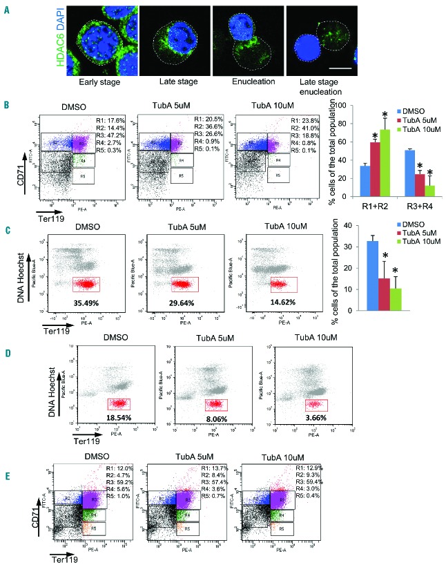
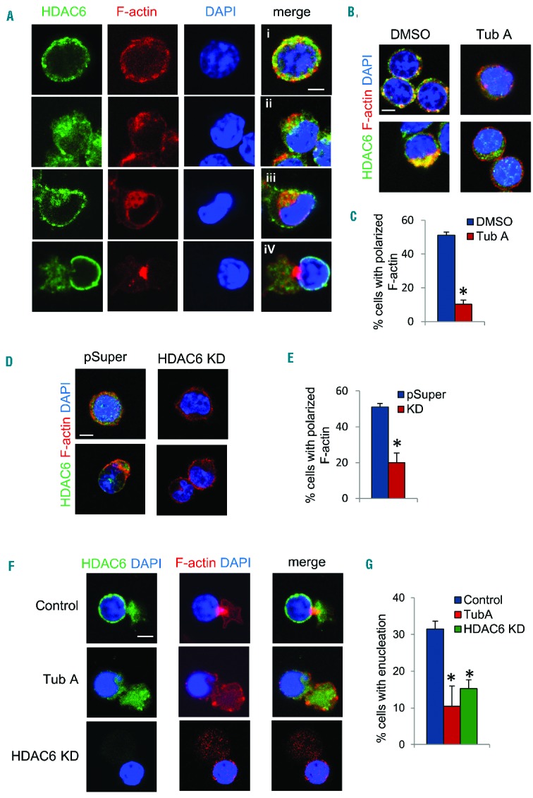
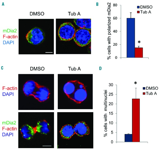
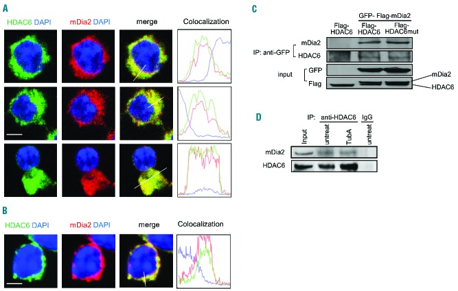
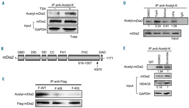
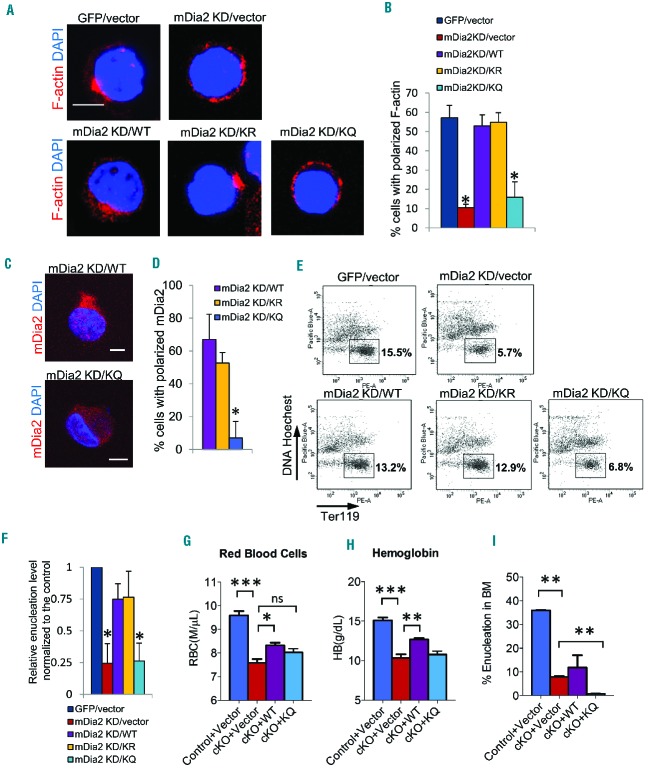
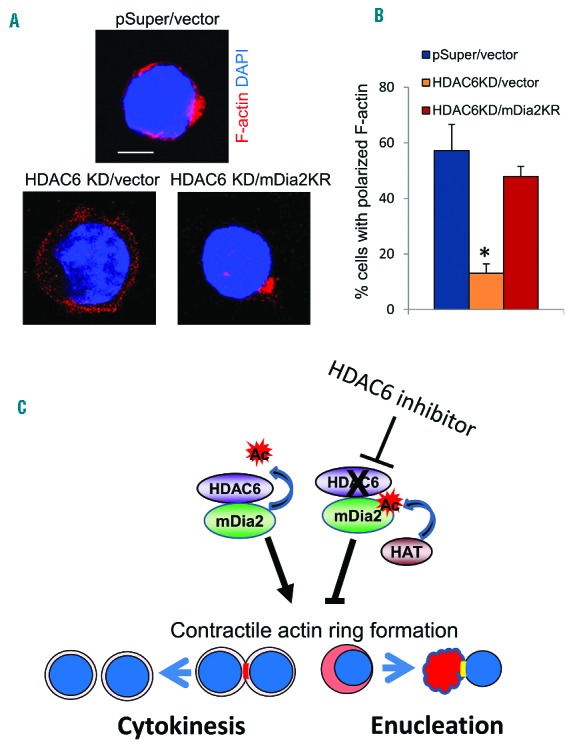
The formin protein mDia2 plays a critical role in a number of cellular processes through its ability to promote nucleation and elongation of actin filaments. In erythroblasts, this includes control of cytokinesis and enucleation by regulating contractile actin ring formation. Here we report a novel mechanism of how mDia2 is regulated: through acetylation and deacetylation at lysine 970 in the formin homology 2 domain. Ectopic expression of an acetyl-mimic mDia2 mutant in mouse erythroblasts is sufficient to abolish contractile actin ring formation at the cleavage furrow and subsequent erythrocyte cytokinesis and enucleation. We also identified that class II histone deacetylase 6 deacetylates and subsequently activates mDia2. Knockdown or inhibition of histone deacetylase 6 impairs contractile actin ring formation, and expression of a non-acetyl-mimic mDia2 mutant restores the contractile actin ring and rescues the impairment of enucleation. In addition to revealing a new step in mDia2 regulation, this study may unveil a novel regulatory mechanism of formin-mediated actin assembly, since the K970 acetylation site is conserved among Dia proteins.


Figures

Figure 1.
Figure 1.
Inhibition of HDAC6 impairs cytokinesis and erythrocyte enucleation. (A) Confocal microscopy analysis of HDAC6 cellular distribution during Ter119-negative mouse fetal progenitor cell differentiation. The Ter119-negative erythroid progenitor cells were harvested from E13.5 mouse fetal livers, and cultured in vitro with erythropoietin for 24 h, and then the culture continued for an additional 24 h without erythropoietin. The cells were fixed at 0, 6 h, 12 h, and 48 h during the culture and immunostained with anti-HDAC6 conjugated with Alexa Fluor 488 and DAPI. Scale bar is 5 μm. The dashed line indicates the cell boundary. (B) Flow cytometric analysis of cultured Ter119-negative mouse fetal progenitors. DMSO, TubA (5 μM) or TubA (10 μM) was added at the time of erythropoietin induction and the cells were treated for 48 h. Cells were stained with Ter119 and CD71, and analyzed by FACS. The percentages of cells in an undifferentiated state (R1+R2) and differentiated state (R3+R4) were analyzed. (C) Flow cytometric analysis of induced Ter119-negative mouse fetal progenitors. DMSO, TubA (5 μM) or TubA (10 μM) was added at the time of erythropoietin induction and the cells were treated for 48 h. Cells were stained with Ter119 and Hoechst 33342, and analyzed by FACS. The percentage of the gated cells (Ter119-positive and Hoechst-negative cells), which represent the pool of enucleated reticulocytes, was analyzed. (D and E) DMSO, TubA (5 μM) or TubA (10 μM) was added 36 h after erythropoietin induction and the cells were treated for an additional 12 h. Cells were stained with (D) Ter119 and Hoechst 33342, or (E) Ter119 and CD71, and analyzed by FACS. The error bars represent mean +SD (n=3), *P<0.05 compared to DMSO treatment.
Figure 2.
Figure 2.
Inhibition of HDAC6 blocks contractile actin ring formation. (A) Immunostaining of HDAC6 and F-actin in cultured Ter119-negative mouse fetal progenitors. i–iv indicate progressive stages during enucleation. (B) Immunostaining of HDAC6 and F-actin in differentiating Ter119-negative mouse fetal progenitors treated with DMSO or TubA. (C) Quantification of cells with polarized F-actin [as shown in stages ii to iv in (A)] in cells treated with DMSO or TubA. (D) Immunostaining of HDAC6 and F-actin in Ter119-negative mouse fetal progenitors infected with retrovirus harboring pSuper vector or HDAC6 shRNA. (E) Quantification of polarized F-actin in cells with HDAC6 knockdown. (F) Immunostaining of HDAC6 and F-actin in cultured Ter119-negative mouse fetal progenitors treated with TubA or HDAC6 shRNA. (G) Quantification of enucleated cells in induced mouse fetal liver progenitor cells with HDAC6 shRNA or treated with TubA. Scale bar is 5 μM. The error bars represent mean +SD (n=3), *P<0.05 compared to DMSO.
Figure 3.
Figure 3.
HDAC6 is required for recruitment of mDia2 at the contractile actin ring. (A) Immunostaining of mDia2 and F-actin in cultured erythropoietin-induced Ter119-negative mouse fetal progenitors treated with DMSO or TubA. (B) Quantification of cells with polarized mDia2. (C) Immunostaining of mDia2 and F-actin in cultured uninduced Ter119-negative mouse fetal progenitors treated with DMSO or TubA. (D) Quantification of multi-nuclear cells as described in (C). Scale bar is 5 μm. The error bars represent mean +SD (n=3), *P<0.05 compared to DMSO.
Figure 4.
Figure 4.
mDia2 interacts with HDAC6 during erythropoiesis. Confocal microscopy analysis of HDAC6 and mDia2 co-localization in cultured Ter119-negative mouse fetal progenitors treated with (A) DMSO or (B) TubA. The florescent intensity was measured using Volocity imaging software. Scale bar is 5 μm. (C) 293T cells were transfected with GFP-Flag-tagged mDia2 and Flag-tagged HDAC6 or HDAC6 mutant as indicated. The cell lysate was immunoprecipitated with GFP antibody. Associated proteins were detected by western blotting with antibodies as indicated. (D) Endogenous HDAC6 in MEL cells treated with TubA and immunoprecipitated with HDAC6 antibody. Associated mDia2 was detected by western blotting using anti-mDia2 antibody.
Figure 5.
Figure 5.
mDia2 is acetylated in vivo and HDAC6 is responsible for deacetylation of mDia2. (A) Total acetylated protein was immunoprecipitated by anti-acetyl-K antibody from the cell extracts treated with or without TSA in Ter119-negative mouse fetal progenitors. mDia2 protein was detected by specific anti-mDia2 antibody using western blotting. (B) Schematic representation of mDia2 protein structure. K970 is the acetylation site in the FH2 domain of mDia2. (C) 293T cells were transfected with Flag-tagged mDia2 WT (F-WT), mDia2 K970R (F-KR), or mDia2 K970Q (F-KQ). The cell extracts were incubated with anti-Flag antibody and acetylated mDia2 and total Flag-tagged mDia2 were detected by anti-acetyl-lysine (anti-acetyl-K) and anti-Flag antibodies. (D) Total acetylated protein was immunoprecipitated by anti-acetyl-K antibody from the cell extracts treated with or without inhibitors in MEL cells as indicated. Acetylated mDia2 protein was detected by anti-mDia2 antibody. The numbers indicate the relative density of the bands. (E) Total acetylated protein was immunoprecipitated by anti-acetyl-K antibody from the cell extracts of stable HDAC6 knockdown (KD) or scramble control MEL cells. mDia2 protein was detected by anti-mDia2 antibody. The numbers indicate the relative density of the bands. Each experiment was repeated three times.
Figure 6.
Figure 6.
Acetylation of mDia2 impairs enucleation. (A) Ter119-negative mouse fetal progenitors were infected with retrovirus carrying GFP or mDia2 shRNA (KD), and shRNA resistant mDia2 WT, mDia2 K970R (KR), mDia2 K970Q (KQ), or control vector as indicated. Cells were fixed and subjected to immunostaining at 48 h after erythropoietin induction. Scale bar is 5 μm. (B) Quantification of cells with polarized F-actin in cells described in (A). The error bars represent mean +SD (n=3), *P<0.05 compared to the GFP/vector control. (C) Immunostaining of mDia2 in cultured Ter119-negative mouse fetal progenitors infected with mDia2 shRNA (KD) and shRNA resistant mDia2 WT, or mDia2 K970Q (KQ). Representative images are shown. Scale bar is 5 μm. (D) Quantification of cells with polarized mDia2 in cells described in (C). The error bars represent mean +SD (n=3), *P<0.05 compared to mDia2 KD/WT. (E) Flow cytometric analysis of cultured Ter119-negative mouse fetal progenitors described in (A). Cells were harvested 48 h after erythropoietin induction and GFP and CD4-positive cells, which harbor mDia2 shRNA and mDia2 KD, were collected and subjected to FACS analysis. The percentage of enucleated reticulocytes from a representative experiment is indicated. (F) Quantification of enucleation in cultured Ter119-negative mouse fetal progenitors described in (E) at 48 h. The enucleation rate was normalized to the GFP/vector control. The error bars represent mean +SD (n=3), *P<0.05 compared to the vector control. (G to I) c-Kit-positive stem/progenitor cells from control or mDia2 conditional knockout (cKO) mice were infected with empty retroviral vector or retrovirus encoding mDia2 WT and KQ mutant as indicated. The cells were then transplanted into lethally irradiated CD45.1-positive recipient mice. One month after bone marrow transplantation, mice were sacrificed for analysis of (G) circulating red blood cell count; (H) hemoglobin level and (I) flow cytometry analysis of enucleation in bone marrow cells by Ter119/Hoechst 33342 staining. Results are representative of three experiments. Data were shown as mean ± SEM. *P<0.05; **P<0.01 and ***P<0.001.
Figure 7.
Figure 7.
mDia2 rescues enucleation in HDAC6 knockdown mouse fetal progenitors. (A) Immunostaining of actin in cultured Ter119-negative mouse fetal progenitors infected with HDAC6 shRNA (KD) and mDia2 K970R or control viruses. Representative images are shown. Scale bar is 5 μm. (B) Quantification of cells with polarized F-actin in cells described in (A). (C) Schematic representation of the HDAC6-mDia2 pathway in cytokinesis and enucleation. Deacetylation of mDia2 by HDAC6 promotes formation of the CAR and subsequent cytokinesis and enucleation. Inhibition of HDAC6 could cause the accumulation of acetylated mDia2, which impairs CAR formation and subsequent cytokinesis and enucleation processes.

Similar articles

Cited by

References

    1. Bogdan S, Schultz J, Grosshans J. Formin’ cellular structures: physiological roles of Diaphanous (Dia) in actin dynamics. Commun Integr Biol. 2013;6(6):e27634. - PMC - PubMed
    1. Wallar BJ, Alberts AS. The formins: active scaffolds that remodel the cytoskeleton. Trends Cell Biol. 2003;13(8):435–446. - PubMed
    1. Watanabe N, Madaule P, Reid T, et al. p140mDia, a mammalian homolog of Drosophila diaphanous, is a target protein for Rho small GTPase and is a ligand for profilin. EMBO J. 1997;16(11):3044–3056. - PMC - PubMed
    1. Watanabe S, Ando Y, Yasuda S, et al. mDia2 induces the actin scaffold for the contractile ring and stabilizes its position during cytokinesis in NIH 3T3 cells. Mol Biol Cell. 2008;19(5):2328–2338. - PMC - PubMed
    1. Wallar BJ, Deward AD, Resau JH, Alberts AS. RhoB and the mammalian Diaphanous-related formin mDia2 in endosome trafficking. Exp Cell Res. 2007;313(3):560–571. - PubMed

Publication types