. 2017 May;38(5):915-922.
doi: 10.3174/ajnr.A5102. Epub 2017 Mar 2.

Clinical and Imaging Characteristics of Diffuse Intracranial Dolichoectasia

Affiliations

Clinical and Imaging Characteristics of Diffuse Intracranial Dolichoectasia

W Brinjikji et al. AJNR Am J Neuroradiol. 2017 May.

Abstract

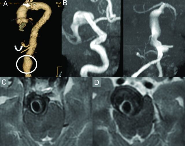
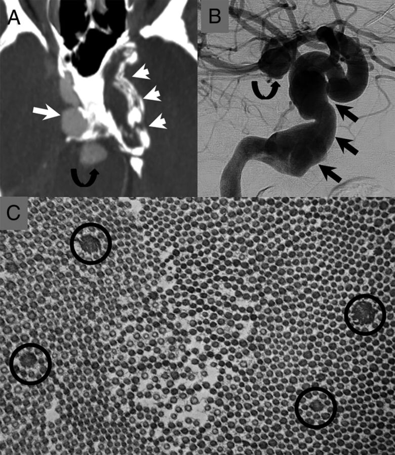
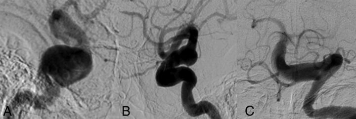
Background and purpose: Among patients with vertebrobasilar dolichoectasia is a subset of patients with disease affecting the anterior circulation as well. We hypothesized that multivessel intracranial dolichoectasia may represent a distinct phenotype from single-territory vertebrobasilar dolichoectasia. The purpose of this study was to characterize clinical characteristics and angiographic features of this proposed distinct phenotype termed "diffuse intracranial dolichoectasia" and compare them with those in patients with isolated vertebrobasilar dolichoectasia.

Materials and methods: We retrospectively reviewed a consecutive series of patients with diffuse intracranial dolichoectasia and compared their demographics, vascular risk factors, additional aneurysm prevalence, and clinical outcomes with a group of patients with vertebrobasilar dolichoectasia. "Diffuse intracranial dolichoectasia" was defined as aneurysmal dilation of entire vascular segments involving ≥2 intracranial vascular beds. Categoric and continuous variables were compared by using χ2 and Student t tests, respectively.

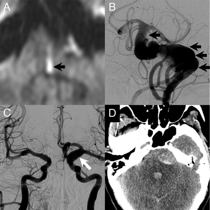
Results: Twenty-five patients had diffuse intracranial dolichoectasia, and 139 had vertebrobasilar dolichoectasia. Patients with diffuse intracranial dolichoectasia were older than those with vertebrobasilar dolichoectasia (70.9 ± 14.2 years versus 60.4 ± 12.5 years, P = .0002) and had a higher prevalence of abdominal aortic aneurysms (62.5% versus 14.3%, P = .01), other visceral aneurysms (25.0% versus 0%, P < .0001), and smoking (68.0% versus 15.9%, P < .0001). Patients with diffuse intracranial dolichoectasia were more likely to have aneurysm growth (46.2% versus 21.5%, P = .09) and rupture (20% versus 3.5%, P = .007) at follow-up. Patients with diffuse intracranial dolichoectasia were less likely to have good neurologic function at follow-up (24.0% versus 57.6%, P = .004) and were more likely to have aneurysm-related death (24.0% versus 7.2%, P = .02).

Conclusions: The natural history of patients with diffuse intracranial dolichoectasia is significantly worse than that in those with isolated vertebrobasilar dolichoectasia. Many patients with diffuse intracranial dolichoectasia had additional saccular and abdominal aortic aneurysms. These findings suggest that diffuse intracranial dolichoectasia may be a distinct vascular phenotype secondary to a systemic arteriopathy affecting multiple vascular beds.


Figures

Fig 1.
Fig 1.
A 67-year-old man who is a former smoker with a history of a penetrating atheromatous ulcer of the aortic arch (white arrow), a 5.6-cm abdominal aortic aneurysm (circle), and a celiac artery aneurysm (curved arrow, A). The patient had an episode of dizziness and headache and underwent a noncontrast CT of the head, which demonstrated enlarged intracranial arteries. An MRA demonstrated fusiform aneurysmal dilation of the entire right M1 segment measuring 10 mm in maximum diameter and a largely thrombosed fusiform aneurysm of the basilar artery, which measured 18 mm in maximum diameter (B and C). Approximately 9 months later, the aneurysm grew to 25 mm in diameter and started causing obstructive hydrocephalus (D). The patient also had a new perforator pontine infarct at the time (not shown). A programmable ventriculoperitoneal shunt was placed; however, the patient died due to complications of hydrocephalus 3 months later.
Fig 2.
Fig 2.
The patient is a 47-year-old man. He presented in his 20s with 2 large fusiform aneurysms of his cavernous carotid arteries. He later developed an aneurysm of his basilar artery and had a subarachnoid hemorrhage from a dissecting aneurysm of his PICA (not shown). A, CTA image demonstrates a large fusiform aneurysm of the right cavernous carotid artery (long white arrow) and a thrombosed/calcified aneurysm of the left cavernous carotid artery (short white arrows). There is also an aneurysm of the basilar tip (curved black arrow). B, Right ICA cerebral angiogram shows a large fusiform aneurysm of the right petrocavernous carotid artery (straight black arrows) and a basilar tip aneurysm (curved black arrow). C, There was suspicion for underlying connective tissue disease. The patient underwent a skin biopsy. Electron microscopy of the skin biopsy shows multiple abnormally enlarged collagen fibers (black circles) consistent with collagen flowers. These are typically seen in Ehlers-Danlos syndrome. The patient later underwent genetic testing for Loeys-Dietz, Ehlers-Danlos, and Marfan syndromes. The findings of all tests were negative.
Fig 3.
Fig 3.
A 51-year-old man with a long history of headaches with associated nausea and vomiting. He had acute-onset left-sided weakness with a prominent left facial droop along with left face, arm, and leg numbness and slurred speech. The patient had no family history of cerebral aneurysms, though his father had an abdominal aortic aneurysm. A, MR imaging at the time of the initial evaluation showed a medial left pontine infarct (black arrow). There was evidence of a large dolichoectatic aneurysm of the basilar artery on MR imaging, and the patient underwent cerebral angiography for further evaluation. B, Cerebral angiography demonstrated a fusiform-type aneurysm of the basilar artery with a filling defect that was consistent with thrombus (black arrows). The patient also had diffuse arteriomegaly with dilation of the right supraclinoid ICA to 6 mm and dilation of the left supraclinoid ICA to 10 mm (white arrow, C). D, The day following the angiography, the patient had a 10/10 headache. Noncontrast CT at the time showed diffuse subarachnoid hemorrhage with most of the blood products surrounding the basilar artery aneurysm. He died the next day.
Fig 4.
Fig 4.
An 87-year-old man with a history of a right third-nerve palsy. A, Right ICA cerebral angiogram demonstrates a 20-mm cavernous carotid fusiform aneurysm with associated dilation of the supraclinoid ICA as well. B, Left ICA cerebral angiogram shows dilation of the left supraclinoid ICA to approximately 10 mm. C, Left vertebral artery cerebral angiogram shows diffuse dilation and tortuosity of the basilar artery measuring 9 mm in maximum diameter. The cause of the third-nerve palsy was thought to be the right cavernous aneurysm.
Fig 5.
Fig 5.
Vertebrobasilar dolichoectasia in a 67-year-old man. A, Right and left ICA cerebral angiograms demonstrate normal-caliber internal carotid arteries, MCAs, and anterior cerebral arteries bilaterally. B, Left vertebral artery cerebral angiogram demonstrates an irregular dolichoectatic and fusiform aneurysm involving the entirety of the basilar artery.

References

    1. Dispensa BP, Saloner DA, Acevedo-Bolton G, et al. Estimation of fusiform intracranial aneurysm growth by serial magnetic resonance imaging. J Magn Reson Imaging 2007;26:177–83 10.1002/jmri.20944 - DOI - PubMed
    1. Flemming KD, Wiebers DO, Brown RD Jr, et al. The natural history of radiographically defined vertebrobasilar nonsaccular intracranial aneurysms. Cerebrovasc Dis 2005;20:270–79 10.1159/000087710 - DOI - PubMed
    1. Passero SG, Rossi S. Natural history of vertebrobasilar dolichoectasia. Neurology 2008;70:66–72 10.1212/01.wnl.0000286947.89193.f3 - DOI - PubMed
    1. Sacho RH, Saliou G, Kostynskyy A, et al. Natural history and outcome after treatment of unruptured intradural fusiform aneurysms. Stroke 2014;45:3251–56 10.1161/STROKEAHA.114.006292 - DOI - PubMed
    1. Mizutani T. A fatal, chronically growing basilar artery: a new type of dissecting aneurysm. J Neurosurg 1996;84:962–71 10.3171/jns.1996.84.6.0962 - DOI - PubMed

LinkOut - more resources