. 2017 Mar 3:7:43650.
doi: 10.1038/srep43650.

Nucleophosmin Interacts with PIN2/TERF1-interacting Telomerase Inhibitor 1 (PinX1) and Attenuates the PinX1 Inhibition on Telomerase Activity

Affiliations

Nucleophosmin Interacts with PIN2/TERF1-interacting Telomerase Inhibitor 1 (PinX1) and Attenuates the PinX1 Inhibition on Telomerase Activity

Derek Hang-Cheong Cheung et al. Sci Rep. .

Abstract

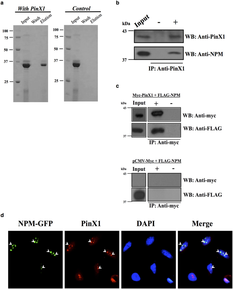
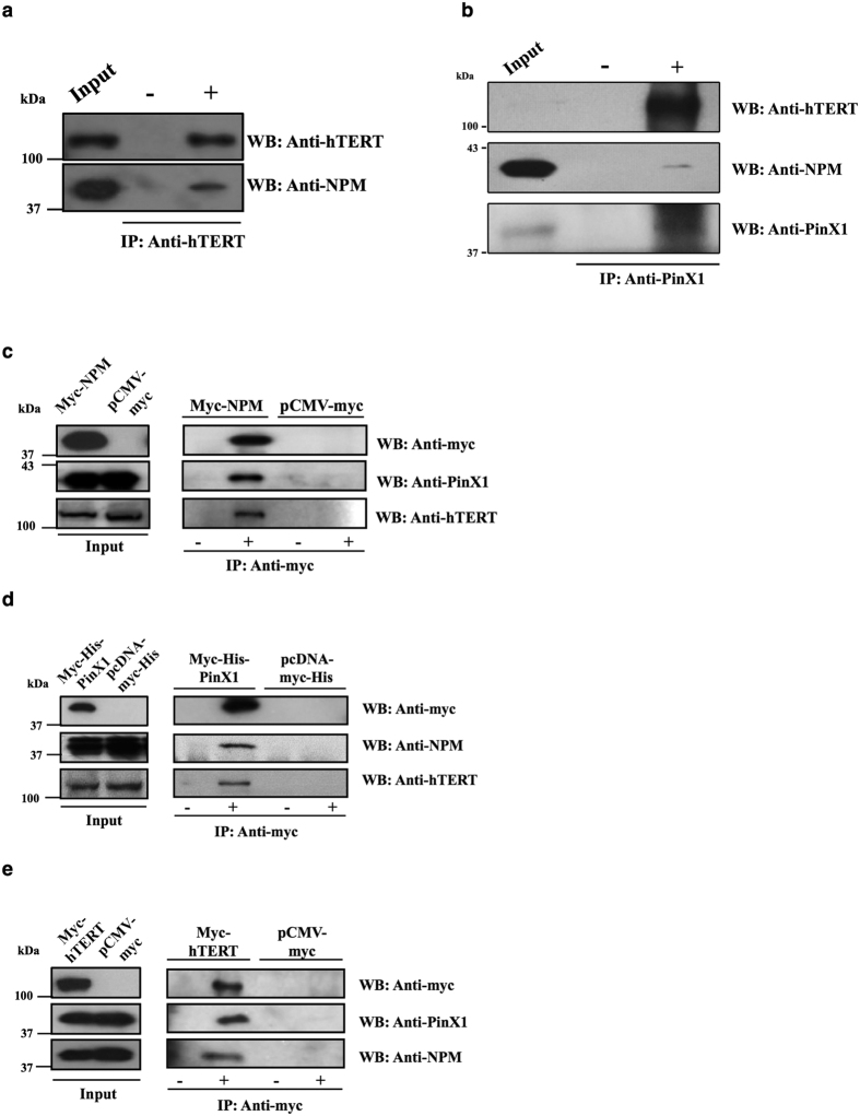
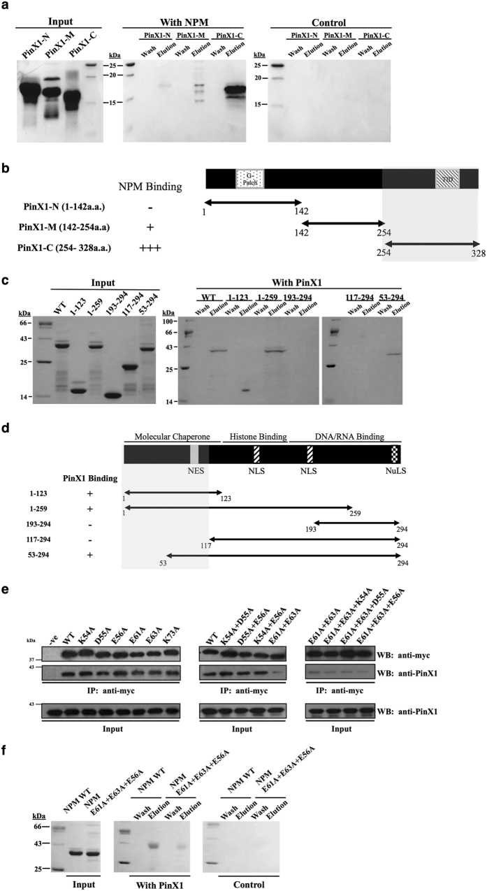
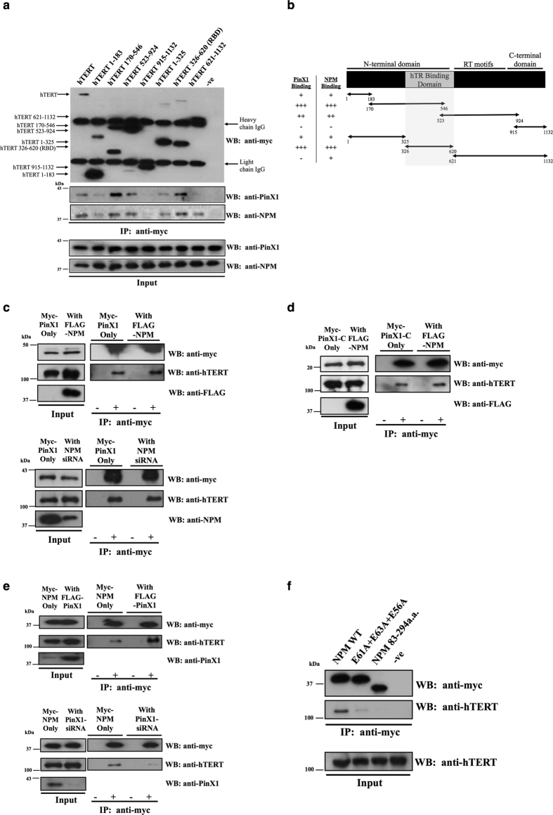
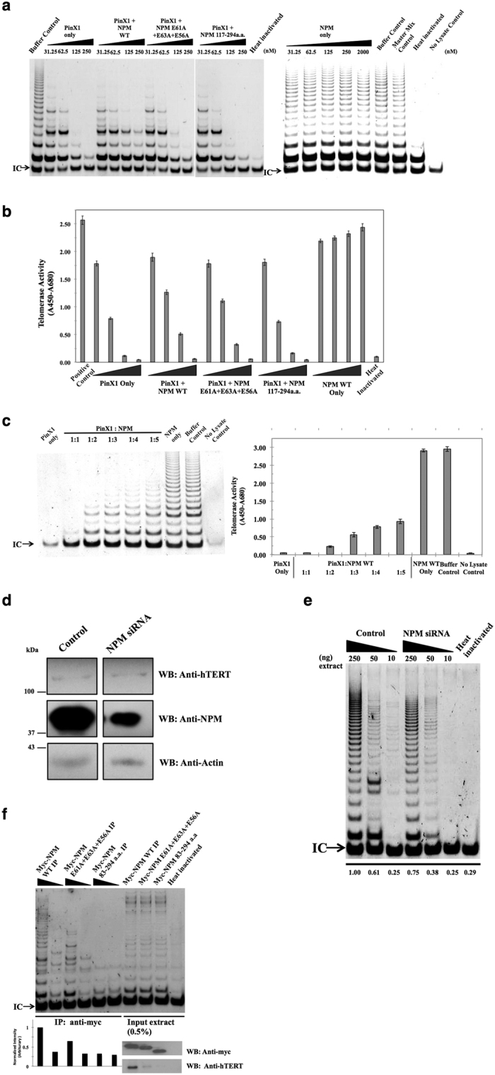
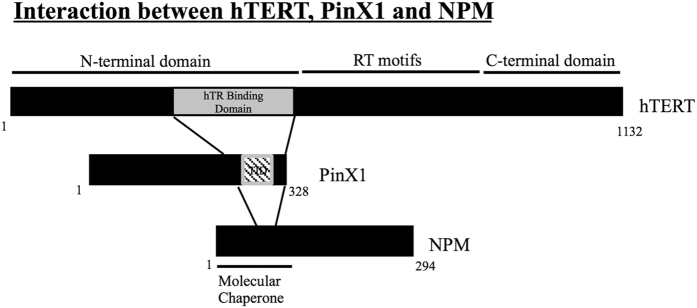
Telomerase activation and telomere maintenance are critical for cellular immortalization and transformation. PIN2/TERF1-interacting telomerase inhibitor 1 (PinX1) is a telomerase regulator and the aberrant expression of PinX1 causes telomere shortening. Identifying PinX1-interacting proteins is important for understanding telomere maintenance. We found that PinX1 directly interacts with nucleophosmin (NPM), a protein that has been shown to positively correlate with telomerase activity. We further showed that PinX1 acts as a linker in the association between NPM and hTERT, the catalytic subunit of telomerase. Additionally, the recruitment of NPM by PinX1 to the telomerase complex could partially attenuate the PinX1-mediated inhibition on telomerase activity. Taken together, our data reveal a novel mechanism that regulates telomerase activation through the interaction between NPM, PinX1 and the telomerase complex.


Conflict of interest statement

The authors declare no competing financial interests.

Figures

Figure 1
Figure 1. Interaction between PinX1 and nucleophosmin in vitro and in cells.
(a) Direct interaction of PinX1 and NPM in vitro. 3 mg of purified PinX1 was immobilized on an NHS column and 1 mg of purified NPM was loaded and allowed to interact with PinX1 in the column. A control column was prepared using the same immobilization procedures but without the addition of PinX1. (b) Endogenous PinX1 in HEK293T cells was precipitated by anti-PinX1 and protein A beads. The ‘−’ lane shows the antibody negative control. The western blots were analyzed using anti-PinX1 (~42 kDa) and anti-NPM (~37 kDa) antibodies. (c) Myc-PinX1 and FLAG-NPM were co-transfected into HEK293T cells. An anti-myc antibody (9B11) and protein A beads were used to isolate the immunoprecipitates. The empty vector pCMV-myc and FLAG-NPM were co-transfected as a control for the anti-myc antibody. The western blots were analyzed using anti-myc and anti-FLAG antibodies. (d) Immunofluorescence showing the co-localization of PinX1 and GFP-tagged NPM in HeLa cells. Endogenous PinX1 (Red) and NPM-GFP (Green) were detected by observing the immunofluorescence in HeLa cells by fluorescence microscopy. The DAPI (Blue) panel shows nuclei staining. The merged panel shows the superimposed PinX1 and NPM images.
Figure 2
Figure 2. Co-immunoprecipitation showing that PinX1, NPM and hTERT are associated as a complex inside the cell.
(a) Endogenous hTERT was immunoprecipitated by the anti-hTERT antibody from an HEK293T lysate. The presence of NPM in the precipitate was detected by an anti-NPM antibody. Antibody negative controls were included. The input controls are shown in the left panels. (b) Endogenous PinX1 was immunoprecipitated by an anti-PinX1 antibody from an HEK293T lysate. The presence of PinX1, hTERT and NPM in the precipitate was detected by anti-PinX1, anti-hTERT and anti-NPM antibodies. Antibody negative controls were included. The input controls are shown in the left panels. 5% of total lysates was loaded for SDS-PAGE. (c) Immunoprecipitation experiments were carried out in FLAG-PinX1- and pCI-neo hTERT-transfected HEK293T lysates with Myc-NPM as bait. Anti-PinX1 and anti-hTERT antibodies were used to detect the corresponding proteins. Antibody negative and empty vector controls were included in each set of experiment. The input controls are shown in the left panels. (d) Immunoprecipitation experiments were carried out in FLAG-NPM- and pCI-neo hTERT-transfected HEK293T lysates with Myc-His-PinX1 as bait. Anti-NPM and anti-hTERT antibodies were used to detect the corresponding proteins. Antibody negative and empty vector controls were included in each set of experiment. The input controls are shown in the left panels. (e) Immunoprecipitation experiments were carried out in FLAG-PinX1- and FLAG-NPM-transfected HEK293T lysates with Myc-hTERT as bait. Anti-PinX1 and anti-NPM antibodies were used to detect the corresponding proteins. Antibody negative and empty vector controls were included in each set of experiment. The input controls are shown in the left panels.
Figure 3
Figure 3. Mapping of the interaction between PinX1 and NPM.
(a) The mapping of the NPM interaction site was carried out by an in vitro pull-down assay on an NHS column by using 2 mg of purified NPM as bait. A control column was prepared using the same immobilization procedures but without the addition of NPM. 1 mg of PinX1 truncations was used as ligands. (b) The schematic diagram shows the ability of the truncated PinX1 proteins to bind NPM, where ‘−’ represents a lack of specific binding and the number of ‘+’ is proportional to the strength of NPM binding. The shaded region indicates the NPM interacting region. (c) PinX1 interacts with the N-terminal region of NPM. The mapping of the PinX1 interaction site was carried out by an in vitro pull-down assay using a 3 mg purified PinX1-immobilized NHS column. The input purified NPM variants (1 mg) are shown on the left panel. (d) Map showing the ability of the truncated NPM proteins to bind PinX1, where ‘−‘ represents a lack of specific binding and ‘+’ indicates specific binding with PinX1. (e) Systematic charge-to-alanine mutations were made on the mapped NPM region and their binding abilities were evaluated by co-immunoprecipitation. The PinX1 binding abilities of single point mutant (left), double point mutants (middle) and triple point mutants (right) were tested. The NPM mutations E61A + E63A + E56A were shown to be critical in PinX1 binding. The negative control was performed by using the cell lysate expressing WT NPM but without adding the anti-myc antibody. (f) In vitro pull-down assay between purified PinX1 and NPM E61A + E63A + E56A. 3 mg of PinX1 was immobilized on an NHS-column, and the ability of PinX1 to pull down the wild-type NPM and the mutant NPM E61A + E63A + E56A variant was compared. 1 mg of NPM variant proteins was used as ligands. A control column was prepared with the same immobilization procedures but without the addition of PinX1.
Figure 4
Figure 4. NPM does not compete with PinX1 for hTERT binding, and PinX1 acts as linker between NPM and hTERT association.
(a) Immunoprecipitation was carried out by transfecting the myc-tagged truncations of hTERT and FLAG-PinX1 into HEK293T cells. The presence of PinX1 and endogenous NPM in the myc-precipitates was analyzed by western blotting. A negative control was performed using cell lysate expressing full-length hTERT but without adding the anti-myc antibody. (b) Map showing the hTERT truncations for PinX1 and NPM binding, where ‘−’ represents a lack of hTERT binding and the number of ‘+’ indicates the relative strength of PinX1 or NPM binding. The shaded region indicates the region of that interacts with both PinX1 and NPM. (c) The effect of NPM level on the PinX1/hTERT association was investigated in myc-PinX1- transfected lysates by western blotting. The overexpression (upper panel) and down-regulation (lower panel) of NPM were achieved by the transfection of FLAG-NPM and NPM siRNA into HEK293T, respectively. An anti-myc antibody was added to precipitate the myc-PinX1-containing complex and the relative amount of hTERT was detected by an anti-hTERT antibody. The input lysates with overexpressed or down-regulated NPM are shown in the left panel. (d) The effect of NPM overexpression on the binding of PinX1-C to hTERT was analyzed by western blotting. The myc-PinX1-C-containing complex was precipitated by an anti-myc antibody, and the amount of hTERT was detected by an anti-hTERT antibody. (e) The effect of PinX1 level on the NPM/hTERT association was investigated in myc-NPM- transfected lysates by western blotting. The overexpression (upper panel) and down-regulation (lower panel) of PinX1 were achieved by the transfection of FLAG-PinX1 or PinX1 siRNA into HEK293T cells, respectively. An anti-myc antibody was added to precipitate the myc-NPM-containing complex, and the relative amount of hTERT was detected by an anti-hTERT antibody. The input lysate with overexpressed or down-regulated PinX1 are shown in the left panel. (f) Immunoprecipitation of wild-type NPM, NPM E61A + E63A + E56A variant and NPM 83–294a.a. against transfected hTERT in HEK293T cells. The relative amount of hTERT was detected by an anti-hTERT antibody.
Figure 5
Figure 5. PinX1/NPM interaction is critical for NPM recruitment to active telomerase and attenuates the PinX1 inhibitory effect on telomerase activity.
(a) The telomerase activities upon the addition of exogenous purified proteins were measured by TRAP assay. The exogenous proteins were incubated with HEK293T cell lysates on ice for 15 minutes prior to telomerase primer extension. The respective final protein concentrations in the reaction mixtures are labeled. (b) Telomerase activity was quantified by TRAP-ELISA as ΔA (A450-A680). The exogenous proteins were added to the reaction mixture at final concentrations of 31.25 nM, 62.5 nM, 125 nM, and 250 nM respectively prior to telomerase primer extension. (c) Left panel: The effect of NPM on the PinX1-mediated inhibition of telomerase activity was measured by TRAP assay. Right panel: Telomerase activity was quantified by TRAP-ELISA as ΔA (A450-A680). Increasing concentrations of wild-type NPM were incubated with a fixed amount of PinX1 (500 nM) and HEK293T lysates on ice prior to telomerase primer extension. (d) The effect of the down-regulation of NPM expression in HeLa cells was analyzed by western blotting. The hTERT, NPM, and actin signals are shown. (e) Decreased telomerase activity in NPM siRNA-transfected HeLa cells was demonstrated in a TRAP assay. The bottom panel indicates the relative band intensities in each lane against 250 ng of control extract (first lane) quantified using ImageJ software. (f) Telomerase activities in myc-immunoprecipitates of wild-type NPM, a mutant NPM with a disrupted PinX1 interaction site, and the NPM variant with the PinX1 binding site deleted were compared using a TRAP assay. The anti-myc and anti-hTERT input signals are shown in the lower right panel. The lower left bar chart shows the relative band intensities in each lane against the higher concentration myc-wild-type NPM immunoprecipitate (first lane) quantified using ImageJ software. The TRAP profiles shown in this figure are representative of more than three replicate experiments. IC: internal control for the PCR amplification. Buffer control: with the protein buffer. Master mix control: without purified proteins or additional buffers. Heat-inactivated control: master mix was heated at 95 °C prior to extension. No-lysate control: without the addition of cell extract.
Figure 6
Figure 6. Summary of findings.
Interaction between hTERT, PinX1 and NPM. The C-terminal end (including the TID domain) of PinX1 interacts with the hTR-binding domain of hTERT and links hTERT and NPM. The N-terminal domain of NPM interacts with the C-terminal domain of PinX1.

Similar articles

Cited by

References

    1. Chakhparonian M. & Wellinger R. J. Telomere maintenance and DNA replication: how closely are these two connected? Trends Genet. 19, 439–446 (2003). - PubMed
    1. Colgin L. & Reddel R. Telomere biology: a new player in the end zone. Curr. Biol. 14, R901–902 (2004). - PubMed
    1. Kim S. H., Kaminker P. & Campisi J. TIN2, a new regulator of telomere length in human cells. Nat. Genet. 23, 405–412 (1999). - PMC - PubMed
    1. Smith S., Giriat I., Schmitt A. & de Lange T. Tankyrase, a poly(ADP-ribose) polymerase at human telomeres. Science 282, 1484–1487 (1998). - PubMed
    1. van Steensel B. & de Lange T. Control of telomere length by the human telomeric protein TRF1. Nature 385, 740–743 (1997). - PubMed

Publication types

LinkOut - more resources