Review
. 2017 Feb;31(1):31-39.
doi: 10.1055/s-0037-1598191.

Orbital Fracture Repair

Affiliations
Review

Orbital Fracture Repair

Seanna Grob et al. Semin Plast Surg. 2017 Feb.

Abstract

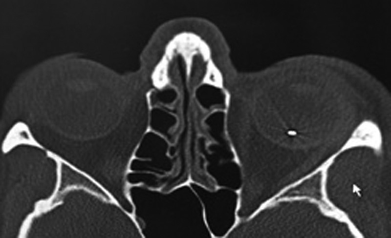
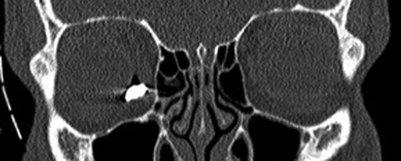
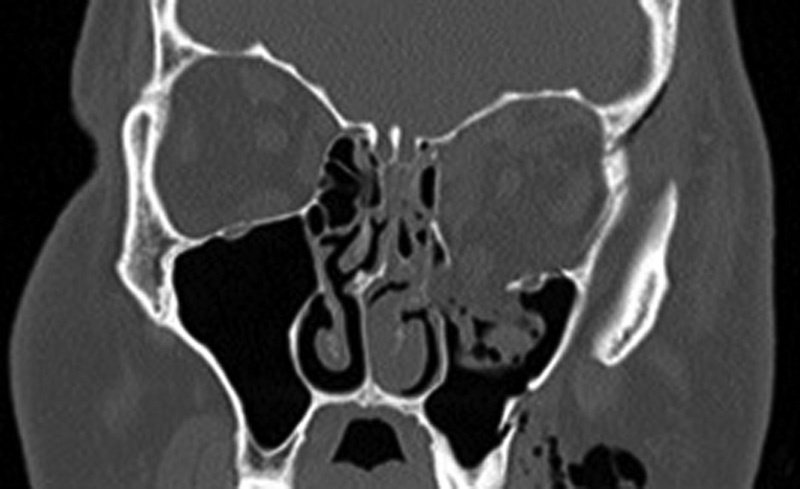
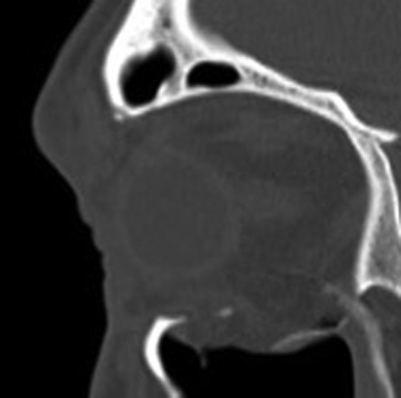
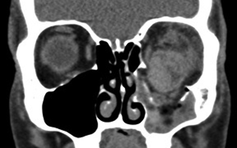
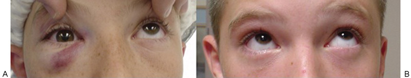
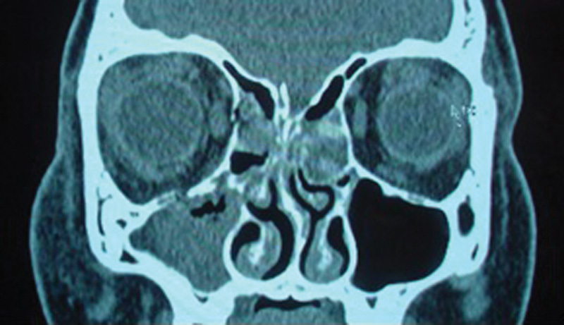
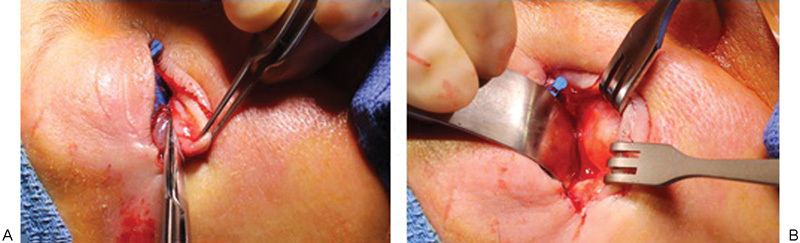
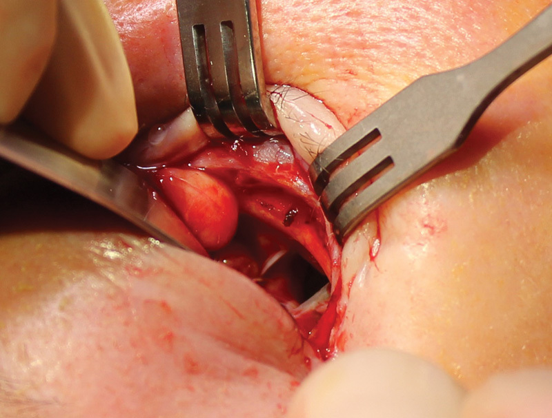
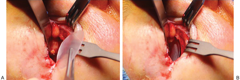
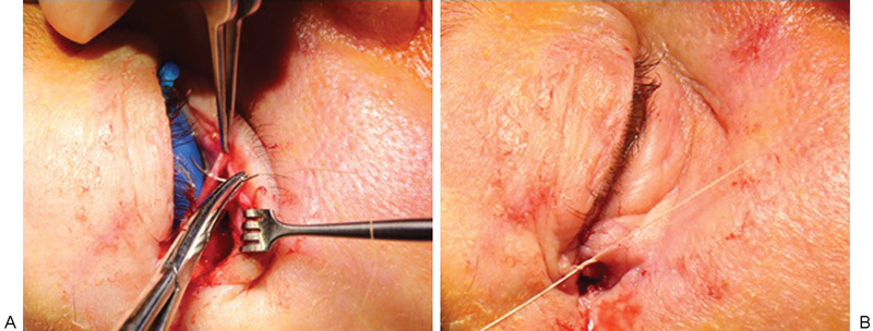
Orbital fractures are very common after facial trauma. The assessment of a patient with a suspected orbital wall injury includes a detailed oculofacial examination as well as radiologic imaging. Surgical repair with or without an implant may be indicated for diplopia, enophthalmos, or both. Cicatricial eyelid malposition is an iatrogenic complication commonly due to poor orbitotomy technique. Optimal repair involves direct exposure of the perimeter of the fractures' site through surgical planes that minimally scar the eyelids. A wide variety of implant options exist; however, thin, pliable, nonadherent materials such as nylon foil may offer several advantages. The authors describe the evaluation and management of orbital wall fractures.

Keywords: blow-out; enophthalmos; entrapment; medial orbital wall; orbit fracture; orbit implant; orbital floor.


Figures

Fig. 1
Fig. 1
Coronal computed tomography images of the obits showing a left inferior orbital wall fracture (A) and irregularity of the nasal globe contour of the left eye (B) suggesting a globe rupture.
Fig. 2
Fig. 2
Photographs of a patient with a left inferior orbital wall fracture showing restriction of both upgaze (A) and downgaze (B).
Fig. 3
Fig. 3
Coronal computed tomography image of the patient from Fig. 2 showing a left inferior orbital wall fracture and displacement of the inferior rectus muscle, which is also shown to be caught along the medial edge of the fracture site.
Fig. 4
Fig. 4
Photograph (A) showing evidence of enophthalmos on the left that is suggested clinically by the smaller palpebral fissure and deepened superior sulcus. Coronal computed tomography image (B) showing an inferior orbital wall fracture and an increase in the orbital volume compared with the noninjured right orbit.
Fig. 5
Fig. 5
Axial computed tomography image showing an intraocular foreign body of the left eye in the setting of an open globe injury from a ceiling fan falling.
Fig. 6
Fig. 6
Coronal computed tomography image showing an intraorbital foreign body of the right orbit adjacent to the globe. The patient had been hammering a nail.
Fig. 7
Fig. 7
Coronal computed tomography image showing an inferior orbital wall fracture on the left.
Fig. 8
Fig. 8
Sagittal computed tomography image showing the inferior rectus herniating in the location of the inferior orbital wall fracture.
Fig. 9
Fig. 9
Coronal computed tomography image showing significant left globe displacement inferiorly through the inferior orbital wall fracture and into the maxillary sinus.
Fig. 10
Fig. 10
Photographs of a patient with a white-eyed blowout fracture on the right, showing restriction in upgaze on the right prior to surgical repair (A) and improvement in upgaze after surgical repair (B).
Fig. 11
Fig. 11
Coronal computed tomography image showing the inferior orbital wall fracture on the right with minimal bone displacement.
Fig. 12
Fig. 12
(A) The lower eyelid is distracted inferiorly after canthotomy/cantholysis and a conjunctiva/retractor band incision is created. (B) With blunt manipulation, a malleable distracts orbit tissue posteriorly, while Senn retractors keep anterior lamella tissues out of the way of the subsequent periosteal incision.
Fig. 13
Fig. 13
Lifting the periosteum exposes the inferior wall fracture.
Fig. 14
Fig. 14
(A) A nylon foil implant is cut to approximate the size of the floor fracture. (B) The implant is place to cover the fracture with stable bone for peripheral support.
Fig. 15
Fig. 15
(A) The fornix incision is closed with a buried 6–0 chromic suture. (B) The lateral tarsus is secured to the lateral rim periosteum with a 4–0 Vicryl suture.

References

    1. Grove A S Jr New diagnostic techniques for the evaluation of orbital trauma Trans Sect Ophthalmol Am Acad Ophthalmol Otolaryngol 197783(4 Pt 1):626–640. - PubMed
    1. Grove A S Jr. Orbital trauma and computed tomography. Ophthalmology. 1980;87(5):403–411. - PubMed
    1. Grove A S Jr. Computed tomography in the management of orbital trauma. Ophthalmology. 1982;89(5):433–440. - PubMed
    1. Grove A S Jr Tadmor R New P F momose K J Orbital fracture evaluation by coronal computed tomography Am J Ophthalmol 197885(5 Pt 1):679–685. - PubMed
    1. Raflo G T. Blow-in and blow-out fractures of the orbit: clinical correlations and proposed mechanisms. Ophthalmic Surg. 1984;15(2):114–119. - PubMed