. 2017:2017:4671607.
doi: 10.1155/2017/4671607. Epub 2017 Feb 1.

Brain White Matter Impairment in Patients with Spinal Cord Injury

Affiliations

Brain White Matter Impairment in Patients with Spinal Cord Injury

Weimin Zheng et al. Neural Plast. 2017.

Abstract

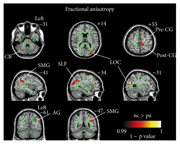
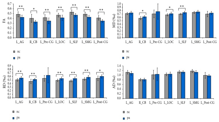
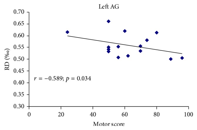
It remains unknown whether spinal cord injury (SCI) could indirectly impair or reshape the white matter (WM) of human brain and whether these changes are correlated with injury severity, duration, or clinical performance. We choose tract-based spatial statistics (TBSS) to investigate the possible changes in whole-brain white matter integrity and their associations with clinical variables in fifteen patients with SCI. Compared with the healthy controls, the patients exhibited significant decreases in WM fractional anisotropy (FA) in the left angular gyrus (AG), right cerebellum (CB), left precentral gyrus (PreCG), left lateral occipital region (LOC), left superior longitudinal fasciculus (SLF), left supramarginal gyrus (SMG), and left postcentral gyrus (PostCG) (p < 0.01, TFCE corrected). No significant differences were found in all diffusion indices between the complete and incomplete SCI. However, significantly negative correlation was shown between the increased radial diffusivity (RD) of left AG and total motor scores (uncorrected p < 0.05). Our findings provide evidence that SCI can cause not only direct degeneration but also transneuronal degeneration of brain WM, and these changes may be irrespective of the injury severity. The affection of left AG on rehabilitation therapies need to be further researched in the future.


Conflict of interest statement

The authors declare no competing financial interests.

Figures

Figure 1
Figure 1
Differences in fractional anisotropy (FA) between the SCI patients and healthy controls based on tract-based spatial statistics (TBSS) (p < 0.01, corrected using threshold-free cluster enhancement). Hot color represents 1 − p values. It is overlaid on the gyrus skeleton (green) and the MNI 152 template. Significant decreases in FA following SCI occurred in the left angular gyrus (AG), right cerebellar (CB), left precentral gyrus (PreCG), left lateral occipital region (LOC), left superior longitudinal fasciculus (SLF), left supramarginal gyrus (SMG), and left postcentral gyrus (PostCG).
Figure 2
Figure 2
Differences in diffusion metrics between the SCI patients and healthy controls based on region of interest (ROI) analysis. The ROIs were extracted based on the findings of TBSS. ∗∗ represents statistical significance with FDR q < 0.05; represents statistical significance with unadjusted p < 0.05. The error bar indicated standard deviation (SD). AG: angular gyrus, PreCG: precentral gyrus, LOC: lateral occipital region, SLF: superior longitudinal fasciculus, SMG: supramarginal gyrus, PostCG: postcentral gyrus, CB: cerebellar, FA: fractional anisotropy, MD: mean diffusivity, RD: radial diffusivity, AD: axial diffusivity.
Figure 3
Figure 3
The correlation between diffusion metrics and clinical scores in SCI patients. Pearson correlation showed negative association between the RD of left AG and motor score of ASIA. (r = −0.589, p = 0.034; uncorrected). RD: radial diffusivity, AG: angular gyrus, ASIA: American Spinal Injury Association, SCI: spinal cord injury.

References

    1. Pernet U., Hepp Reymond M. C. Retrograde degeneration of the pyramidal tract cells in the motor cortex of apes (Macaca fascicularis) Acta Anatomica. 1975;91(4):552–561. doi: 10.1159/000144414. - DOI - PubMed
    1. Feringa E. R., Vahlsing H. L. Labeled corticospinal neurons one year after spinal cord transection. Neuroscience Letters. 1985;58(3):283–286. doi: 10.1016/0304-3940(85)90067-9. - DOI - PubMed
    1. Hains B. C., Black J. A., Waxman S. G. Primary cortical motor neurons undergo apoptosis after axotomizing spinal cord injury. Journal of Comparative Neurology. 2003;462(3):328–341. doi: 10.1002/cne.10733. - DOI - PubMed
    1. Lee B. H., Lee K. H., Kim U. J., et al. Injury in the spinal cord may produce cell death in the brain. Brain Research. 2004;1020(1-2):37–44. doi: 10.1016/j.brainres.2004.05.113. - DOI - PubMed
    1. Kim B. G., Dai H.-N., McAtee M., Vicini S., Bregman B. S. Remodeling of synaptic structures in the motor cortex following spinal cord injury. Experimental Neurology. 2006;198(2):401–415. doi: 10.1016/j.expneurol.2005.12.010. - DOI - PubMed

Publication types

LinkOut - more resources