Review
. 2017;5(1):67-78.
doi: 10.1007/s40139-017-0127-9. Epub 2017 Jan 25.

Retinal Degeneration and Regeneration-Lessons From Fishes and Amphibians

Affiliations
Review

Retinal Degeneration and Regeneration-Lessons From Fishes and Amphibians

Divya Ail et al. Curr Pathobiol Rep. 2017.

Abstract

Purpose of review: Retinal degenerative diseases have immense socio-economic impact. Studying animal models that recapitulate human eye pathologies aids in understanding the pathogenesis of diseases and allows for the discovery of novel therapeutic strategies. Some non-mammalian species are known to have remarkable regenerative abilities and may provide the basis to develop strategies to stimulate self-repair in patients suffering from these retinal diseases.

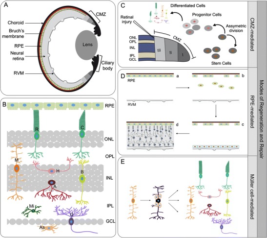
Recent findings: Non-mammalian organisms, such as zebrafish and Xenopus, have become attractive model systems to study retinal diseases. Additionally, many fish and amphibian models of retinal cell ablation and cell lineage analysis have been developed to study regeneration. These investigations highlighted several cellular sources for retinal repair in different fish and amphibian species. Moreover, major differences in repair mechanisms have been reported in these animal models.

Summary: This review aims to emphasize first on the importance of zebrafish and Xenopus models in studying the pathogenesis of retinal diseases and, second, on the different modes of regeneration processes in these model organisms.

Keywords: Ciliary marginal zone; Müller glial cells; Retinal degeneration; Retinal pigment epithelium; Retinal regeneration; Retinal stem cells.


Conflict of interest statement

Conflict of Interest

Divya Ail and Muriel Perron declare that they have no conflict of interest.

Human and Animal Rights and Informed Consent

This article does not contain any studies with human or animal subjects performed by any of the authors

Figures

Fig. 1
Fig. 1
a Structure of the retina. Schematic representation of a cross section of the zebrafish or Xenopus eye showing the ciliary marginal zone (CMZ), retinal pigment epithelium (RPE), neural retina, choroid, Bruch’s membrane, and the retinal vascular membrane (RVM). b Cell types of the retina. The retina is composed of different cell types: the nuclei of the two types of photoreceptors, rods (R) and cones (C), form the outer nuclear layer (ONL), whereas the Müller cells (M), horizontal cells (H), bipolar cells (B), and amacrine cells (A) are present in the inner nuclear layer (INL), and the ganglion cells (G) in the ganglion cell layer (GCL). The axons of these neurons and glial cells form synaptic connections in the outer and inner plexiform layers (OPL and IPL). The astrocytes (As) are located near the blood vessels whereas the microglia (Mi) are mostly located in the plexiform layers but can be distributed through the different layers. c–e Modes of regeneration and repair. CMZ-mediated (c): In the constantly growing retinas of zebrafish and Xenopus, the spatial cellular gradient in the CMZ recapitulates embryonic retinogenesis with zone I, the most peripheral part of the CMZ, where stem cells reside, zone II encompassing retinal progenitor cells, and zone III consisting of late retinal progenitors including post-mitotic retinoblasts. The stem cells divide asymmetrically to self-renew and generate one progenitor cell, and this mode of asymmetric division is retained even in the case of retinal injury. RPE-mediated (d): In Xenopus following partial retinectomy, wherein the RPE and RVM are left intact (a), a subset of RPE cells (green oval cells) detach from the Bruch’s membrane (in red) and migrate to the RVM (b). When they adhere to the RVM and form a distinct layer of cells (blue cells), they start proliferating (c) and regenerate the whole neural retina (d), while RPE cells that remained attached to the Bruch’s membrane renew the RPE layer (d). Müller-cell mediated (e): In zebrafish upon retinal injury, a subset of Müller cells (orange) undergo asymmetric division (black) to renew themselves and generate multipotent progenitor cells that can actively divide and regenerate all major retinal cell types (color figure online)

References

    1. Bainbridge JW, Mehat MS, Sundaram V, Robbie SJ, Barker SE, Ripamonti C, et al. Long-term effect of gene therapy on Leber’s congenital amaurosis. N Engl J Med. 2015;372(20):1887–1897. - PMC - PubMed
    1. Dejneka NS, Surace EM, Aleman TS, Cideciyan AV, Lyubarsky A, Savchenko A, et al. In utero gene therapy rescues vision in a murine model of congenital blindness. Mol Ther. 2004;9(2):182–188. - PubMed
    1. Fahim AT, Daiger SP, Weleber RG (2013) Retinitis pigmentosa overview. In: Pagon RA, Adam MP, Ardinger HH, Wallace SE, Amemiya A, Bean LJH, Bird TD, Ledbetter N, Mefford HC, Smith RJH, Stephens K (eds) GeneReviews. University of Washington, Seattle - PubMed
    1. Lambert NG, ElShelmani H, Singh MK, Mansergh FC, Wride MA, Padilla M, et al. Risk factors and biomarkers of age-related macular degeneration. Prog Retin Eye Res. 2016;54:64–102. - PMC - PubMed
    1. Seddon JM, Chen CA. The epidemiology of age-related macular degeneration. Int Ophthalmol Clin. 2004;44(4):17–39. - PubMed

LinkOut - more resources