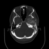
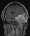
Calcified Middle Cranial Fossa Mass
- PMID: 28255529
- PMCID: PMC5332251
- DOI: 10.1055/s-0037-1598112
Calcified Middle Cranial Fossa Mass
Abstract
A 21-year-old male presented for evaluation of transient loss of consciousness and was found to have a hyperdense mass in the left middle fossa. He underwent craniotomy for tumor resection. Intra- and extradural invasion was noted. Gross total resection was achieved. Pathology demonstrated a densely cellular neoplasm with predominately spindle cell morphology in a collagen-containing stroma, areas of vascular proliferation, focal mineralization, and regions of cartilage formation. High mitotic index and regions of necrosis were seen. Based on the final diagnosis of osteosarcoma, the patient was referred for chemotherapy and radiation. Intracranial osteosarcoma is a nonmeningiomatous mesenchymal tumor. Most osteosarcomas are meningeal-based and supratentorial. Presentation depends on tumor location and may include focal neurologic deficits, cranial neuropathy, seizures, or symptoms of increased intracranial pressure. Given the relative rarity of intracranial osteosarcoma, there are no established guidelines for treatment, and therapy is guided by experience with systemic osteosarcoma. Gross total resection is recommended whenever feasible. Both chemotherapy and radiation therapy are used as adjuvant therapy. Regardless of treatment, osteosarcoma remains a highly aggressive malignancy with a poor prognosis. Morbidity and mortality may be the result of local recurrence or development of pulmonary or skeletal metastasis.
Keywords: middle fossa; neoplasms of connective and soft tissue; osteosarcoma.
Figures


References
-
- Paulus W, Scheithauer B W, Perry A. Lyon, France: International Agency for Research on Cancer; 2007. Mesenchymal, non-meningothelial tumours; pp. 173–176.
-
- Cihan Y B, Kaplan B, Deniz K et al.Primary calvarial osteosarcoma: a case report. J Neurol Sci (Turkish) 2012;1:122–128.
-
- Prayson R A. Philadelphia, PA: Elsevier; 2012. Non-glial tumors; pp. 518–521.
-
- Ashkan K, Pollock J, D'Arrigo C, Kitchen N D. Intracranial osteosarcomas: report of four cases and review of the literature. J Neurooncol. 1998;40(01):87–96. - PubMed
-
- Setzer M, Lang J, Turowski B, Marquardt G.Primary meningeal osteosarcoma: case report and review of the literature Neurosurgery 20025102488–492., discussion 492 - PubMed
Publication types
LinkOut - more resources
Full Text Sources
Other Literature Sources

