. 2017 Feb 22;4(3):e334.
doi: 10.1212/NXI.0000000000000334. eCollection 2017 May.

Microstructural visual system changes in AQP4-antibody-seropositive NMOSD

Affiliations

Microstructural visual system changes in AQP4-antibody-seropositive NMOSD

Frederike C Oertel et al. Neurol Neuroimmunol Neuroinflamm. .

Abstract

Objective: To trace microstructural changes in patients with aquaporin-4 antibody (AQP4-ab)-seropositive neuromyelitis optica spectrum disorders (NMOSDs) by investigating the afferent visual system in patients without clinically overt visual symptoms or visual pathway lesions.

Methods: Of 51 screened patients with NMOSD from a longitudinal observational cohort study, we compared 6 AQP4-ab-seropositive NMOSD patients with longitudinally extensive transverse myelitis (LETM) but no history of optic neuritis (ON) or other bout (NMOSD-LETM) to 19 AQP4-ab-seropositive NMOSD patients with previous ON (NMOSD-ON) and 26 healthy controls (HCs). Foveal thickness (FT), peripapillary retinal nerve fiber layer (pRNFL) thickness, and ganglion cell and inner plexiform layer (GCIPL) thickness were measured with optical coherence tomography (OCT). Microstructural changes in the optic radiation (OR) were investigated using diffusion tensor imaging (DTI). Visual function was determined by high-contrast visual acuity (VA). OCT results were confirmed in a second independent cohort.

Results: FT was reduced in both patients with NMOSD-LETM (p = 3.52e-14) and NMOSD-ON (p = 1.24e-16) in comparison with HC. Probabilistic tractography showed fractional anisotropy reduction in the OR in patients with NMOSD-LETM (p = 0.046) and NMOSD-ON (p = 1.50e-5) compared with HC. Only patients with NMOSD-ON but not NMOSD-LETM showed neuroaxonal damage in the form of pRNFL and GCIPL thinning. VA was normal in patients with NMOSD-LETM and was not associated with OCT or DTI parameters.

Conclusions: Patients with AQP4-ab-seropositive NMOSD without a history of ON have microstructural changes in the afferent visual system. The localization of retinal changes around the Müller-cell rich fovea supports a retinal astrocytopathy.


Figures

Figure 1
Figure 1. Flowchart of cohort selection
AQP4 = aquaporin-4; MOG = myelin oligodendrocyte glycoprotein; NMOSD = neuromyelitis optica spectrum disorder; OCT = optical coherence tomography.
Figure 2
Figure 2. OCT results
Boxplots of mean OCT values with values of individual eyes (jitter) in HC (left, white), NMOSD-LETM (middle, light blue), NMOSD-ON (right, dark blue), and for each confirmatory cohort (without color) for (A) FT values (µm); (B) pRNFL thickness (µm); (C) GCIPL volume (mm3); (D) FT in a representative macular scan of right eye from an HC; (E) FT changes in a representative macular scan of right eye from a patient with NMOSD-LETM. FT = foveal thickness; GCIPL = combined ganglion cell and inner plexiform layer volume; HC = healthy control; LETM = longitudinally extensive transverse myelitis; NMOSD-LETM = NMOSD patients with a history of LETM but no history of ON; NMOSD-ON = NMOSD patients with a history of ON; OCT = optical coherence tomography; ON = optic neuritis; pRNFL = peripapillary retinal nerve fiber layer thickness.
Figure 3
Figure 3. DTI results 1
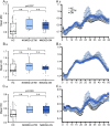
(A) Tract presentation of OR from LGN to V1 for averaged weight-mean DTI values of 50 segments in HC (white), NMOSD-LETM (light blue), and NMOSD-ON (dark blue) for FA (mean ± SEM). (B) Boxplot of mean FA values for middle 3/5 of the OR in HC (left, white), NMOSD-LETM (middle, light blue), and NMOSD-ON (right, dark blue). (C) Example of resulting fibers from tractography analysis. DTI = diffusion tensor imaging; FA = fractional anisotropy; HC = healthy control; LETM = longitudinally extensive transverse myelitis; LGN = lateral geniculate nucleus; NMOSD = neuromyelitis optica spectrum disorder; NMOSD-LETM = NMOSD patients with a history of LETM but no history of ON; NMOSD-ON = NMOSD patients with a history of ON; ON = optic neuritis; OR = optic radiation; V1 = primary visual cortex.
Figure 4
Figure 4. DTI results 2
(A.a–C.a) Boxplots of mean DTI values for middle 3/5 of the OR and (A.b–C.b) Tract presentation of OR from the LGN to V1 for averaged weight-mean DTI values of 50 segments in HC (white), NMOSD-LETM (light blue), and NMOSD-ON (dark blue) for (A) MD, (B) AD, and (C) RD (mean ± SEM for all). AD = axial diffusivity; DTI = diffusion tensor imaging; FA = fractional anisotropy; HC = healthy control; LETM = longitudinally extensive transverse myelitis; LGN = lateral geniculate nucleus; MD = mean diffusivity; NMOSD = neuromyelitis optica spectrum disorder; NMOSD-LETM = NMOSD patients with a history of LETM but no history of ON; NMOSD-ON = NMOSD patients with a history of ON; ON = optic neuritis; OR = optic radiation; RD = radial diffusivity; V1 = primary visual cortex.

References

    1. Jarius S, Wildemann B, Paul F. Neuromyelitis optica: clinical features, immunopathogenesis and treatment. Clin Exp Immunol 2014;176:149–164. - PMC - PubMed
    1. Metz I, Beißbarth T, Ellenberger D, et al. . Serum peptide reactivities may distinguish neuromyelitis optica subgroups and multiple sclerosis. Neurol Neuroimmunol Neuroinflamm 2016;3:e204. doi: 10.1212/NXI.0000000000000204. - DOI - PMC - PubMed
    1. Zekeridou A, Lennon VA. Aquaporin-4 autoimmunity. Neurol Neuroimmunol Neuroinflamm 2015;2:e110. doi: 10.1212/NXI.0000000000000110. - DOI - PMC - PubMed
    1. Waters P, Reindl M, Saiz A, et al. . Multicentre comparison of a diagnostic assay: aquaporin-4 antibodies in neuromyelitis optica. J Neurol Neurosurg Psychiatry 2016;87:1005–1015. - PMC - PubMed
    1. Zamvil SS, Slavin AJ. Does MOG Ig-positive AQP4-seronegative opticospinal inflammatory disease justify a diagnosis of NMO spectrum disorder? Neurol Neuroimmunol Neuroinflamm 2015;2:e62. doi: 10.1212/NXI.0000000000000062. - DOI - PMC - PubMed