. 2017 May;108(5):1049-1057.
doi: 10.1111/cas.13218. Epub 2017 May 11.

Novel small molecule inhibiting CDCP1-PKCδ pathway reduces tumor metastasis and proliferation

Affiliations

Novel small molecule inhibiting CDCP1-PKCδ pathway reduces tumor metastasis and proliferation

Katsuhiko Nakashima et al. Cancer Sci. 2017 May.

Abstract

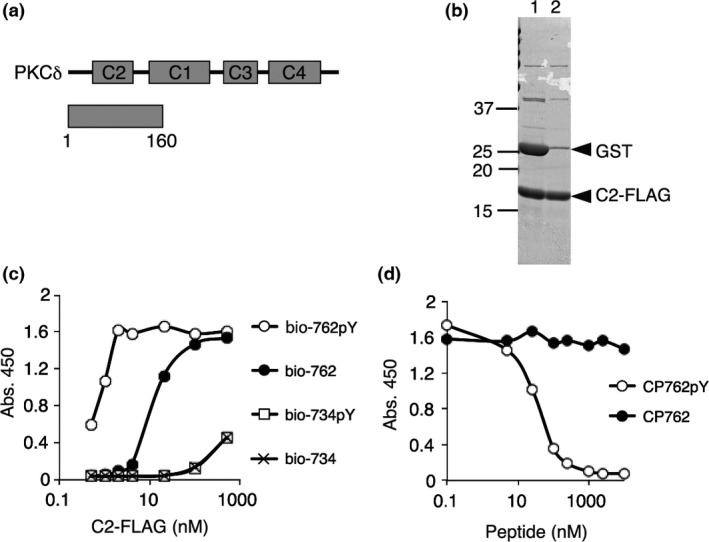
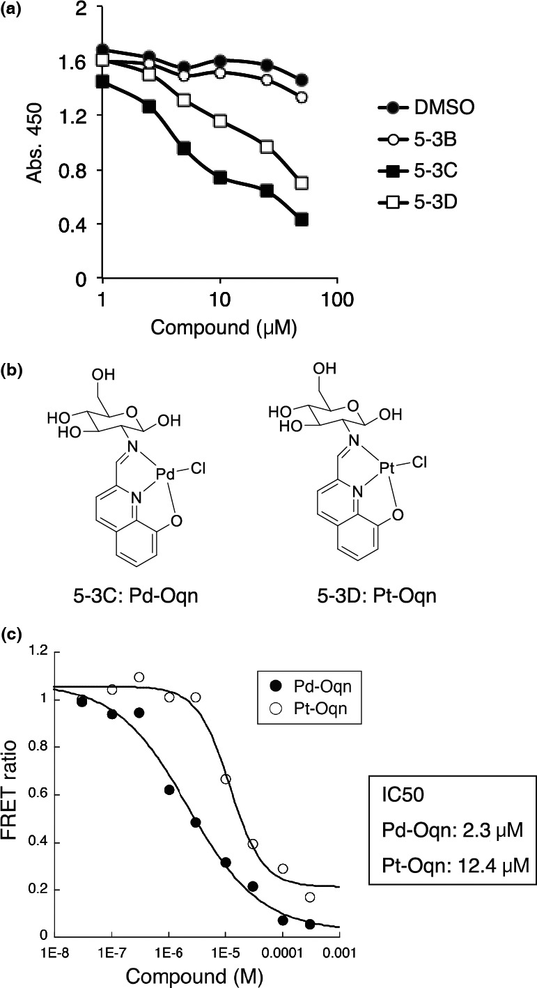
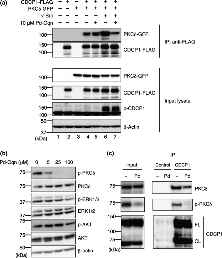
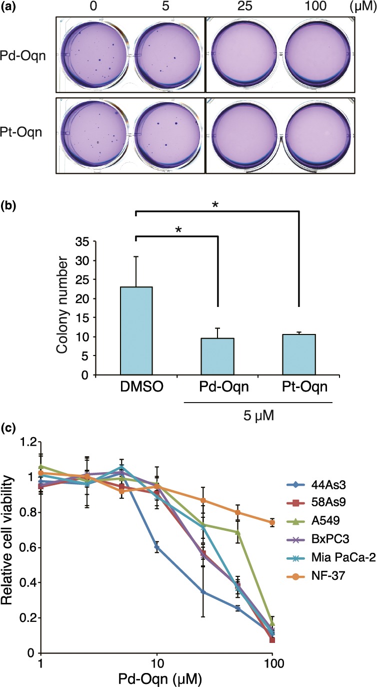
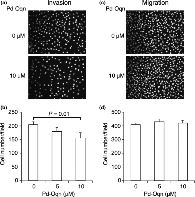
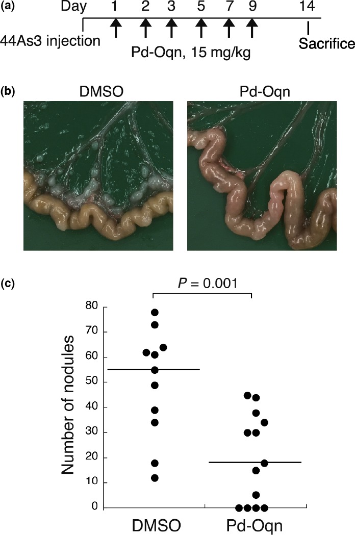
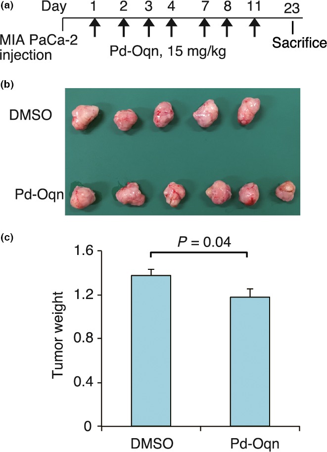
CUB domain-containing protein-1 (CDCP1) is a trans-membrane protein predominantly expressed in various cancer cells and involved in tumor progression. CDCP1 is phosphorylated at tyrosine residues in the intracellular domain by Src family kinases and recruits PKCδ to the plasma membrane through tyrosine phosphorylation-dependent association with the C2 domain of PKCδ, which in turn induces a survival signal in an anchorage-independent condition. In this study, we used our cell-free screening system to identify a small compound, glycoconjugated palladium complex (Pd-Oqn), which significantly inhibited the interaction between the C2 domain of PKCδ and phosphorylated CDCP1. Immunoprecipitation assays demonstrated that Pd-Oqn hindered the intercellular interaction of phosphorylated CDCP1 with PKCδ and also suppressed the phosphorylation of PKCδ but not that of ERK or AKT. In addition, Pd-Oqn inhibited the colony formation of gastric adenocarcinoma 44As3 cells in soft agar as well as their invasion. In mouse models, Pd-Oqn markedly reduced the peritoneal dissemination of gastric adenocarcinoma cells and the tumor growth of pancreatic cancer orthotopic xenografts. These results suggest that the novel compound Pd-Oqn reduces tumor metastasis and growth by inhibiting the association between CDCP1 and PKCδ, thus potentially representing a promising candidate among therapeutic reagents targeting protein-protein interaction.

Keywords: CDCP1; PKCδ; Src; chemical screening; metastasis.


Figures

Figure 1
Figure 1
Establishment of an assay system for the interaction of CDCP1 with PKCδ. (a) Domain structure of PKCδ and the region of the C2 recombinant protein (1–160) that was used for the C2‐CDCP1 binding assay. (b) Purification of C2‐FLAG. GST‐C2‐FLAG protein purified from bacterial lysates was proteolyticaly digested (lane 1) and the GST was removed (lane 2). The proteins were subjected to SDS‐12.5% PAGE and Coomassie brilliant blue (CBB) staining. (c) Selective binding of the C2 domain to the phosphorylated site at Y762 of CDCP1. Biotinylated peptide, bio‐762pY, bio‐762, bio‐734pY, or bio‐734, immobilized on a 96‐well plate coated with streptavidin and C2‐FLAG (0.5–500 nM) was used for the binding assay. (d) Competitive inhibition in the C2‐CDCP1 binding assay by free CP762pY peptide. C2‐FLAG (2 nM) and bio‐762pY were used for the assay and the binding reaction was performed in the presence of 0.1–10 000 nM CP762pY or CP762 peptide that was not biotinylated.
Figure 2
Figure 2
Identification of small molecules that inhibit C2‐CDCP1 binding. (a) Inhibitory effect of small compounds on the C2‐CDCP1 binding assay. The small compounds 5‐3B, 5‐3C, and 5‐3D (1.0–50 μM) or DMSO were used in the C2‐CDCP1 binding assay as inhibitory compounds. (b) Structures of 5‐3C and 5‐3D, identified as inhibitory compounds on C2‐CDCP1 binding. 5‐3C; Pd‐Oqn. 5‐3D; Pt‐Oqn. (c) Inhibition kinetics of Pd‐Oqn and Pt‐Oqn on C2‐CDCP1 binding. HTRF assays with Pd‐Oqn (Pd) or Pt‐Oqn (Pt) were performed and IC50 values were calculated.
Figure 3
Figure 3
Inhibition of the CDCP1‐PKCδ pathway by Pd‐Oqn. (a) Blockage of the CDCP1‐PKCδ interaction by Pd‐Oqn in cells. Immunoprecipitation was performed as described in Materials and Methods. Immunoprecipitated proteins and input lysates were subjected to western blotting with the antibodies as indicated at the right side. The top indicates the transfection plasmids and Pd‐Oqn addition. (b) Inhibition of the phosphorylation of PKCδ by Pd‐Oqn. 44As3 cells were treated with Pd‐Oqn and the lysates were subjected to western blotting with the antibodies as indicated at the right side. (c) Inhibition of the endogenous CDCP1‐PKCδ interaction by Pd‐O qn. 44As3 cells were treated with DMSO (‐) or Pd‐Oqn (Pd) for 24 h and the cell lysates were subjected to immunoprecipitation with anti‐CDCP1 antibody or control IgG. The precipitated proteins (IP) and the cell lysate (input) were subjected to western blotting with the antibodies as indicated at right side. CDCP1 bands showed both full‐length (FL) and cleaved form (CL) at about 135 and 70 kDa respectively. Molecular weight is indicated at the left.
Figure 4
Figure 4
Inhibitory effect of Pd‐Oqn on the growth of cancer cells. (a, b) Inhibition of colony formation of 44As3 cells in soft agar by Pd‐Oqn and Pt‐Oqn. The soft agar colony formation assay was performed as described in Materials and Methods. Representative photos of the colonies in soft agar with DMSO, Pd‐Oqn, or Pt‐Oqn (5, 25, and 100 μM) are shown in (a). The bar graph indicates the number of colonies in soft agar with DMSO, 5 μM Pd‐Oqn, or Pt‐Oqn as the means ± SD of three biological replicates. Statistical significance was determined using the standard Student's t‐test. *P < 0.05. (d) Inhibition of cell proliferation of various cancer cells by Pd‐Oqn. The cells were cultured with Pd‐Oqn (1.0–100 μM) or DMSO (control) and cell viability was determined using the CCK assay after 48 h. The graph indicates the relative value to DMSO treated cells of each cell line as the means ± SD of three biological replicates.
Figure 5
Figure 5
Inhibitory effect of Pd‐Oqn on the invasion of cancer cells. 44As3 cells were subjected to invasion (a, b) and migration (c, d) assays in the presence or absence of Pd‐Oqn. Representative photos of invaded (a) or migrated (c) cells are shown as nuclear staining with DAPI. The graph indicates the cell number of invaded (b) or migrated (d) cells upon DMSO or Pd‐Oqn treatment as the means ± SD of three biological replicates. Statistical significance was determined using the Student's t‐test.
Figure 6
Figure 6
Effect of Pd‐Oqn on the peritoneal dissemination of gastric adenocarcinoma. (a) Schedule of the experiment for injection of Pd‐Oqn or DMSO into the mouse model of peritoneal dissemination using 44As3 cells. (b) Representative photos of the nodules on the mouse mesenterium. (c) The dot plot indicates the number of nodules in each mouse treated with Pd‐Oqn (n = 13) or DMSO (n = 10). The bars in the plot indicate the median value. Statistical significance was determined using the Student's t‐test.
Figure 7
Figure 7
Effect of Pd‐Oqn on pancreatic cancer of orthotopic xenografts. (a) Schedule of the experiment for the injection of Pd‐Oqn or DMSO into the orthotopic xenograft model of pancreatic cancer. (b) Photo of the pancreatic tumor from the dissected mouse. (c) The bar graph indicates the weight of the tumor treated with Pd‐Oqn (n = 6) or DMSO (n = 5) as the means ± SD. Statistical significance was determined using the Student's t‐test.

References

    1. Scherl‐Mostageer M, Sommergruber W, Abseher R, Hauptmann R, Ambros P, Schweifer N. Identification of a novel gene, CDCP1, overexpressed in human colorectal cancer. Oncogene 2001; 20: 4402–8. - PubMed
    1. Uekita T, Jia L, Narisawa‐Saito M, Yokota J, Kiyono T, Sakai R. CUB domain‐containing protein 1 is a novel regulator of anoikis resistance in lung adenocarcinoma. Mol Cell Biol 2007; 27: 7649–60. - PMC - PubMed
    1. Uekita T, Sakai R. Roles of CUB domain‐containing protein 1 signaling in cancer invasion and metastasis. Cancer Sci 2011; 102: 1943–8. - PubMed
    1. Benes CH, Wu N, Elia AE, Dharia T, Cantley LC, Soltoff SP. The C2 domain of PKCdelta is a phosphotyrosine binding domain. Cell 2005; 121: 271–80. - PubMed
    1. Miyazawa Y, Uekita T, Ito Y, Seiki M, Yamaguchi H, Sakai R. CDCP1 regulates the function of MT1‐MMP and invadopodia‐mediated invasion of cancer cells. Mol Cancer Res 2013; 11: 628–37. - PubMed

MeSH terms