. 2017 May;23(5):405-415.
doi: 10.1111/cns.12683. Epub 2017 Mar 2.

GSK-3β inhibitor TDZD-8 reduces neonatal hypoxic-ischemic brain injury in mice

Affiliations

GSK-3β inhibitor TDZD-8 reduces neonatal hypoxic-ischemic brain injury in mice

Sammen Huang et al. CNS Neurosci Ther. 2017 May.

Abstract

Aims: Glycogen synthase kinase 3β (GSK-3β) is activated following hypoxic-ischemic (HI) brain injury. TDZD-8 is a specific GSK-3β inhibitor. Currently, the impact of inhibiting GSK-3β in neonatal HI injury is unknown. We aimed to investigate the effect of TDZD-8 following neonatal HI brain injury.

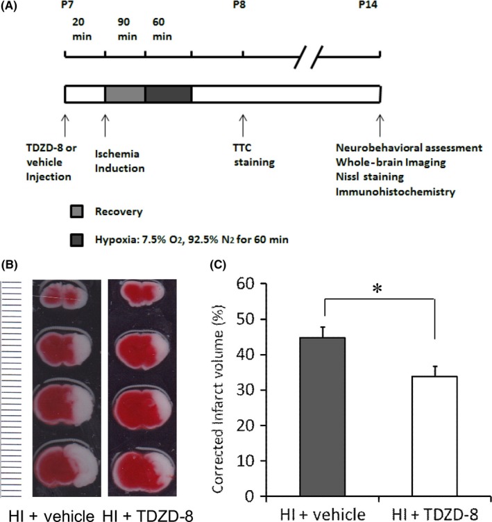
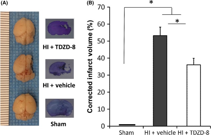
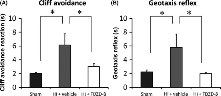
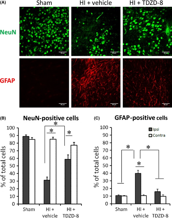
Methods: Unilateral common carotid artery ligation followed by hypoxia was used to induce HI injury in postnatal day 7 mouse pups pretreated with TDZD-8 or vehicle. The infarct volume, whole-brain imaging, Nissl staining, and behavioral tests were used to evaluate the protective effect of TDZD-8 on the neonatal brain and assess functional recovery after injury. Western blot was used to evaluate protein levels of phosphorylated protein kinase B (Akt), GSK-3β, and cleaved caspase-3. Protein levels of cleaved caspase-3, neuronal marker, and glial fibrillary acidic protein were detected through immunohistochemistry.

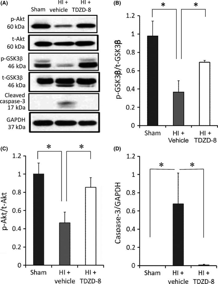
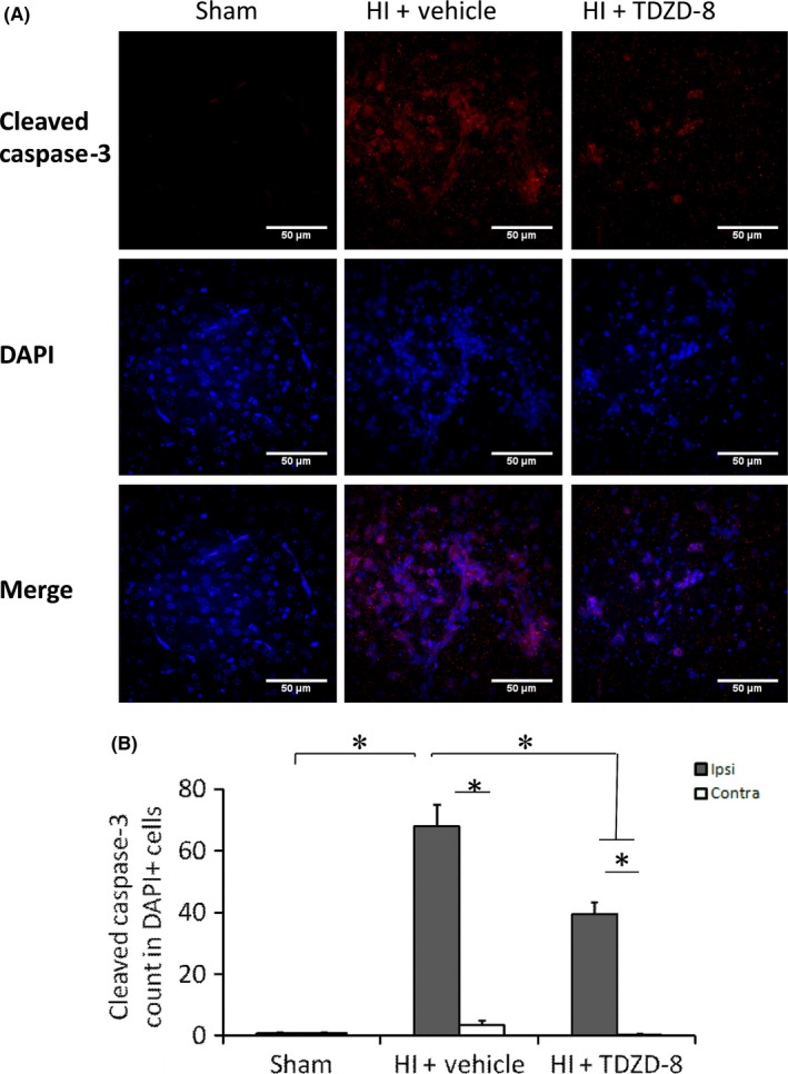
Results: Pretreatment with TDZD-8 significantly reduced brain damage and improved neurobehavioral outcomes following HI injury. TDZD-8 reversed the reduction of phosphorylated Akt and GSK-3β, and the activation of caspase-3 induced by hypoxia-ischemia. In addition, TDZD-8 suppressed apoptotic cell death and reduced reactive astrogliosis.

Conclusion: TDZD-8 has the therapeutic potential for hypoxic-ischemic brain injury in neonates. The neuroprotective effect of TDZD-8 appears to be mediated through its antiapoptotic activity and by reducing astrogliosis.

Keywords: GSK-3β; TDZD-8; neonatal hypoxia-ischemia, neuroprotection.


Conflict of interest statement

The authors declare no conflict of interest.

Figures

Figure 1
Figure 1
TDZD‐8 pretreatment reduced infarct volume 24 hours following neonatal hypoxic‐ischemic brain injury. (A) Timeline of neonatal hypoxic‐ischemic injury and experimental procedures. Seven‐day‐old pups (P7) were randomly selected to be injected with either TDZD‐8 or vehicle solution 20 minutes prior to ischemia induction. This was followed by 90 minutes of recovery and 60 minutes of hypoxia with 7.5% O2 and 92.5% N2. TTC staining was performed 24 hours after HI (P8). Neurobehavioral assessment, whole‐brain imaging, Nissl staining, and immunohistochemistry were performed 7 days after the HI (P14). (B) Effect of TDZD‐8 pretreatment on infarct volume. Representative TTC images of coronal brain slices 24 hours after HI injury show brain damage areas in white color (the dotted line on the left of the image represented “mm” in size for each line). (C) Summary of brain infarction volumes of TTC images. Pretreatment with TDZD‐8 significantly reduced the infarct volume compared to the vehicle‐treated group (*P<.05, Student's t test); HI+vehicle group, n=9; HI+TDZD‐8 group, n=20
Figure 2
Figure 2
TDZD‐8 pretreatment reduced infarct volume 7 days following neonatal hypoxic‐ischemic brain injury. (A) Effect of TDZD‐8 on brain damage. Representative whole‐brain images and Nissl staining taken 7 days after HI injury are shown (the dotted line on the left of the whole‐brain image represented “mm” in size for each line). (B) Analysis of brain damage for whole‐brain images. Hypoxic‐ischemic injury induced significant brain damage in the vehicle‐treated group compared to the sham group. Pretreatment with TDZD‐8 significantly reduced the infarct volume and improved whole‐brain morphology compared to the vehicle‐treated group 7 days after the HI injury (*P<.01 vs sham group or vehicle group; One‐way ANOVA followed by Fisher LSD Method); sham group, n=12; HI+vehicle group, n=18; HI+TDZD‐8 group, n=20
Figure 3
Figure 3
TDZD‐8 pretreatment improved neurobehavioral outcome assessed at 7 days following neonatal hypoxic‐ischemic brain injury. (A) Cliff avoidance test. (B) Geotaxis Reflex test. TDZD‐8 pretreatment significantly improved cliff avoidance and geotaxis test scores in comparison with the vehicle control group but had no difference compared to the sham group (*P<.05 vs sham group or vehicle group; One‐way ANOVA followed by Fisher LSD Method); sham group, n=18; HI+vehicle group, n=12; HI+TDZD‐8 group, n=11
Figure 4
Figure 4
Developmental changes in GSK‐3β and Akt levels. (A) 3D gene expression patterns of GSK‐3β in coronal and sagittal slice in C57BL mice were determined at E18.5, P4, P14, and P28, using ISH. Images were obtained from Website: © 2015 Allen Institute for Brain Science. Allen Developing Mouse Brain Atlas [Internet]. Available from: http://developingmouse.brain-map.org. (B) Western blots showing protein levels of GSK‐3β and Akt at E17, P7, and P14. Analysis of band intensity showing the ratio of (C) p‐GSK3β to GAPDH, (D) t‐GSK3β to GAPDH, and (E) p‐GSK3β to t‐GSK3β. Analysis of band intensity showing the ratio of (F) p‐Akt to GAPDH, (G) t‐Akt to GAPDH, and (H) p‐Akt to t‐Akt. (*P<.05 vs sham group or TDZD‐8 treatment group; One‐way ANOVA followed by Fisher LSD Method). E17 group, n=5; P7 group, n=4; P14 group, n=5
Figure 5
Figure 5
The effects of TDZD‐8 on the expression levels of cell survival and apoptotic markers following HI. Samples were collected 24 hours after HI from the ipsilateral brain hemispheres. (A) Representative Western blot gel showing protein expression levels from the sham, vehicle‐treated HI, and TDZD‐8‐treated HI groups. Analysis of band intensity showing the ratio of (B) p‐GSK‐3β to t‐GSK‐3β, (C) p‐Akt to t‐Akt, and (D) cleaved caspase‐3 to GAPDH. (*P<.05 vs sham group or TDZD‐8 treatment group; One‐way ANOVA followed by Fisher LSD Method). Sham group, n=5; HI+vehicle group, n=5; HI+TDZD‐8 group, n=5
Figure 6
Figure 6
Immunohistochemistry staining showing the effects of TDZD‐8 on cleaved caspase‐3 staining following hypoxic‐ischemic brain injury. Brain samples were collected 7 days after HI and sliced coronally with a thickness of ~50 μm. (A) Representative images for cleaved caspase‐3 and DAPI staining in sham, vehicle, and TDZD‐8 groups following HI. (B) Analysis of cleaved caspase‐3 count in DAPI‐positive cells. (P<.05 for all comparisons; One‐way ANOVA followed by Fisher LSD Method). For ipsilateral brain, sham group, n=5; HI+vehicle group, n=9; HI+TDZD‐8 group, n=7. For contralateral brain, sham group, n=5; HI+vehicle group, n=8; HI+TDZD‐8 group, n=5
Figure 7
Figure 7
Immunohistochemistry staining showing the effects of TDZD‐8 on NeuN‐positive cells and GFAP‐positive cells following hypoxic‐ischemic brain injury. Brain samples were collected 7 days after HI and sliced into coronal sections with a thickness of ~50 μm. (A) Representative images for NeuN and GFAP staining. (B) Analysis of NeuN‐positive cells as a percentage of DAPI‐positive cells in sham, vehicle, and treatment groups. (C) Analysis of GFAP‐positive cells as a percentage of DAPI‐positive cells. (P<.05 for all comparisons; One‐way ANOVA followed by Fisher LSD Method). For ipsilateral brain, sham group, n=9; HI+vehicle group, n=9; HI+TDZD‐8 group, n=10. For contralateral brain, sham group, n=9; HI+vehicle group, n=9; HI+TDZD‐8 group, n=8

References

    1. Woodgett JR. Molecular‐cloning and expression of glycogen‐synthase kinase‐3 factor‐A. EMBO J. 1990;9:2431‐2438. - PMC - PubMed
    1. Yao HB, Shaw PC, Wong CC, et al. Expression of glycogen synthase kinase‐3 isoforms in mouse tissues and their transcription in the brain. J Chem Neuroanat. 2002;23:291‐297. - PubMed
    1. Mukai F, Ishiguro K, Sano Y, et al. Alternative splicing isoform of tau protein kinase I/glycogen synthase kinase 3 beta. J Neurochem. 2002;81:1073‐1083. - PubMed
    1. Hur EM, Zhou FQ. GSK3 signalling in neural development. Nat Rev Neurosci. 2010;11:539‐551. - PMC - PubMed
    1. Shi SH, Cheng T, Jan LY, et al. APC and GSK‐3 beta are involved in mPar3 targeting to the nascent axon and establishment of neuronal polarity. Curr Biol. 2004;14:2025‐2032. - PubMed

MeSH terms