

Melanin, Radiation, and Energy Transduction in Fungi
- PMID: 28256187
- PMCID: PMC11687467
- DOI: 10.1128/microbiolspec.FUNK-0037-2016
Melanin, Radiation, and Energy Transduction in Fungi
Abstract
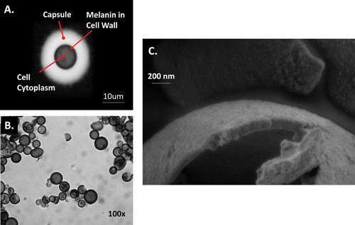
Melanin pigments are found in many diverse fungal species, where they serve a variety of functions that promote fitness and cell survival. Melanotic fungi inhabit some of the most extreme habitats on earth such as the damaged nuclear reactor at Chernobyl and the highlands of Antarctica, both of which are high-radiation environments. Melanotic fungi migrate toward radioactive sources, which appear to enhance their growth. This phenomenon, combined with the known capacities of melanin to absorb a broad spectrum of electromagnetic radiation and transduce this radiation into other forms of energy, raises the possibility that melanin also functions in harvesting such energy for biological usage. The ability of melanotic fungi to harness electromagnetic radiation for physiological processes has enormous implications for biological energy flows in the biosphere and for exobiology, since it provides new mechanisms for survival in extraterrestrial conditions. Whereas some features of the way melanin-related energy transduction works can be discerned by linking various observations and circumstantial data, the mechanistic details remain to be discovered.
Figures
References
-
- Butler MJ, Day AW. 1998. Fungal melanins: a review. Can J Microbiol 44:1115–1136. 10.1139/w98-119 - DOI
Publication types
MeSH terms
Substances
LinkOut - more resources
Full Text Sources
Other Literature Sources
Medical
