. 2017 Mar 3:7:43308.
doi: 10.1038/srep43308.

Vascular Permeability Drives Susceptibility to Influenza Infection in a Murine Model of Sickle Cell Disease

Affiliations

Vascular Permeability Drives Susceptibility to Influenza Infection in a Murine Model of Sickle Cell Disease

Erik A Karlsson et al. Sci Rep. .

Abstract

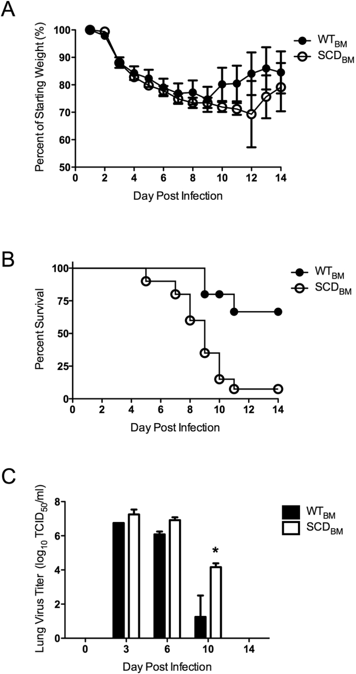
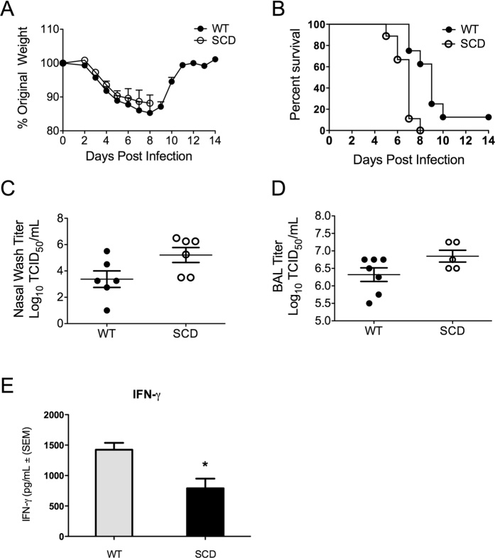
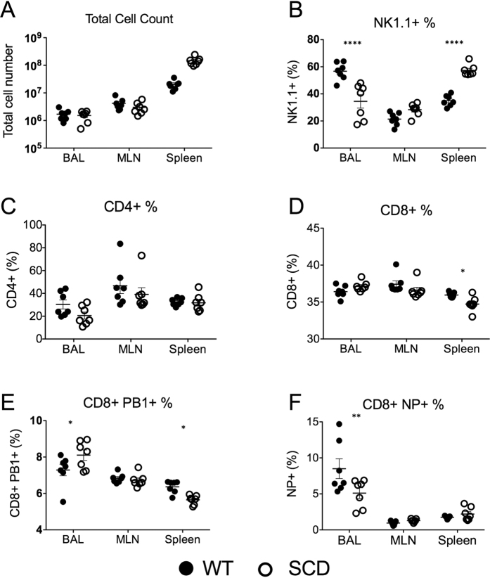
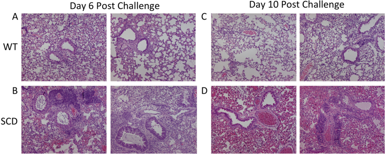
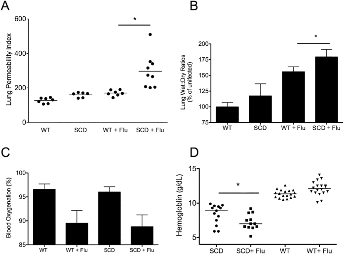
Sickle cell disease (SCD) is a major global health concern. Patients with SCD experience disproportionately greater morbidity and mortality in response to influenza infection than do others. Viral infection is one contributing factor for the development of Acute Chest Syndrome (ACS), a major cause of morbidity and mortality in SCD patients. We determined whether the heightened sensitivity to influenza infection could be reproduced in the two different SCD murine models to ascertain the underlying mechanisms of increased disease severity. In agreement with clinical observations, we found that both genetic and bone marrow-transplanted SCD mice had greater mortality in response to influenza infection than did wild-type animals. Despite similar initial viral titers and inflammatory responses between wild-type and SCD animals during infection, SCD mice continued to deteriorate and failed to resolve the infection, resulting in increased mortality. Histopathology of the lung tissues revealed extensive pulmonary edema and vascular damage following infection, a finding confirmed by heightened vascular permeability following virus challenge. These findings implicate the development of exacerbated pulmonary permeability following influenza challenge as the primary factor underlying heightened mortality. These studies highlight the need to focus on prevention and control strategies against influenza infection in the SCD population.


Conflict of interest statement

The authors declare no competing financial interests.

Figures

Figure 1
Figure 1. Heightened susceptibility in transplanted SCD mice during influenza infection.
(A) Weight loss and (B) survival of transplanted WTBM (n = 20, 15 survivors) and SCDBM (n = 18, 2 survivors) mice after influenza inoculation (p =  < 0.0001). *p < 0.05 by log-rank test. (C) Lung viral titers (n = 3/group) after inoculation. *p < 0.05 by paired t-test. Data and error bars represent the mean and standard error for (A and C).
Figure 2
Figure 2. Susceptibility of BERK SCD mice to influenza.
(A) Weight loss after influenza challenge in BERK SCD mice (SCD) and heterozygous littermate controls (WT). (B) Survival kinetics of mice in A after influenza inoculation (WT: 1 survivor, SCD: no survivors). *p < 0.05 by Log rank test. n = 10 mice per group (C,D) Viral titers recovered from nasal and lung lavages of mice at day 6 post- inoculation. (E) Levels of IFN-γ in the BALF of mice at day 6 post–inoculation. Each data point represents an individual mouse. *p < 0.05 by log rank test. Data and error bars represent the mean and standard error for (A,C,D and E).
Figure 3
Figure 3. Immune cell profiles of BERK SCD mice and heterozygous littermate controls at day-7 post-influenza inoculation.
Immune cells were stained and quantitated by flow cytometry analysis of cells collected from the lungs (BAL), lymph nodes (MLN), and spleen. *p < 0.05, **p < 0.01, ****p < 0.001 by 2-way ANOVA. Black circles are WT mice and white circles are SCD animals. Data and error bars represent the mean and standard error.
Figure 4
Figure 4
Representative images from hematoxylin and eosin staining of the lungs harvested from WT mice at days 6 and 10 post-inoculation (A,C) and SCD mice at days 6 and 10 post-inoculation (B,D).
Figure 5
Figure 5. Pulmonary permeability and heightened anemia following influenza infection.
Uninfected WT and SCD mice had similar levels of pulmonary permeability using Evans Blue assay (A) but SCD displayed greater permeability following challenge compared to infected Het controls (n = 6–9 per group). Wet-dry ratios following infection show a similar pattern whereby the SCD lungs had a significantly greater wet: dry ratio compared to heterozygous controls. All samples were normalized to uninfected WT control mice (wet: dry ratio = 3.13). Following challenge both WT and SCD mice displayed a significant reduction in blood oxygenation but no significant difference between het and SCD was observed following infection (C), n = 7–10 per group. Following influenza challenge SCD had a significant decrease in hemoglobin levels at day 6 post-challenge (D), n = 12–18 per group. For all figures *p < 0.05 by Mann-Whitney. Data and error bars represent the mean and standard error where applicable.
Figure 6
Figure 6. Effectiveness of antivirals in protecting SCD mice from influenza.
Protective capacity of oseltamivir in WT (A) and SCD mice (B). Daily dosage of oseltamivir is indicated in figure legend. *p < 0.05 by Mantel log –rank test compared to vehicle controls. DPI = days post- infection. n = 10 mice per group. Vehicle = no survivors, 20 mg/kg = 1 survivor, 50 mg/kg = 4 survivors).

References

    1. Miller A. C. & Gladwin M. T. Pulmonary complications of sickle cell disease. American Journal of Respiratory and Critical Care Medicine 185, 1154–1165, doi: 10.1164/rccm.201111-2082CI (2012). - DOI - PMC - PubMed
    1. Khoury R. A., Musallam K. M., Mroueh S. & Abboud M. R. Pulmonary complications of sickle-cell disease. Hemoglobin 35, 625–635 (2011). - PubMed
    1. van Tuijn C. F., Nur E., van Beers E. J., Zaaijer H. L. & Biemond B. J. Acute chest syndrome in sickle cell disease due to the new influenza A (H1N1) virus infection. Am J Hematol 85, 303–304, doi: 10.1002/ajh.21638 (2010). - DOI - PubMed
    1. Rees D. C., Williams T. N. & Gladwin M. T. Sickle-cell disease. Lancet 376, 2018–2031 (2010). - PubMed
    1. Vichinsky E. P. et al.. Causes and Outcomes of the Acute Chest Syndrome in Sickle Cell Disease. New England Journal of Medicine 342, 1855–1865, doi: 10.1056/NEJM200006223422502 (2000). - DOI - PubMed

Publication types