. 2017 Mar 3:7:43173.
doi: 10.1038/srep43173.

Nicotinic acid inhibits glioma invasion by facilitating Snail1 degradation

Affiliations

Nicotinic acid inhibits glioma invasion by facilitating Snail1 degradation

Jiejing Li et al. Sci Rep. .

Abstract

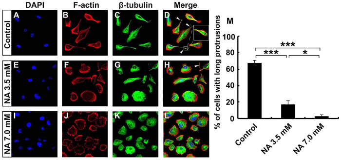
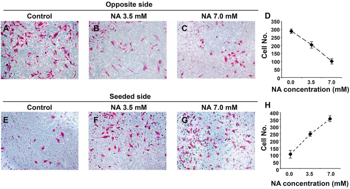
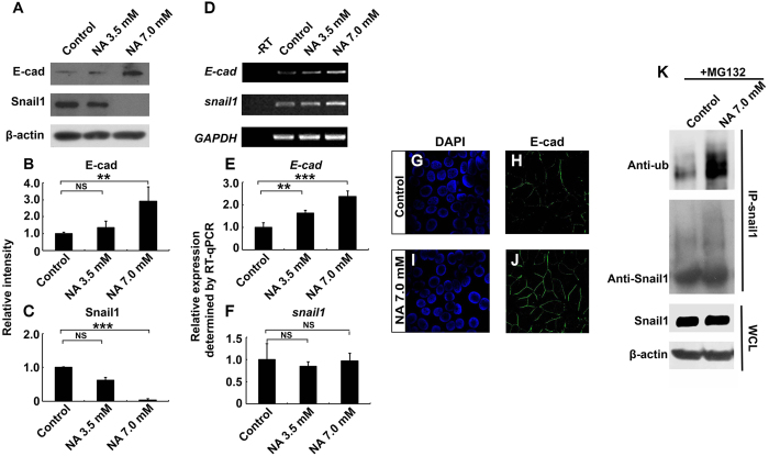
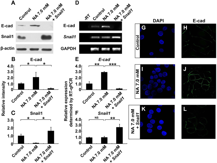
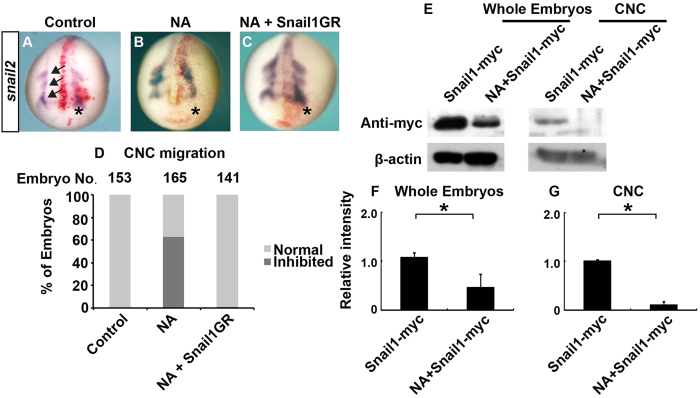
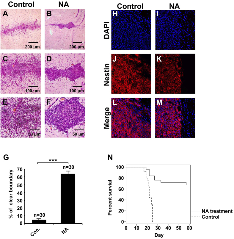
Malignant glioma is a formidable disease that commonly leads to death, mainly due to the invasion of tumor cells into neighboring tissues. Therefore, inhibition of tumor cell invasion may provide an effective therapy for malignant glioma. Here we report that nicotinic acid (NA), an essential vitamin, inhibits glioma cell invasion in vitro and in vivo. Treatment of the U251 glioma cells with NA in vitro results in reduced invasion, which is accompanied by a loss of mesenchymal phenotype and an increase in cell-cell adhesion. At the molecular level, transcription of the adherens junction protein E-cadherin is upregulated, leading to accumulation of E-cadherin protein at the cell-cell boundary. This can be attributed to NA's ability to facilitate the ubiquitination and degradation of Snail1, a transcription factor that represses E-cadherin expression. Similarly, NA transiently inhibits neural crest migration in Xenopus embryos in a Snail1-dependent manner, indicating that the mechanism of action for NA in cell migration is evolutionarily conserved. We further show that NA injection blocks the infiltration of tumor cells into the adjacent brain tissues and improves animal survival in a rat model of glioma. These results suggest that NA treatment may be developed into a potential therapy for malignant glioma.


Conflict of interest statement

The authors declare no competing financial interests.

Figures

Figure 1
Figure 1. U251 cells lose mesenchymal phenotype upon NA treatment.
U251 cells were incubated with PBS (control) or the indicated concentration of NA for 8 hr. Rhodamine phalloidin labeling for F-actin (red), immunocytochemistry for β-tubulin (green) and DAPI labeling for nuclei (blue) were carried out as described in Methods. White arrowheads denote the long protrusions that consist mainly of microtubules, and the inset in (D) shows an amplified image of the indicated protrusion. The percentage of cells with this type of long protrusions was calculated for each treatment and summarized in (F). *P < 0.05; ***P < 0.001.
Figure 2
Figure 2. NA treatment inhibits U251 cell invasion in vitro.
Transwell assays were carried out for U251 cells incubated in PBS (control) or the indicated concentration of NA as described in Methods. Images of cells (stained with Giemsa) that invaded through the matrigel (A–C) or remained on the seeded side (E–G) in a representative experiment are shown, and results of 15 different regions in 3 independent experiments (5 regions per experiment) are summarized in (D) and H for the opposite and seeded sides, respectively.
Figure 3
Figure 3. NA upregulates E-cadherin expression by promoting ubiquitination and degradation of Snail1.
U251 cells were treated with PBS (control) or the indicated concentration of NA for 4 hr. (A–C) Western blot analyses for whole-cell lysates were performed with an anti-E-cadherin antibody. Membranes were stripped and reblotted for Snail1 and β-actin. Representative images of Western blots are shown in A, and relative intensity of E-cadherin and Snail1 normalized against β-actin was calculated and summarized in (B,C), respectively. (D–F) Total RNA was extracted, and RT-PCR was carried out for the transcripts of E-cadherin, snail1, and GAPDH. Representative images of semi-quantitative RT-PCR are shown in (D) and relative expression levels of E-cadherin and snail1 (normalized against GAPDH), as determined by RT-qPCR, are shown in (E,F) respectively. NS, not significant; **P < 0.01; ***P < 0.001. (G–J) Cells were fixed and processed for DAPI staining (blue) and immunocytochemistry for E-cadherin (green). (K) U251 cells were treated with MG132 and NA or PBS (control), IP was carried out for cell lysates with an anti-Snail1 antibody, and Western blot was performed with an anti-ubiquitin antibody. Western blot for whole-cell lysates (WCL) was also performed separately with an anti-Snail1 antibody.
Figure 4
Figure 4. Snail1 reverses the effects of NA on E-cadherin expression.
U251 cells were treated with PBS (control) or 7.0 mM NA for 4 hr, or transfected with a plasmid expressing Snail1 and then treated with 7.0 mM NA for 4 hr. (A) Western blot analyses for whole-cell lysates were performed with an anti-E-cadherin antibody. Membranes were stripped and reblotted for Snail1 and β-actin. Representative images of Western blots are shown in A, and relative intensity of E-cadherin and Snail1 normalized against β-actin was calculated and summarized in (B,C) respectively. (D–F) Total RNA was extracted, and RT-PCR was carried out for the transcripts of E-cadherin, snail1, and GAPDH. Representative images of semi-quantitative RT-PCR are shown in (D), and relative expression levels of E-cadherin and snail1 (normalized against GAPDH), as determined by RT-qPCR, are shown in E and F, respectively. NS, not significant; *P < 0.05; **P < 0.01; ***P < 0.001. (G–L) Cells were fixed and processed for DAPI staining (blue) and immunocytochemistry for E-cadherin (green).
Figure 5
Figure 5. NA inhibits CNC migration in Xenopus embryos by inducing Snail1 degradation.
(A–D) Xenopus embryos were injected in one blastomere at 2-cell stage with PBS (control; A), NA (70 ng; B), or NA combined with mRNA encoding GR-fused Snail1 (C). To induce nuclear translocation of Snail1, dexamethasone was added at stage 10–10.5 (C). Embryos were cultured to stage ~18 and processed for in situ hybridization for snail2. The injected side (on the right and denoted with an asterisk) was labeled with co-injected β-galatosidase (red), and arrows indicate the directions of migration of the three CNC streams. A representative embryo from each group is shown in (A–C), and quantitative results are summarized in (D). (E) Embryos were injected with mRNA encoding myc-tagged Snail1, and cultured with or without NA until stage 15–17 (shortly before CNC migration). Western blot was carried out with an anti-myc antibody for whole-embryo lysates or lysates of dissected CNC. Representative images of Western blots are shown in (E), and relative intensity of myc-tagged Snail1 normalized against β-actin was calculated and summarized in (F) (whole embryos) and (G) (CNC). *P < 0.05.
Figure 6
Figure 6. NA injection inhibits C6 glioma cell invasion in vivo and improves survival in a rat model of glioma.
Rats allografted with C6 cells were injected with PBS (control) or NA as described in Methods. (A–G) Brain slices were collected and H&E staining was carried out. Representative images of brain slices collected from rats injected with PBS or NA are shown in (A–F) (with indicated magnification), and quantitative results from both groups (6 rats/group) are summarized in G. Arrows indicate C6 glioma cells. ***P < 0.001. (H–M) Brain slices were processed for DAPI staining (blue) and immunocytochemistry for Nestin (red). (N) NA significantly improved the survival of C6-allografted rats as compared with control (PBS; n = 25 for each group, P < 0.001).

References

    1. Karunasena E. et al.. ‘Cut from the same cloth’: Shared microsatellite variants among cancers link to ectodermal tissues-neural tube and crest cells. Oncotarget 6, 22038 (2015). - PMC - PubMed
    1. Kim T.-M., Huang W., Park R., Park P. J. & Johnson M. D. A developmental taxonomy of glioblastoma defined and maintained by MicroRNAs. Cancer Research 71, 3387–3399 (2011). - PMC - PubMed
    1. Cuddapah V. A., Robel S., Watkins S. & Sontheimer H. A neurocentric perspective on glioma invasion. Nature Reviews Neuroscience 15, 455–465 (2014). - PMC - PubMed
    1. Louis D. N. et al.. The 2007 WHO classification of tumours of the central nervous system. Acta Neuropathologica 114, 97–109 (2007). - PMC - PubMed
    1. Dandy W. E. Removal of right cerebral hemisphere for certain tumors with hemiplegia: Preliminary report. Journal of the American Medical Association 90, 823–825 (1928).

Publication types