. 2017 Mar 3:7:43160.
doi: 10.1038/srep43160.

Yield Optimisation of Hepatitis B Virus Core Particles in E. coli Expression System for Drug Delivery Applications

Affiliations

Yield Optimisation of Hepatitis B Virus Core Particles in E. coli Expression System for Drug Delivery Applications

Izzat Fahimuddin Bin Mohamed Suffian et al. Sci Rep. .

Abstract

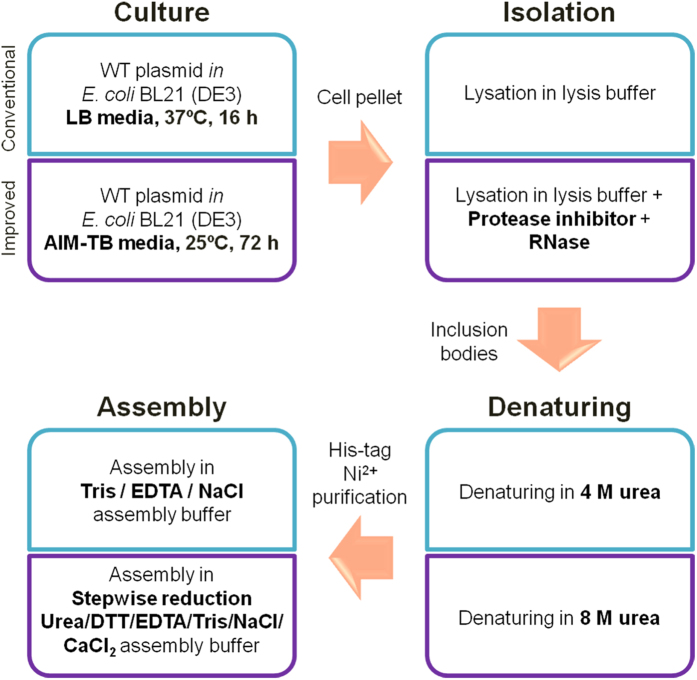
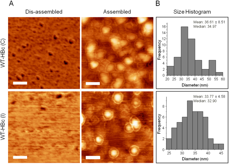
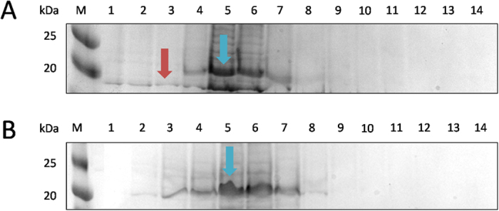
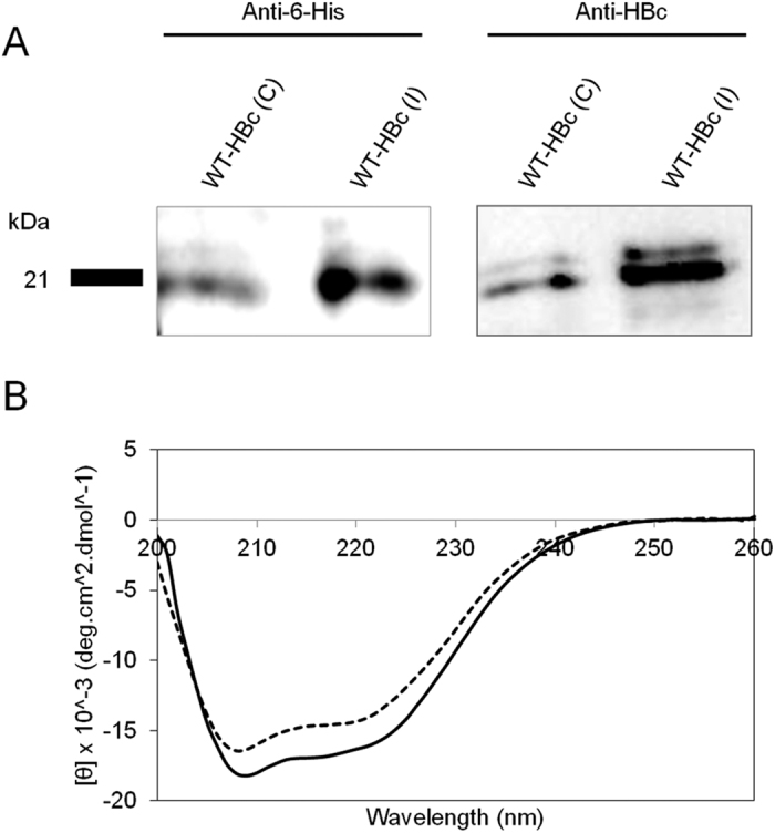
An E. coli expression system offers a mean for rapid, high yield and economical production of Hepatitis B Virus core (HBc) particles. However, high-level production of HBc particles in bacteria is demanding and optimisation of HBc particle yield from E. coli is required to improve laboratory-scale productivity for further drug delivery applications. Production steps involve bacterial culture, protein isolation, denaturation, purification and finally protein assembly. In this study, we describe a modified E. coli based method for purifying HBc particles and compare the results with those obtained using a conventional purification method. HBc particle morphology was confirmed by Atomic Force Microscopy (AFM). Protein specificity and secondary structure were confirmed by Western Blot and Circular Dichroism (CD), respectively. The modified method produced ~3-fold higher yield and greater purity of wild type HBc particles than the conventional method. Our results demonstrated that the modified method produce a better yield and purity of HBc particles in an E. coli-expression system, which are fully characterised and suitable to be used for drug delivery applications.


Conflict of interest statement

The authors declare no competing financial interests.

Figures

Figure 1
Figure 1. A scheme summarising WT-HBc particles production in E. coli expression system.
WT-HBc particles were prepared using one of the two methods; the “conventional” or “improved” method.
Figure 2
Figure 2. Morphological analysis of purified HBc core particles with Atomic Force Microscopy (AFM).
(A) AFM images using tapping mode AFM (TM-AFM) and (B) Histogram analysis of assembled HBc particles. HBc particles were deposited on the mica substrates and measurements were carried out in air at 25 °C, using a Bruker Dimension ICON with Scan Assist. Dis-assembled HBc were achieved by dilution in distilled water at 40 °C for 10 min. Core shell structure was observed for the assembled particles for both formulations. Histograms were obtained for n = 50, analysed using WSxM v5.0 Developed 6.2 and Origin 7.5 software. Scan size is 300 nm × 300 nm. Scale bars are 60 nm.
Figure 3
Figure 3. Elution profile of WT-HBc monomers from Ni2+-chelate affinity chromatography column (cropped gels).
A column with 6 ml of cOmplete™ His-Tag Purification Resin was equilibrated with 3-times bed-volume (18 ml) of the dissociation buffer. The column was loaded with the protein probe and washed with 18 ml dissociation buffer. Bound proteins were eluted with 14 ml of elution buffer and collected in 1 ml fractions. The aliquots of each fractions were subjected to SDS-PAGE gel electrophoresis and stained with Coomassie Brilliant Blue. M, marker in kDa; Numbers 1–14 represent aliquots of the respective elution fractions for (A) WT-HBc (C) or (B) WT-HBc (I) monomers. Blue arrows represent WT-HBc monomers (21 kDa). Higher number of fractions (7 fractions, lane 2–8) were collected in case of WT-HBc (I) compared to WT-HBc (C) (5 fractions, lane 4–8). Some unknown protein bands were observed at molecular weight lower than 20 kDa (red arrow).
Figure 4
Figure 4. Protein specificity and secondary structure analysis of HBc particles.
(A) Western blotting (cropped blot) and (B) Circular Dichroism (CD) analysis of WT-HBc particles. Denatured HBc samples were subjected to SDS-PAGE followed by immuno-blotting using anti-6-His and anti-HBc antibodies. Results confirmed the presence of specific protein bands at 21 kDa. CD graph shows the overall conformation of far-UV CD analysis of WT-HBc core particles. WT-HBc (C) (solid line) and WT-HBc (I) (dashed line) exhibited spectra typical of α-helix-containing proteins with minima at 220 and 208 nm. The CD spectra are typical of WT-HBc core monomer secondary structure.

References

    1. Ferrari M. Cancer nanotechnology: opportunities and challenges. Nature reviews. Cancer 5, 161–171, doi: 10.1038/nrc1566 (2005). - DOI - PubMed
    1. Hoption Cann S. A., van Netten J. P. & van Netten C. Dr William Coley and tumour regression: a place in history or in the future. Postgraduate medical journal 79, 672–680 (2003). - PMC - PubMed
    1. Anderson R. J. & Schneider J. Plasmid DNA and viral vector-based vaccines for the treatment of cancer. Vaccine 25 Suppl 2, B24–34, doi: 10.1016/j.vaccine.2007.05.030 (2007). - DOI - PubMed
    1. Li L. et al.. Evaluation of specific delivery of chimeric phi29 pRNA/siRNA nanoparticles to multiple tumor cells. Molecular bioSystems 5, 1361–1368, doi: 10.1039/b903428e (2009). - DOI - PMC - PubMed
    1. Yamashita I., Iwahori K. & Kumagai S. Ferritin in the field of nanodevices. Biochimica et biophysica acta 1800, 846–857, doi: 10.1016/j.bbagen.2010.03.005 (2010). - DOI - PubMed

Publication types

MeSH terms

LinkOut - more resources