. 2017 Mar 3:7:43487.
doi: 10.1038/srep43487.

The Rotavirus NSP4 Viroporin Domain is a Calcium-conducting Ion Channel

Affiliations

The Rotavirus NSP4 Viroporin Domain is a Calcium-conducting Ion Channel

Thieng Pham et al. Sci Rep. .

Abstract

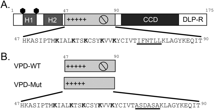
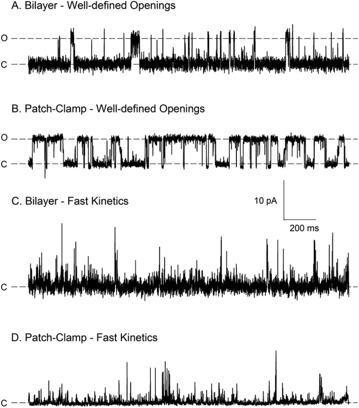
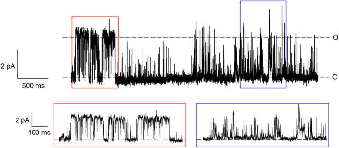
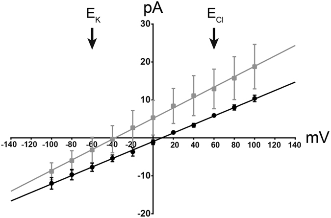
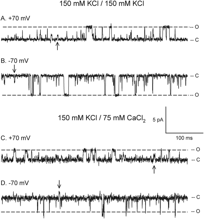
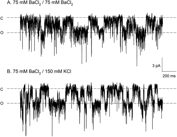
Viroporins are small virus-encoded ion channel proteins. Most viroporins are monovalent selective cation channels, with few showing the ability to conduct divalent cations, like calcium (Ca2+). Nevertheless, some viroporins are known to disrupt host cell Ca2+ homeostasis, which is critical for virus replication and pathogenesis. Rotavirus nonstructural protein 4 (NSP4) is an endoplasmic reticulum transmembrane glycoprotein that has a viroporin domain (VPD), and NSP4 viroporin activity elevates cytosolic Ca2+ in mammalian cells. The goal of this study was to demonstrate that the NSP4 VPD forms an ion channel and determine whether the channel can conduct Ca2+. Using planar lipid bilayer and liposome patch clamp electrophysiology, we show that a synthetic peptide of the NSP4 VPD has ion channel activity. The NSP4 VPD was selective for cations over anions and channel activity was observed to have both well-defined "square top" openings as well as fast current fluctuations, similar to other viroporins. Importantly, the NSP4 VPD showed similar conductance of divalent cations (Ca2+ and Ba2+) as monovalent cations (K+), but a viroporin defective mutant lacked Ca2+ conductivity. These data demonstrate that the NSP4 VPD is a Ca2+-conducting viroporin and establish the mechanism by which NSP4 disturbs host cell Ca2+ homeostasis.


Conflict of interest statement

The authors declare no competing financial interests.

Figures

Figure 1
Figure 1. Illustration of NSP4 and VPD peptides.
(A) Linear schematic of NSP4 and primary sequence of the viroporin domain (VPD, aa47–90) highlighting the pentalysine domain (+++++) with five conserved lysines (bold) and the amphipathic domain (∅) with the residues targeted by the VPD-Mut mutation (underlined). H1 and H2, hydrophobic domains 1 and 2, respectively; CCD, coiled-coil domain; DLP-R, double-layered particle receptor domain. The hexagon symbols represent the two N-linked glycosylation sites (aa8, 18). (B) Linear schematic of the two VPD peptides corresponding to wild-type (VPD-WT) or viroporin deficient (VPD-Mut) sequences. The primary sequence of the VPD-Mut peptide details the mutations (underlined) in the amphipathic domain.
Figure 2
Figure 2. Electrophysiological signatures of VPD-WT in patch-clamp and planar lipid bilayer experiments.
Representative traces of VPD-WT in either planar lipid bilayer or patch-clamp, as indicated, show defined opening events with discrete levels of conductance (A and B) or rapid current fluctuations (C and D). For planar lipid bilayer experiments, either 2.5 μg (A) or 12.5 μg (C) of VPD-WT dissolved in DMSO was added to the cis side of the bilayer. For patch-clamp experiments, VPD-WT dissolved in DMSO was reconstituted into multilamellar liposomes using the rehydration/dehydration method at either a 1:3000 (B) or 1:200 (D) protein-to-lipid ratio (w:w). Symmetric buffer A and buffer PA were used in the patch-clamp or planar lipid bilayer experiments shown, respectively. The applied voltage was +90 mV for all traces. The labels “c” and “o” denote the closed and open current levels, respectively. For panels C and D, discrete levels of conductance are not easily observed.
Figure 3
Figure 3. Shift in VPD-WT kinetics within the same recording.
VPD-WT dissolved in DMSO was reconstituted into multilamellar liposomes at a 1:200 protein-to-lipid ratio (w:w) using the cloud method. The trace was obtained at +50 mV in patch-clamp in symmetric buffer A. The “c” denotes the closed level of conductance, and the “o” represents the open level of the channel obtained from well-defined openings. The red and blue boxes indicate the regions of the trace that are expanded below. The expanded segment in the red box has a conductance level of 50 pS. However, the expanded segment in the blue box has ill-defined conductance levels due to the fast channel kinetics.
Figure 4
Figure 4. Cation selectivity of VPD-WT.
VPD-WT dissolved in water was reconstituted into giant unilamellar liposomes used for patch clamp on a Port-a-Patch device to determine the current-voltage relationship in symmetric 650 mM KCl/650 mM KCl (black circles) or asymmetric 65 mM KCl/650 mM KCl (grey squares) buffers. The average single channel current (pA) is plotted for each voltage (mV) from −100 mV to +100 mV in 10 mV steps. The lines represent the linear regressions through the data points. The equilibrium potential for K+(EK) and Cl (ECl) were calculated using the Nernst equation and are indicated (arrows). They represent the reversal potentials expected for a channel solely selective for K+or Cl, respectively. The graph represents data from n = 3 liposomes (error bars represent standard deviation) from one preparation. The experiment was repeated with five separate liposome preparations with similar results.
Figure 5
Figure 5. VPD-WT can conduct K+ and Ca2+ in patch-clamp experiments.
The peptide was dissolved in DMSO and reconstituted with the dehydration/rehydration method at a protein:lipid ratio of 1:3000 (w:w). The traces were obtained at the indicated voltages in symmetric buffer A or asymmetric conditions of buffer A in the pipette and buffer CA in the bath, in the same patch. The conductance of the main state was 42 pS in symmetric buffer A (A,B), and 29 pS with buffer CA in the bath (C,D), as determined from I/V plots made from the well-defined openings observed in this experiment (Supplemental Fig. 1). The conductance of the substate (arrow) was 13 pS in both conditions. The labels “c” and “o” denote the closed and open current levels, respectively.
Figure 6
Figure 6. VPD-WT can conduct Ba2+.
The peptide was dissolved in water and reconstituted with the cloud method at a protein:lipid ratio of 1:20 (w:w). The traces were obtained with patch-clamp in the indicated buffer conditions at −50 mV. The labels “c” and “o” denote the closed and open current levels, respectively. By convention, opening transitions are in the downward directions at negative voltages.
Figure 7
Figure 7. Viroporin-deficient VPD-Mut cannot conduct Ca2+.
(A) Recombinant VPD-WT and VPD-Mut constructs were tested in the E. coli lysis assay. The optical density at 630 nm (OD630) of uninduced (-IPTG, black and grey lines) and VPD-WT (red line) or VPD-Mut (blue line) expressing cultures were determined at 10 min intervals for 90 min and presented as the percent OD630 relative to that at the time of induction. Traces are the average of 3 separate experiments and error bars represent the standard deviation. (B) Immunoblot of NSP4 expression for VPD-WT (top) and VPD-Mut (bottom) in the cultures measured in panel A. Samples for immunoblot were collected at the time post-induction indicated and detected using an anti-StrepII epitope antibody. Full-length blots are included in Supplementary Figure 3A. (C) Immunoblot analysis of bacterially-expressed NSP4 in total cell lysate [T], soluble proteins [S], peripheral membrane protein [P], and integral membrane protein [I] fractions. The upper panel shows NSP4 (47–146) and NSP4 (95–146) that were used as membrane-localized and soluble controls, respectively. The lower panel shows NSP4 VPD-WT and NSP4 VPD-Mut membrane localization. The upper panel was detected using a peptide antisera directed to NSP4 aa120–147, in the coiled-coil domain and the lower panel was detected with an anti-StrepII monoclonal antibody to detect the C-terminal epitope tags. Full-length blots are included in Supplementary Figure 3B. (D) Liposomes containing the fluorescent Ca2+ indicator Fluo-5N were suspended in HEPES buffered saline (see Materials and Methods) containing 2 mM CaCl2 and treated with water alone (vehicle, black) or 15 μM VPD-WT (red) or VPD-Mut (blue) dissolved in water. The change in Fluo-5N fluorescence was monitored over 20 min. The graph is a representative of 3 separate experiments. Traces are the average ± the standard deviation of 3 replicates.

Similar articles

Cited by

References

    1. Hyser J. M. & Estes M. K. Pathophysiological Consequences of Calcium-Conducting Viroporins. Annu. Rev. Virol. 2, 473–496 (2015). - PMC - PubMed
    1. Royle J., Dobson S. J., Muller M. & Macdonald A. Emerging Roles of Viroporins Encoded by DNA Viruses: Novel Targets for Antivirals? Viruses 7, 5375–5387 (2015). - PMC - PubMed
    1. Scott C. & Griffin S. Viroporins: structure, function and potential as antiviral targets. J Gen. Virol. 96, 2000–2027 (2015). - PubMed
    1. Hyser J. M. Viroporins in Electrophysiology of Unconventional Channels and Pores(ed. Delcour A. H.) 153–181 (Springer, Switzerland, 2015).
    1. Tate J. E. et al.. Global, Regional, and National Estimates of Rotavirus Mortality in Children <5 Years of Age, 2000–2013. Clin Infect Dis. 62, Suppl 2, S96–S105 (2016). - PMC - PubMed

Publication types

MeSH terms

LinkOut - more resources