. 2017 Apr;90(1072):20170062.
doi: 10.1259/bjr.20170062. Epub 2017 Mar 3.

MR imaging in patients with male-to-female sex reassignment surgery: postoperative anatomy and complications

Affiliations

MR imaging in patients with male-to-female sex reassignment surgery: postoperative anatomy and complications

Michele Bertolotto et al. Br J Radiol. 2017 Apr.

Abstract

Objective: To investigate the role of MRI in the evaluation of both the new female anatomy and complications in male-to-female sex reassignment surgery (MtF-SRS).

Methods: 71 consecutive patients with MtF-SRS had 74 MRI [age range, 21-63 years; mean (±standard deviation) age, 36 ± 10 years; median age, 37 years]. In 47 patients, MRI was performed to rule out early post-operative complications after gender conversion (n = 40), vaginoplasty (n = 6) or remodelling of the labia majora (n = 1). In 27 patients, MRI was performed 1-20 years after MtF-SRS for late post-operative complications, pain or dysuria, inflammatory changes or poor cosmetic outcome. Three patients had MRI both before and after the operation.

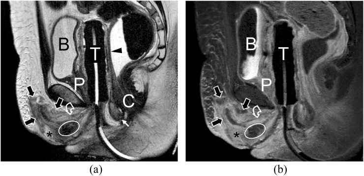
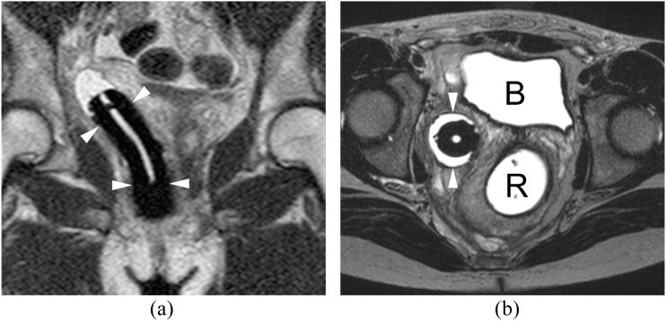
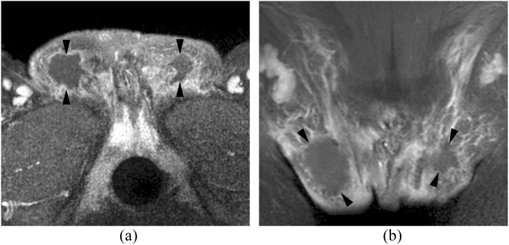
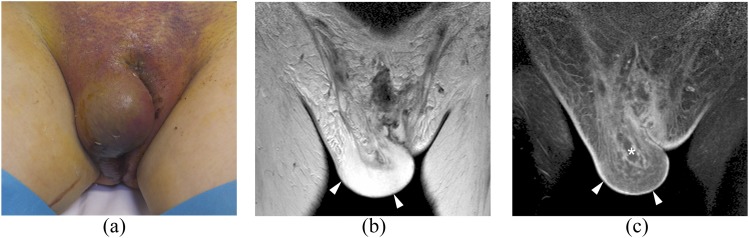
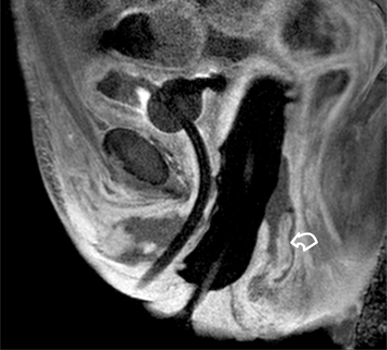
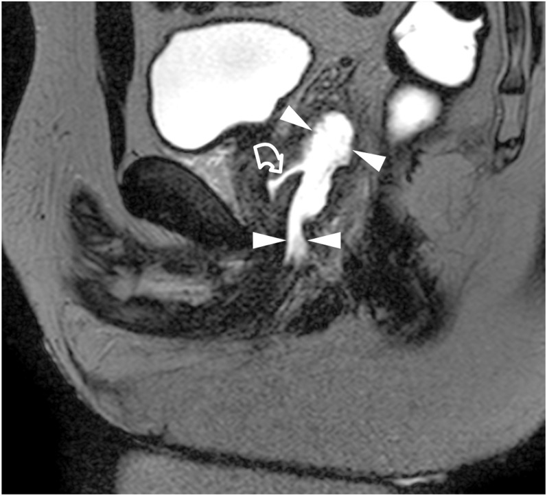
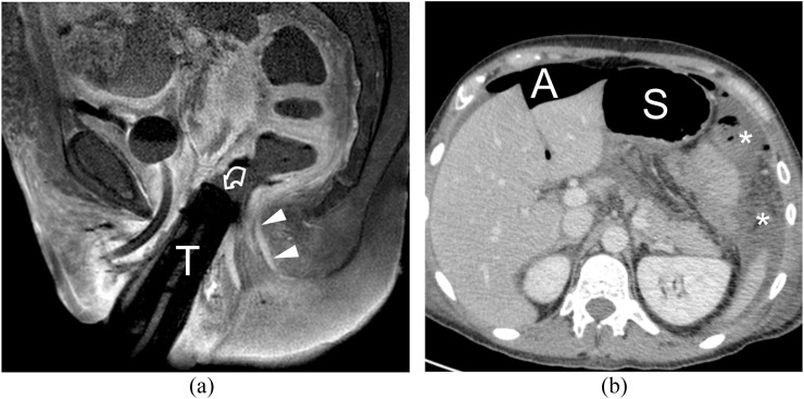
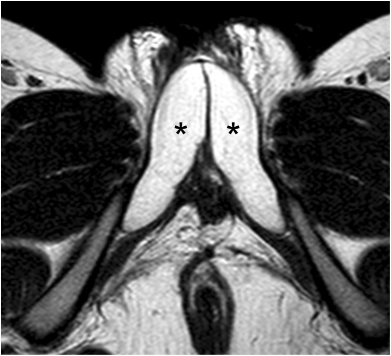
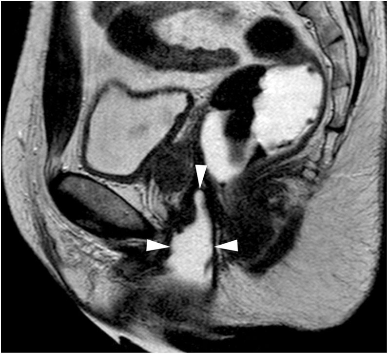
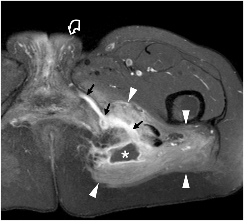
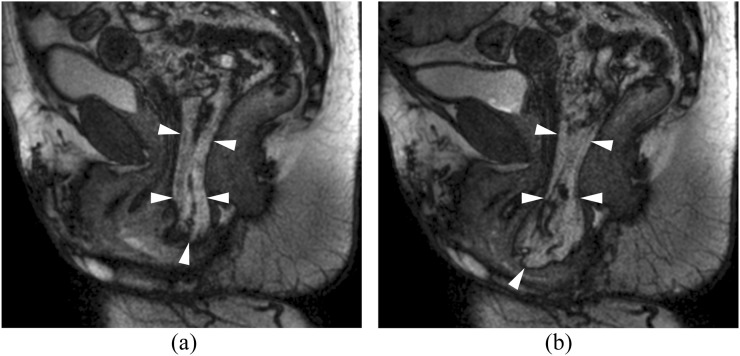
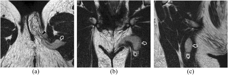
Results: MRI allowed investigation of the new female anatomy in all cases. Soon after MtF-SRS, a small amount of blood was identified in all patients around the neoclitoris, urethral plaque and labia. Post-operative complications were clinically significant fluid collections (n = 5), labial abscesses (n = 2), severe cellulitis (n = 3), partial neovaginal prolapse (n = 3), focal necrosis and dehiscence of the vaginal wall (n = 2) and hypovascularization of the neoclitoris (n = 1). After ileal vaginoplasty, three patients developed clinically insignificant haematomas, one a large rectovaginal fistula with dehiscence of the intestinal anastomosis and bowel perforation (n = 1). In the 27 patients investigated 1-20 years after MfF-SRS, MRI demonstrated cavernosal remnants (n = 10), spared testis (n = 1) neovaginal strictures (n = 8), fistulas and abscesses (n = 3) and prolapse (n = 2). Three of these patients also had fibrotic changes. In the remaining three patients, no pathological features were identified.

Conclusion: After genital reconfiguration, MRI allows assessment of the post-operative anatomy and of post-operative complications. Advances in knowledge: Imaging features of the new anatomy and of surgical complications after SRS are discussed and illustrated.


Figures

Figure 1.
Figure 1.
Evaluation of the new female anatomy in a 30-year-old male-to-female patient investigated 9 days after sex reassignment surgery: the rectum is distended with gel, and tutor is inserted in the neovagina. (a) Midsagittal T2 weighted MR image showing the neovagina with the tutor inside (T), urethral plaque (curved arrow), neurovascular bundle folding in the mons veneris (thick arrows), neoclitoris (asterisk), rectovaginal septum (arrowhead). The bulbocavernosus muscle (thin arrow) has been used to reinforce the neovaginal introitus in front of the anal canal (C). A small haematoma (ellipse) is visible near the neoclitoris. (b) The midsagittal fat-suppressed T1 weighted MR image obtained after gadolinium contrast administration shows the same anatomical features demonstrating vascularization of the neoclitoris (asterisk), urethral plaque (curved arrow) and of the neovaginal wall around the tutor. B, bladder; P, prostate.
Figure 2.
Figure 2.
A 37-year-old patient with male-to-female sex reassignment surgery before 10 days: coronal (a) and axial (b) T2 weighted images showing right deviation of the neovagina (arrowheads). B, bladder; R, rectum.
Figure 3.
Figure 3.
Abscess formation in the labia majora of a 22-year-old male-to-female patient investigated 9 days after sex reassignment surgery: axial (a) and coronal (b) fat-suppressed T1 weighted MR images obtained after gadolinium contrast administration show fluid collections within the labia (arrowheads) and peripheral rim of enhancement consistent with abscesses. The patient was treated with systemic antibiotics and subsequently underwent successful percutaneous drainage on the right side.
Figure 4.
Figure 4.
Cellulitis of the right labium in a 35-year-old male-to-female patient investigated 9 days after sex reassignment surgery presenting with fever and marked swelling and redness of the right labium: (a) Photograph showing a clinically obvious cellulitis. (b, c) Coronal T2 weighted (b) and fat-suppressed T1 weighted images obtained after gadolinium contrast administration (c) show thickening with oedema and hyperaemia of the right labium (arrowheads) and a small fluid collection (asterisk) not requiring drainage. The patient healed with a course of antibiotics within 1 week.
Figure 5.
Figure 5.
Partial prolapse in a 50-year-old male-to-female patient investigated 5 days after sex reassignment surgery: the midsagittal fat-suppressed T1 weighted MR image obtained after gadolinium contrast agent shows the prolapsed, well-vascularized posterior neovaginal wall (curved arrow).
Figure 6.
Figure 6.
Dehiscence in the anterior neovaginal wall of a 25-year-old patient who had male-to-female sex reassignment surgery 9 days before: the neovagina (arrowheads) and the rectum were distended with gel. The midsagittal T2 weighted image is showing extravasation of gel from the anterior wall of the neovagina (curved arrow).
Figure 7.
Figure 7.
A 43-year-old patient who had male-to-female sex reassignment surgery 11 years before: the patient developed acute abdomen 4 days after secondary ileal vaginoplasty. (a) The midsagittal fat-suppressed T1 weighted image is showing protrusion in the rectum (curved arrow) of the tutor inserted in the neovagina (T), immediately above the anal canal (arrowheads). (b) The CT image obtained after i.v. iodinated contrast injection shows extraluminal air (A) and intraperitoneal fluid containing air bubbles (asterisks), consistent with peritonitis. Direct visualization of the discontinuity of the bowel wall was not obtained. An emergency laparotomy was performed, which identified a 3-cm rectovaginal fistula and dehiscence of the ileal anastomosis. S, stomach.
Figure 8.
Figure 8.
Evaluation of the new female anatomy in a 28-year-old male-to-female patient operated before 10 years complaining of discomfort and dyspareunia during penetration: the T2 weighted axial image obtained after ultrasound-guided intracavernosal Prostaglandin E1 injection showing large cavernosal stumps (asterisks). The clitoris was not manufactured.
Figure 9.
Figure 9.
A 51-year-old male-to-female patient operated 13 years before, who developed neovaginal stenosis: the T2 weighted midsagittal image obtained after distension with gel shows a short, funnel-like neovagina (arrowheads).
Figure 10.
Figure 10.
A 33-year-old male-to-female patient operated 6 months before presenting with clinically suspicious abscess in the left gluteal region: the patient complained of pus discharge from a fistula between the left majus and minus labium. The axial fat-saturated T1 weighted image obtained after gadolinium contrast injection shows left gluteus abscess (asterisk) with markedly hyperaemic gluteus maximus and adductor magnus muscles arrowheads. A fistula (small arrows) spreads from the abscess towards the labia (curved arrow).
Figure 11.
Figure 11.
Neovaginal prolapse in a 39-year-old patient who had male-to-female sex reassignment surgery and sigmoid neovagina 16 years before; the midsagittal balanced Fast Field Echo image obtained during pelvic squeeze (a) and strain (b). Neovaginal protrusion is already present during pelvic squeeze (arrowheads) and increases during pelvic strain.
Figure 12.
Figure 12.
A 41-year-old male-to-female patient operated in South America 22 years before: during sex reassignment, the left testis was spared. The patient complained of a large left labium majus and of experiencing severe left perineal pain during vigorous intercourse. On medical examination, the testis was not palpable. T2 weighted MR images on axial (a), coronal (b) and sagittal planes (c) show the testis (curved arrows) buried in the musculature of the left upper leg. The spermatic cord [arrowhead in (a)] runs front–rear within the left labium majus. The asterisks indicate an epididymal cyst.

References

    1. Krege S, Bex A, Lümmen G, Rübben H. Male-to-female transsexualism: a technique, results and long-term follow-up in 66 patients. BJU Int 2001; 88: 396–402. - PubMed
    1. Rossi Neto R, Hintz F, Krege S, Rübben H, Vom Dorp F. Gender reassignment surgery—a 13 year review of surgical outcomes. Int Braz J Urol 2012; 38: 97–107. doi: https://doi.org/10.1590/s1677-55382012000100014 - DOI - PubMed
    1. Cova M, Mosconi E, Liguori G, Bucci S, Trombetta C, Belgrano E, et al. . Value of magnetic resonance imaging in the evaluation of sex-reassignment surgery in male-to-female transsexuals. Abdom Imaging 2003; 28: 728–32. doi: https://doi.org/10.1007/s00261-002-0100-9 - DOI - PubMed
    1. Trombetta C, Liguori G, Bucci S. Radiological evaluation of vaginal width and depth in male-to-female transsexuals by the use of magnetic resonance imaging. World J Urol 2004; 22: 405–8. doi: https://doi.org/10.1007/s00345-004-0422-z - DOI - PubMed
    1. Brunocilla E, Soli M, Franceschelli A, Schiavina R, Borghesi M, Gentile G, et al. . Radiological evaluation by magnetic resonance of the “new anatomy” of transsexual patients undergoing male to female sex reassignment surgery. Int J Impot Res 2012; 24: 206–9. - PubMed