. 2017 Mar 3;12(3):e0173174.
doi: 10.1371/journal.pone.0173174. eCollection 2017.

Dietary protein-induced hepatic IGF-1 secretion mediated by PPARγ activation

Affiliations

Dietary protein-induced hepatic IGF-1 secretion mediated by PPARγ activation

Xiaojuan Wan et al. PLoS One. .

Abstract

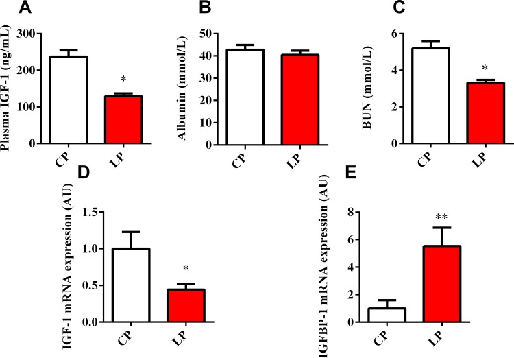
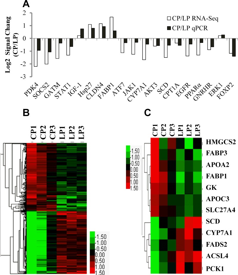
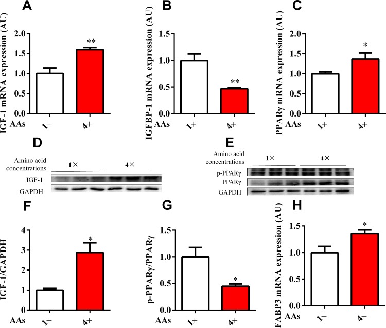
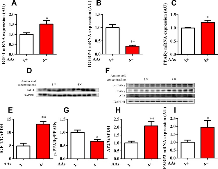
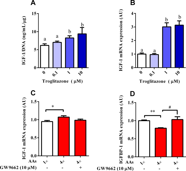
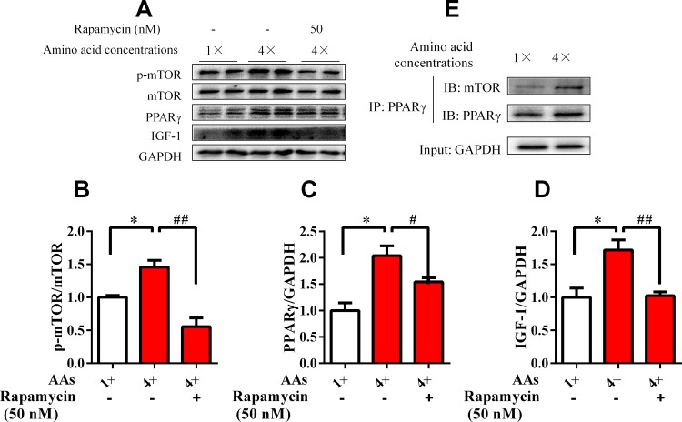
Dietary protein or amino acid (AA) is a crucial nutritional factor to regulate hepatic insulin-like growth factor-1 (IGF-1) expression and secretion. However, the underlying intracellular mechanism by which dietary protein or AA induces IGF-1 expression remains unknown. We compared the IGF-1 gene expression and plasma IGF-1 level of pigs fed with normal crude protein (CP, 20%) and low-protein levels (LP, 14%). RNA sequencing (RNA-seq) was performed to detect transcript expression in the liver in response to dietary protein. The results showed that serum concentrations and mRNA levels of IGF-1 in the liver were higher in the CP group than in the LP group. RNA-seq analysis identified a total of 1319 differentially expressed transcripts (667 upregulated and 652 downregulated), among which the terms "oxidative phosphorylation", "ribosome", "gap junction", "PPAR signaling pathway", and "focal adhesion" were enriched. In addition, the porcine primary hepatocyte and HepG2 cell models also demonstrated that the mRNA and protein levels of IGF-1 and PPARγ increased with the increasing AA concentration in the culture. The PPARγ activator troglitazone increased IGF-1 gene expression and secretion in a dose dependent manner. Furthermore, inhibition of PPARγ effectively reversed the effects of the high AA concentration on the mRNA expression of IGF-1 and IGFBP-1 in HepG2 cells. Moreover, the protein levels of IGF-1 and PPARγ, as well as the phosphorylation of mTOR, significantly increased in HepG2 cells under high AA concentrations. mTOR phosphorylation can be decreased by the mTOR antagonist, rapamycin. The immunoprecipitation results also showed that high AA concentrations significantly increased the interaction of mTOR and PPARγ. In summary, PPARγ plays an important role in the regulation of IGF-1 secretion and gene expression in response to dietary protein.


Conflict of interest statement

Competing Interests: The authors have declared that no competing interests exist.

Figures

Fig 1
Fig 1. Effects of dietary protein regulated the serum index and IGF expression in porcine liver.
Serum IGF-1 (A), albumin (B), and urea nitrogen (C) levels were detected in 63 day-old piglets (n = 6) fed with 20% crude protein diet (CP) and 14% crude protein diet (LP) using commercial kits. Total RNA was harvested and analyzed by qPCR for IGF-1 (D) and IGFBP-1 (E) mRNA expression in liver tissue (n = 6). Data represent the mean ± SEM. * P < 0.05, ** P < 0.01 vs. LP.
Fig 2
Fig 2. Validation of microarray results by qPCR and hierarchical cluster analysis of differentially expressed genes.
(A) Comparison of expression ratios (log 2, y-axis; genes, x-axis) measured by qPCR and microarray in the 19 selected genes. Ratios by microarray and qPCR were averaged for triplicates. (B) The levels of differentially expressed genes were calculated by log2 and compared between 20% crude protein diet (CP, n = 3) and 14% crude protein diet (LP, n = 3) groups. (C) Differentially expressed genes in PPAR signaling pathway. The red color denotes high expression, whereas the green color indicates low expression.
Fig 3
Fig 3. Effects of AA regulated the IGF-1, IGFBP-1, and PPARγ expression in porcine primary hepatocytes.
Porcine primary hepatocytes in media with standard (1×) and four fold (4×) physiological AA concentrations were cultured for 24 h. Cellular mRNAs isolated from each treatment were subjected to qPCR analyses (n = 6). (A–C) IGF-1 (A), IGFBP-1 (B), PPARγ (C) and FABP3 (H) mRNA expression relative to β-actin in porcine primary hepatocytes. (D–G) The protein expression level of IGF-1 and PPARγ were assessed using Western blot. All results contain three replicates (n = 3). The results are expressed as mean ± SEM. * P < 0.05, ** P < 0.01 compared with cells treated with the standard (1×) group.
Fig 4
Fig 4. Effects of AA regulated the expression of IGF-1, IGFBP-1 and PPARγ in HepG2 cells.
HepG2 cells were culture in media with the standard (1×) and 4 times (4×) physiological AA concentrations for 24 h. IGF-1 (A), IGFBP-1 (B), PPARγ (C) and FABP3 (I) mRNA expression was assessed by qPCR. (D–H) Protein expression levels of IGF-1, GAPDH, PPARγ, p-PPARγ and AP2 were assessed by Western blot analysis. All results were obtained from three replicates (n = 6). Results were expressed as mean ± SEM. * P < 0.05, ** P < 0.01 vs. cells treated with the standard group.
Fig 5
Fig 5. Effects of AA on IGF-1 and IGFBP-1 mRNA expression were mediated by PPARγ.
IGF-1 secretion (A) and IGF-1 mRNA expression (B) were measured after treatment with the PPARγ agonist troglitazone. Values with different letters were significantly different (P < 0.05, n = 6). (C and D) Cells treated with HepG2 in media with 1× or 4× physiological AA concentrations, which contained 10 μM of the PPARγ inhibitor GW9662. IGF-1 and IGFBP-1 mRNA expression were analyzed by qPCR. *P < 0.05 (n = 6). Results were expressed as mean ± SEM.
Fig 6
Fig 6. mTOR was involved in the AA-induced activation of PPARγ.
HepG2 cells were cultured in media with 1× and 4× physiological AA concentrations for 48 h. One fraction of the total protein was used to determine the total and phosphorylated levels of the mTOR (A and B), GAPDH, PPARγ (C), and IGF-1 (D) proteins by Western blot analysis. All results contained three replicates (n = 3). The other total protein extracts were analyzed by immunoprecipitation (IP) with anti-PPARγ (E) capture antibodies. Data were expressed as the mean ± SEM. Values with different letters were significantly different (P < 0.05).

References

    1. Daughaday WH, Rotwein P. Insulin-like growth factors I and II. Peptide, messenger ribonucleic acid and gene structures, serum, and tissue concentrations. Endocr Rev. 1989;10(1):68–91. Epub 1989/02/01. 10.1210/edrv-10-1-68 - DOI - PubMed
    1. Ohlsson C, Mohan S, Sjogren K, Tivesten A, Isgaard J, Isaksson O, et al. The role of liver-derived insulin-like growth factor-I. Endocr Rev. 2009;30(5):494–535. PubMed Central PMCID: PMC2759708. 10.1210/er.2009-0010 - DOI - PMC - PubMed
    1. Boisclair YR, Rhoads RP, Ueki I, Wang J, Ooi GT. The acid-labile subunit (ALS) of the 150 kDa IGF-binding protein complex: an important but forgotten component of the circulating IGF system. The Journal of endocrinology. 2001;170(1):63–70. Epub 2001/06/30. - PubMed
    1. Firth SM, Baxter RC. Cellular actions of the insulin-like growth factor binding proteins. Endocr Rev. 2002;23(6):824–54. Epub 2002/12/06. 10.1210/er.2001-0033 - DOI - PubMed
    1. Berryman DE, Christiansen JS, Johannsson G, Thorner MO, Kopchick JJ. Role of the GH/IGF-1 axis in lifespan and healthspan: lessons from animal models. Growth Horm IGF Res. 2008;18(6):455–71. PubMed Central PMCID: PMC2631405. 10.1016/j.ghir.2008.05.005 - DOI - PMC - PubMed

MeSH terms