

Quantitative analysis of the therapeutic effect of magnolol on MPTP-induced mouse model of Parkinson's disease using in vivo 18F-9-fluoropropyl-(+)-dihydrotetrabenazine PET imaging
- PMID: 28257461
- PMCID: PMC5336287
- DOI: 10.1371/journal.pone.0173503
Quantitative analysis of the therapeutic effect of magnolol on MPTP-induced mouse model of Parkinson's disease using in vivo 18F-9-fluoropropyl-(+)-dihydrotetrabenazine PET imaging
Abstract
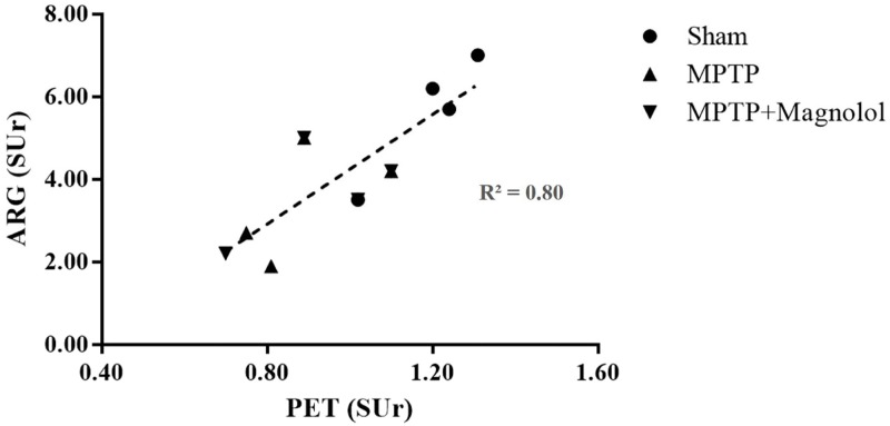
18F-9-Fluoropropyl-(+)-dihydrotetrabenazine [18F-FP-(+)-DTBZ] positron emission tomography (PET) has been shown to detect dopaminergic neuron loss associated with Parkinson's disease (PD) in human and neurotoxin-induced animal models. A polyphenol compound, magnolol, was recently proposed as having a potentially restorative effect in 1-methyl-4-phenyl-1,2,3,6-tetrahydropyridine (MPTP)- or 6-hydroxydopamine-treated animal models. In this study, 18F-FP-(+)-DTBZ PET was used to determine the therapeutic efficacy of magnolol in an MPTP-PD mouse model that was prepared by giving an intraperitoneally (i.p.) daily dose of 25 mg/kg MPTP to male C57BL/6 mice for 5 consecutive days. Twenty-minute static 18F-FP-(+)-DTBZ PET scans were performed before MPTP treatment and 5 days after the termination of MPTP treatment to set up the baseline control. Half of the MPTP-treated mice then received a daily dose of magnolol (10 mg/kg dissolved in corn oil, i.p.) for 6 days. 18F-FP-(+)-DTBZ PET imaging was performed the day after the final treatment. All 18F-FP-(+)-DTBZ PET images were analysed and the specific uptake ratio (SUr) was calculated. Ex vivo autoradiography (ARG) and corresponding immunohistochemistry (IHC) studies were conducted to confirm the distribution of dopaminergic terminals in the striatum. The striatal SUr ratios of 18F-FP-(+)-DTBZ PET images for the Sham, the MPTP, and the MPTP + Magnolol-treated groups were 1.25 ± 0.05, 0.75 ± 0.06, and 1.00 ± 0.11, respectively (n = 4 for each group). The ex vivo 18F-FP-(+)-DTBZ ARG and IHC results correlated favourably with the PET imaging results. 18F-FP-(+)-DTBZ PET imaging suggested that magnolol post-treatment may reverse the neuronal damage in the MPTP-lesioned PD mice. In vivo imaging of the striatal vesicular monoamine transporter type 2 (VMAT2) distribution using 18F-FP-(+)-DTBZ animal PET is a useful method to evaluate the efficacy of therapeutic drugs i.e., magnolol, for the management of PD.
Conflict of interest statement
Figures


Similar articles
-
MicroPET imaging of vesicular monoamine transporter 2 revealed the potentiation of (+)-dihydrotetrabenazine on MPTP-induced degeneration of dopaminergic neurons.Nucl Med Biol. 2021 May-Jun;96-97:9-18. doi: 10.1016/j.nucmedbio.2021.02.004. Epub 2021 Feb 20. Nucl Med Biol. 2021. PMID: 33647803
-
PET imaging with [18F]FP-(+)-DTBZ in 6-OHDA-induced partial and full unilaterally-lesioned model rats of Parkinson's disease and the correlations to the biological data.Nucl Med Biol. 2020 Nov-Dec;90-91:1-9. doi: 10.1016/j.nucmedbio.2020.08.002. Epub 2020 Aug 15. Nucl Med Biol. 2020. PMID: 32861175
-
[18F]FP-(+)-DTBZ PET study in a lactacystin-treated rat model of Parkinson disease.Ann Nucl Med. 2017 Aug;31(7):506-513. doi: 10.1007/s12149-017-1174-3. Epub 2017 Apr 27. Ann Nucl Med. 2017. PMID: 28451991
-
(+)-2-Hydroxy-3-isobutyl-9-(3-[18F]fluoropropoxy)-10-methoxy-1,2,3,4,6,7-hexahydro-11bH-benzo[a]quinolizine.2007 Feb 12 [updated 2010 Oct 14]. In: Molecular Imaging and Contrast Agent Database (MICAD) [Internet]. Bethesda (MD): National Center for Biotechnology Information (US); 2004–2013. 2007 Feb 12 [updated 2010 Oct 14]. In: Molecular Imaging and Contrast Agent Database (MICAD) [Internet]. Bethesda (MD): National Center for Biotechnology Information (US); 2004–2013. PMID: 20641662 Free Books & Documents. Review.
-
Classics in Neuroimaging: Radioligands for the Vesicular Monoamine Transporter 2.ACS Chem Neurosci. 2019 Jan 16;10(1):25-29. doi: 10.1021/acschemneuro.8b00429. Epub 2018 Sep 10. ACS Chem Neurosci. 2019. PMID: 30198706 Review.
Cited by
-
Computational and Experimental Assessments of Magnolol as a Neuroprotective Agent and Utilization of UiO-66(Zr) as Its Drug Delivery System.ACS Omega. 2021 Sep 15;6(38):24382-24396. doi: 10.1021/acsomega.1c02555. eCollection 2021 Sep 28. ACS Omega. 2021. PMID: 34604621 Free PMC article.
-
Selecting the Best Animal Model of Parkinson's Disease for Your Research Purpose: Insight from in vivo PET Imaging Studies.Curr Neuropharmacol. 2023;21(5):1241-1272. doi: 10.2174/1570159X21666230216101659. Curr Neuropharmacol. 2023. PMID: 36797611 Free PMC article.
-
Rational Design of Thermosensitive Hydrogel to Deliver Nanocrystals with Intranasal Administration for Brain Targeting in Parkinson's Disease.Research (Wash D C). 2021 Nov 19;2021:9812523. doi: 10.34133/2021/9812523. eCollection 2021. Research (Wash D C). 2021. PMID: 34888525 Free PMC article.
-
Effectiveness of Magnolol, a Lignan from Magnolia Bark, in Diabetes, Its Complications and Comorbidities-A Review.Int J Mol Sci. 2021 Sep 17;22(18):10050. doi: 10.3390/ijms221810050. Int J Mol Sci. 2021. PMID: 34576213 Free PMC article. Review.
-
Magnolol extends lifespan and improves age-related neurodegeneration in Caenorhabditis elegans via increase of stress resistance.Sci Rep. 2024 Feb 7;14(1):3158. doi: 10.1038/s41598-024-53374-9. Sci Rep. 2024. PMID: 38326350 Free PMC article.
References
-
- Gelb DJ, Oliver E, Gilman S (1999) Diagnostic criteria for Parkinson disease. Arch Neurol 56: 33–39. - PubMed
-
- Jenner P, Olanow CW (2006) The pathogenesis of cell death in Parkinson’s disease. Neurology 66: S24–S36. - PubMed
-
- Ungerstedt U (1968) 6-Hydroxy-dopamine induced degeneration of central monoamine neurons. Eur J Pharmacol 5: 107–110. - PubMed
-
- Schwarting RK, Huston JP (1996) Unilateral 6-hydroxydopamine lesions of meso-striatal dopamine neurons and their physiological sequelae. Prog Neurobiol 49: 215–266. - PubMed
MeSH terms
Substances
LinkOut - more resources
Full Text Sources
Other Literature Sources
Medical
Molecular Biology Databases
