. 2017 May;26(5):1037-1048.
doi: 10.1002/pro.3150. Epub 2017 Apr 20.

Structural basis for dimerization and RNA binding of avian infectious bronchitis virus nsp9

Affiliations

Structural basis for dimerization and RNA binding of avian infectious bronchitis virus nsp9

Tingting Hu et al. Protein Sci. 2017 May.

Abstract

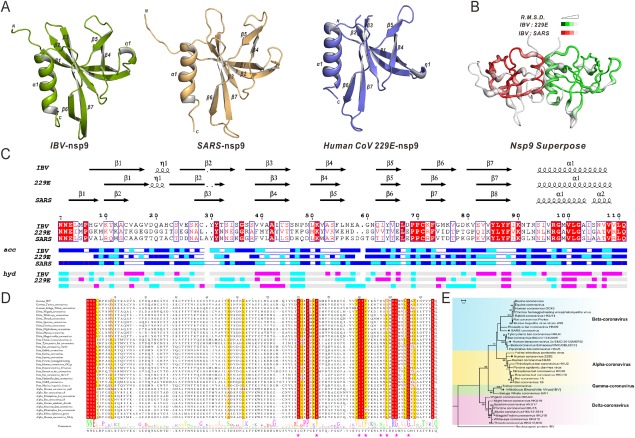
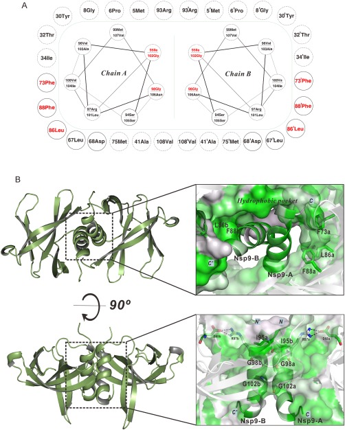
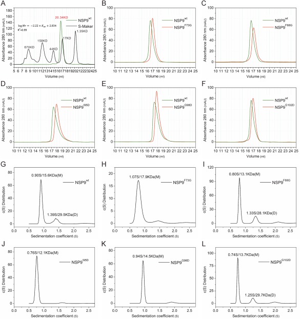
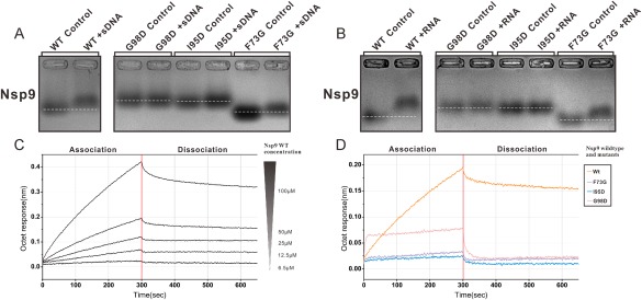
The potential for infection by coronaviruses (CoVs) has become a serious concern with the recent emergence of Middle East respiratory syndrome and severe acute respiratory syndrome (SARS) in the human population. CoVs encode two large polyproteins, which are then processed into 15-16 nonstructural proteins (nsps) that make significant contributions to viral replication and transcription by assembling the RNA replicase complex. Among them, nsp9 plays an essential role in viral replication by forming a homodimer that binds single-stranded RNA. Thus, disrupting nsp9 dimerization is a potential anti-CoV therapy. However, different nsp9 dimer forms have been reported for alpha- and beta-CoVs, and no structural information is available for gamma-CoVs. Here we determined the crystal structure of nsp9 from the avian infectious bronchitis virus (IBV), a representative gamma-CoV that affects the economy of the poultry industry because it can infect domestic fowl. IBV nsp9 forms a homodimer via interactions across a hydrophobic interface, which consists of two parallel alpha helices near the carboxy terminus of the protein. The IBV nsp9 dimer resembles that of SARS-CoV nsp9, indicating that this type of dimerization is conserved among all CoVs. This makes disruption of the dimeric interface an excellent strategy for developing anti-CoV therapies. To facilitate this effort, we characterized the roles of six conserved residues on this interface using site-directed mutagenesis and a multitude of biochemical and biophysical methods. We found that three residues are critical for nsp9 dimerization and its abitlity to bind RNA.

Keywords: Nsp9; coronaviruses; dimerization; infectious bronchitis virus; nonstructural proteins.


Figures

Figure 1
Figure 1
Nsp9 crystal structures(monomer), structural superimposition, multiple sequence alignment, and phylogenetic tree. (A) Ribbon representation of IBV nsp9(green), HCoV‐229E nsp9(orange), and SARS‐CoV nsp9(purple). (B) Superposition of IBV, HCoV‐229E, and SARS‐CoV nsp9. IBV and SARS‐CoV(red) are superimposed(RMSD = 0.875Å), IBV and HCoV‐229E(green) are superimposed(RMSD = 1.409Å). (C) Secondary structure elements and multiple sequence alignments of IBV, HCoV‐229E, and SARS‐CoV. (D) Multiple sequence alignment of the nsp9 protein with representatives from all four CoV genera. The seven residues(F73, L86, F88, I95, G98, and G102) are marked with red asterisks. (E) Phylogenetic analysis of the CoV nsp9 family is presented.
Figure 2
Figure 2
Mutational analysis of the IBV nsp9 protein. (A) Sketch map for the IBV nsp9 parallel α‐helices and the hydrophobic pocket. The green dash represents the hydrophobic contact interface. Circles connected by spirals represent parallel α‐helices. Circles outside the green dash represent hydrophobic residues and critical polar residues surrounding the parallel α‐helices. Solid circles indicate conserved residues. Dashed circles indicate non‐conserved residues. Mutant residues are colored red in the parallel α‐helices and surrounding pocket. (B) Parallel α‐helices and the hydrophobic region in the crystal structure of the nsp9 dimer. Surface and cartoon representations are colored according to the level of residue hydrophobicity from white (polar) to green (hydrophobic). The side chains of conserved hydrophobic residues (F73, L86, and F88) are shown. Hydrophobic interactions in parallel α‐helices highlighting residues I95, G98, and G102 are shown.
Figure 3
Figure 3
Biochemical characterization of IBV nsp9 dimerization. (A) The SEC assay is used to evaluate the molecular weight of wild‐type nsp9. The standard equation “ logMr=2.22×Kav+2.834”, Adj.R2 is used (Adjusted R2 is 0.99). The black line is from commercial molecular weight standards, and the wild‐type nsp9 chromatogram is colored green. (B‐F) Comparison of SEC results for wild‐type nsp9 and different mutants. (G‐L) Sedimentation velocity ultracentrifugation results for wild‐type nsp9 and nsp9 mutants. Sedimentation coefficients (S) and molecular weight (MW) are indicated. “M” and “D” stand for the monomer and dimer.
Figure 4
Figure 4
Oligonucleotide binding analysis of wild‐type and mutant IBV nsp9. (A,B) EMSA illustrating wild‐type and mutants (F73G, I95D, and G98D) IBV nsp9 association with ssDNA/RNA(20‐mer). The white dotted lines represent the position of the bands of IBV nsp9 wild‐type and mutants (F73G, I95D, and G98D) without reaction with ssDNA/RNA. (C) Biolayer interferometry analysis of wild‐type nsp9 with RNA. Biotinylated 20‐mer RNA is immobilized on SA biosensors. Binding curves show association and dissociation for different protein concentrations. (D) Sensorgrams obtained using biosensors loaded with nsp9 mutants. The variation of the response is recorded with mutants at a concentration of 50 uM.

References

    1. Spaan WJM, Cavanagh D (2004) Virus Taxonomy VIIIth Report of the ICTV. London: Elsevier/Academic Press; 7: 945–962.
    1. Knipe DM, Fields BN. Fields virology In: Knipe DM, Ed. (2001) Lippincott Williams & Wilkins.
    1. Hudson CB, Beaudette FR (1932) Infection of the Cloaca with the virus of infectious nronchitis. Science 76:34. - PubMed
    1. Siddell SG, Ziebuhr J, Snijder EJ (2005) Topley and Wilson's microbiology and microbia infections. Virology 10:823–856.
    1. Pereira HG. Reoviridae. Andrewes' Viruses of Vertebrates In: Porterfield JS, Ed. (1989) London: BaiIlibre Tindall; pp 42–57.

Publication types

MeSH terms