. 2017 Jun;23(6):897-905.
doi: 10.1016/j.bbmt.2017.02.018. Epub 2017 Feb 28.

Human Mesenchymal Stem Cell-Educated Macrophages Are a Distinct High IL-6-Producing Subset that Confer Protection in Graft-versus-Host-Disease and Radiation Injury Models

Affiliations

Human Mesenchymal Stem Cell-Educated Macrophages Are a Distinct High IL-6-Producing Subset that Confer Protection in Graft-versus-Host-Disease and Radiation Injury Models

Myriam N Bouchlaka et al. Biol Blood Marrow Transplant. 2017 Jun.

Abstract

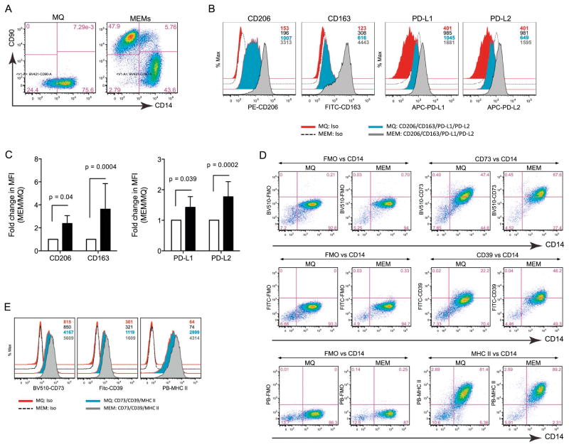
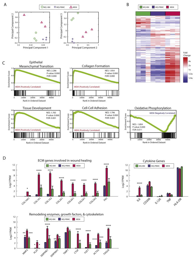
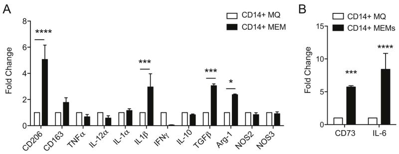
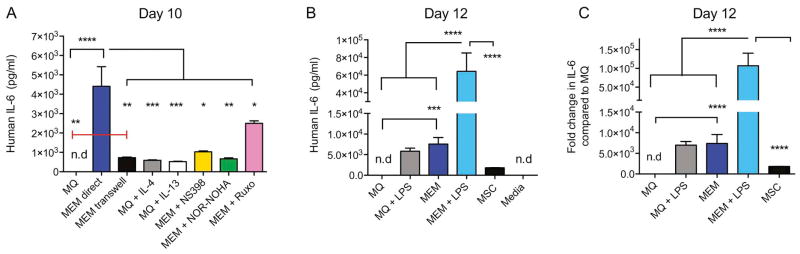
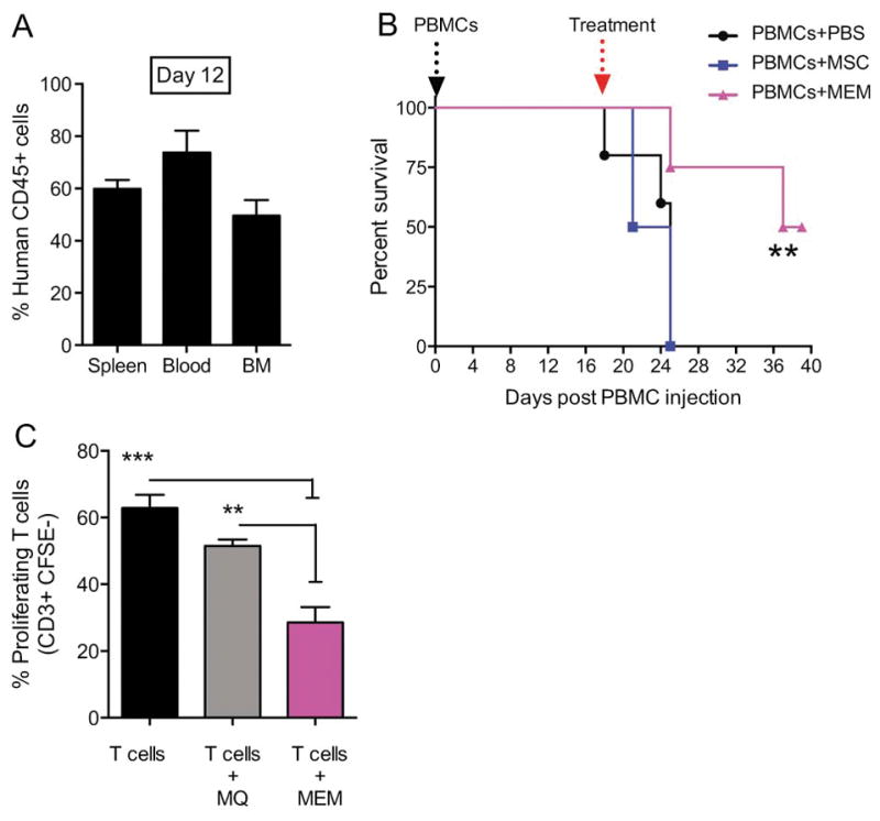
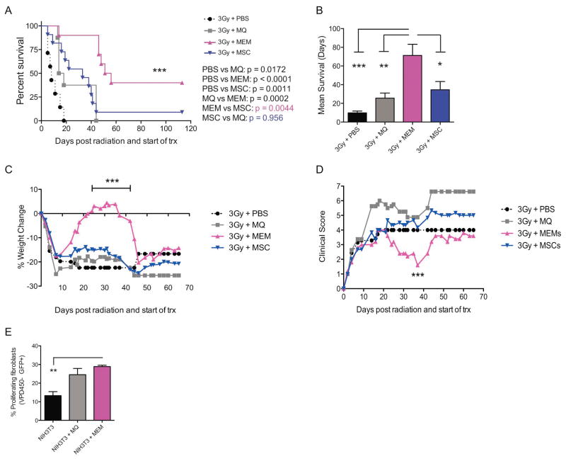
Mesenchymal stem cells (MSCs) have immunosuppressive and tissue repair properties, but clinical trials using MSCs to prevent or treat graft-versus-host disease (GVHD) have shown mixed results. Macrophages (MØs) are important regulators of immunity and can promote tissue regeneration and remodeling. We have previously shown that MSCs can educate MØs toward a unique anti-inflammatory immunophenotype (MSC-educated MØs [MEMs]); however, their implications for in vivo models of inflammation have not been studied yet. We now show that in comparison with MØs, MEMs have increased expression of the inhibitory molecules PD-L1, PD-L2, in addition to markers of alternatively activated MØs: CD206 and CD163. RNA-Seq analysis of MEMs, as compared with MØs, show a distinct gene expression profile that positively correlates with multiple pathways important in tissue repair. MEMs also show increased expression of IL-6, transforming growth factor-β, arginase-1, CD73, and decreased expression of IL-12 and tumor necrosis factor-α. We show that IL-6 secretion is controlled in part by the cyclo-oxygenase-2, arginase, and JAK1/STAT1 pathway. When tested in vivo, we show that human MEMs significantly enhance survival from lethal GVHD and improve survival of mice from radiation injury. We show these effects could be mediated in part through suppression of human T cell proliferation and may have attenuated host tissue injury in part by enhancing murine fibroblast proliferation. MEMs are a unique MØ subset with therapeutic potential for the management of GVHD and/or protection from radiation-induced injury.

Keywords: Graft-versus-host disease (GVHD); IL-6; MSC-educated macrophages (MEMs); Macrophages; Mesenchymal stem cells (MSCs); Radiation injury.


Figures

Figure 1
Figure 1. MEMs express higher levels of CD206, CD163, PD-L1, PD-L2, CD39 and CD73
On day +10 of ex vivo expansion, MØ or MEMs were analyzed by flow cytometry for CD90 and CD14. (A) CD14+CD90 cells are MØs. CD14CD90+ cells are MSCs. (B) Cell surface expression of CD206, CD163, PD-L1 and PD-L2 were determined on the CD14+CD90 population in MØ and MEM cultures by MFI (Mean fluorescent intensity) of isotypes (iso), MØs and MEMs, respectively. Numbers denote the MFIs for each group with color-matched histograms. (C) CD206, CD163, PD-L1/L2 fold change in MFI in MEM over MØ from 6 donors. (D) Expression of CD73, CD39, MHC II (HLA-DR) of fluorescent minus one (FMO) vs. CD14 on MØ and MEMs. (E) MFI of iso, MØs and MEMs for CD73, CD39 or MHC II. Numbers in plots denote MFI for each group. Bar graph statistics (mean ± SEM) by Two-way ANOVA with Bonferroni multiple comparisons.
Figure 2
Figure 2. MEMs express a unique gene expression profile that distinguishes them from macrophages cultured from peripheral blood or bone marrow
Human MØs were generated from CD14+ monocytes from PBMCs or BM cells for 1 week. MEMs were generated by incubating PBMC-derived MØ (MØ-PBMC) with MSCs at a 10:1 ratio for 3 days. On day +10 of culture, CD14+ macrophages were re-sorted from the MØ-PBMCs, MØ-BM and MEM cultures and RNA was isolated for RNA-Seq. (A) Principal component analysis was applied to RNASeq gene expression of 9 samples, 3 each of MEM (red triangle), MØ-BM (green circle) and MØ-PBMC (blue cross) populations. Each symbol represents a unique sample. Samples are plotted based on their coefficients in principal component space. (B) Each column of the heatmap is a unique sample (red: MEM, green: MØ-BM, blue: MØ-PBMC). Genes shown are those with at least a 2-fold difference and false discovery rate<0.15 between any of the group comparisons. (C) Gene set enrichment analysis was run comparing MEMs (N=3) to other macrophages (MØ-BM and MØ-PBMC, N=6). Genes in each set are indicated by black lines along the bottom tracks. The enrichment score across the list is plotted in green. NES= Normalized enrichment score, FDR= false discovery rate. (D) Log2 transformed FPKM RNAseq gene expression, averaged across the 3 replicates for each population (red: MEM, green: MØ-BM, blue: MØ-PBMC). Error bars show SEM.
Figure 3
Figure 3. Decreased pro-inflammatory cytokine profile and increased expression of anti-inflammatory genes by MEMs
On day +10 of ex vivo expansion, CD14+ sorted MØ or CD14+ sorted MEMs were collected for RT-PCR. (A–B) Fold change in mRNA expression of genes normalized to GAPDH housekeeping gene. N=3 and set up in triplicate. Mean ± SEM analyzed by Two-way ANOVA with Bonferroni’s multiple comparison. ****P<0.0001, ***P<0.001 and *P<0.05.
Figure 4
Figure 4. MEMs secrete higher IL-6 constitutively or after LPS stimulation, and is dependent on direct contact with MSCs, via JAK1/JAK2, arginase and COX2 pathways
(A) IL-6 production by ELISA was measured in MØs exposed to media alone (MQ), in direct contact with MSCs (MEM direct), in MØ with MSCs added to the upper chamber of a Transwell (MEM transwell), in MØ with rh-IL4 (MQ+IL-4) or rh-IL13 (MQ+IL-13), or in MEMs exposed to NS398 (MEM + NS398), NOR-NOHA (MEM + NOR-NOHA), or Ruxolitinib (MEM + Ruxo). Each group was set up in triplicate, n=3–8 donors. (B–C) IL-6 production by ELISA was measured in MØs sorted from MØs + media or MØs + MSCs (MEMs) and re-plated in fresh media with or without LPS for 2 days. N= 3 donors and each group was set up in triplicate. (B) Human IL-6 concentration and (C) fold change in human IL-6 production by each group compared to MØ in media alone is shown. Mean ± SEM analyzed by one-way ANOVA with Tukey’s multiple comparisons. Data representative of 3 experiments with reproducible results. n.d: not detected.
Figure 5
Figure 5. Treatment of xenogeneic GVHD with MEMs allow for increased survival & inhibit T cells proliferation in vitro
(A) Day +0, NSG mice received 30x106 PBMCs i.v to induce a xenogeneic GVHD in the absence of total body irradiation. (A) On Day +12 post transplant, human CD45+ engraftment in spleen, blood and bone marrow (BM) was assessed. (B) On day +18, when mice showed clinical evidence of GVHD, mice were randomized to receive PBS, 5x10⁵ MSCs or MEMs i.v to treat GVHD and monitored for survival. N= 5 mice/group, one representative experiment of 3 performed. Survival curves compared by log rank analysis, **P<0.001 (C) Allogeneic CD3+ sorted CFSE-labeled T cells were cultured in triplicate with anti-CD3 and anti-CD28 antibodies either alone (T cells), with MØ (T cells + MØ), or MEM (T cells + MEM) at a 2:1 ratio for 5 days. Cells were then collected from each group and stained for CD3+ to determine the percentage of proliferating cells, which are CD3+CFSE. Mean ± SEM calculated by one-way ANOVA.
Figure 6
Figure 6. Treatment of lethal radiation with MEMs allows for increased survival and improved weight loss and clinical scores
(A–D) On day 0, NSG mice received lethal total body irradiation (3Gy) followed by (3 hours later) PBS, 5x10⁵ MØs, 5x10⁵ MSCs or 5x10⁵ MEMs treatment i.v. N= 8–11 mice/group. (A) Survival curve compared by log rank analysis. (B) Median survival in days for each group.*** p= 0.0003, **p= 0.0066, *p= 0.02, mean ± SEM by one-way ANOVA of analysis with Bonferroni multiple comparison post test. (C) Percent weight change compared to day 0 for each group, ***p< 0.0001, Two-way ANOVA with Tukey’s multiple comparisons post test. (D) Overall clinical score (weight loss, posture, activity, skin and fur texture). On day 37: *p=0.015 MEM vs PBS, *p= 0.011 MEM vs MSC and ***p= 0.0002 MEM vs MØ, two-way ANOVA with Tukey’s multiple comparisons post test. Data representative of one of 2 experiments with similar results. (E) MØ or MEM were co-cultured at 25:1 ratio with NIH-3T3-GFP+ cells pre-labeled with Violet proliferation dye 450. On day 7, the percentage of proliferating NIH-3T3 fibroblasts was assessed by flow cytometry by gating on GFP+ VPD450 cells and compared to NIH-3T3-GFP cells cultured alone. Mean ± SEM calculated by two-way ANOVA with Tukey’s multiple comparisons post test. Data representative of one of 2 experiments with similar results.

Comment in

References

    1. Lange C, Brunswig-Spickenheier B, Cappallo-Obermann H, et al. Radiation rescue: mesenchymal stromal cells protect from lethal irradiation. PLoS One. 2011;6:e14486. - PMC - PubMed
    1. Yang X, Balakrishnan I, Torok-Storb B, Pillai MM. Marrow Stromal Cell Infusion Rescues Hematopoiesis in Lethally Irradiated Mice despite Rapid Clearance after Infusion. Adv Hematol. 2012;2012:142530. - PMC - PubMed
    1. Hoffmann P, Ermann J, Edinger M, Fathman CG, Strober S. Donor-type CD4(+)CD25(+) regulatory T cells suppress lethal acute graft-versus-host disease after allogeneic bone marrow transplantation. The Journal of experimental medicine. 2002;196:389–399. - PMC - PubMed
    1. Martelli MF, Di Ianni M, Ruggeri L, et al. HLA-haploidentical transplantation with regulatory and conventional T-cell adoptive immunotherapy prevents acute leukemia relapse. Blood. 2014;124:638–644. - PubMed
    1. Sawitzki B, Brunstein C, Meisel C, et al. Prevention of graft-versus-host disease by adoptive T regulatory therapy is associated with active repression of peripheral blood Toll-like receptor 5 mRNA expression. Biol Blood Marrow Transplant. 2014;20:173–182. - PMC - PubMed