. 2017 Jun:34:47-54.
doi: 10.1016/j.cellsig.2017.02.022. Epub 2017 Feb 28.

Functional interaction of PkcA and PldB regulate aggregation and development in Dictyostelium discoideum

Affiliations

Functional interaction of PkcA and PldB regulate aggregation and development in Dictyostelium discoideum

Sean Singh et al. Cell Signal. 2017 Jun.

Abstract

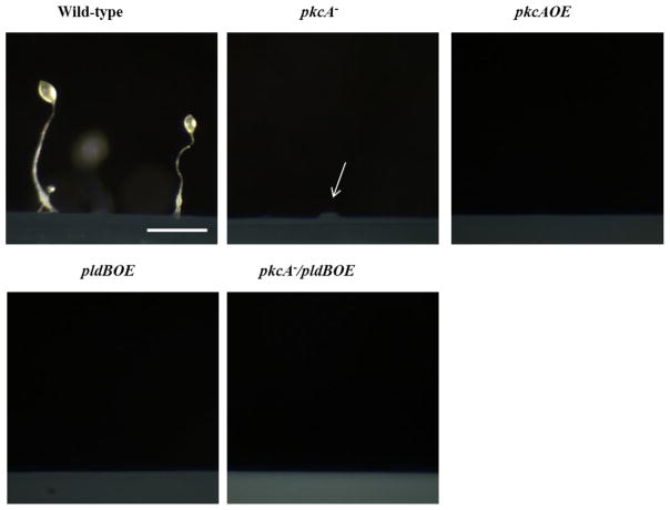
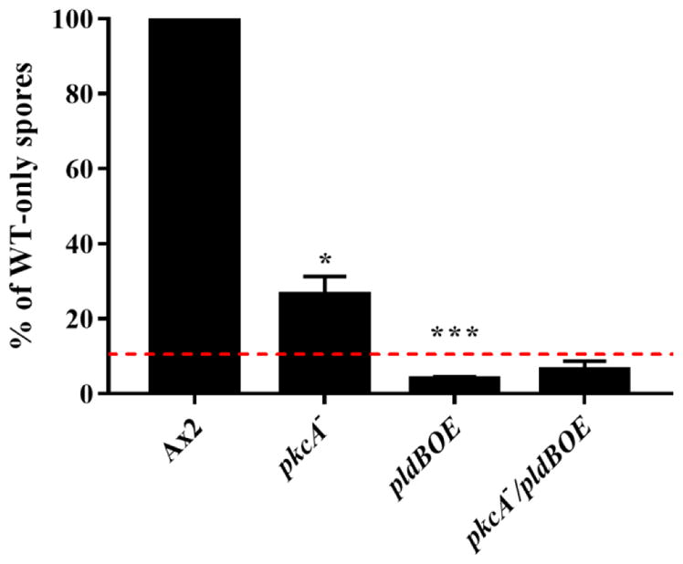
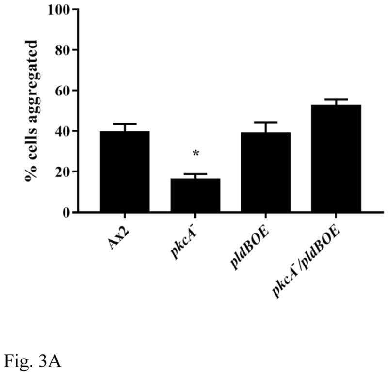
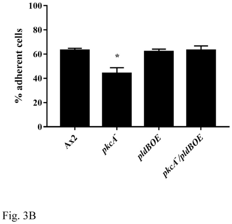
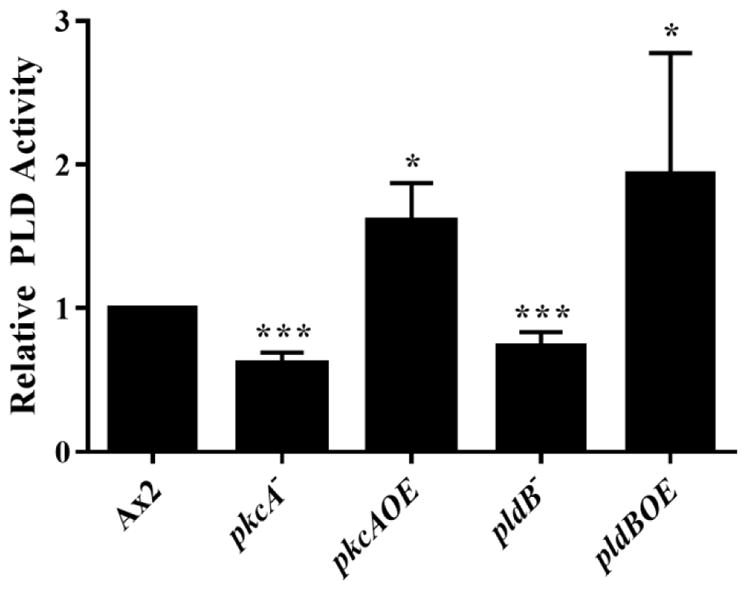
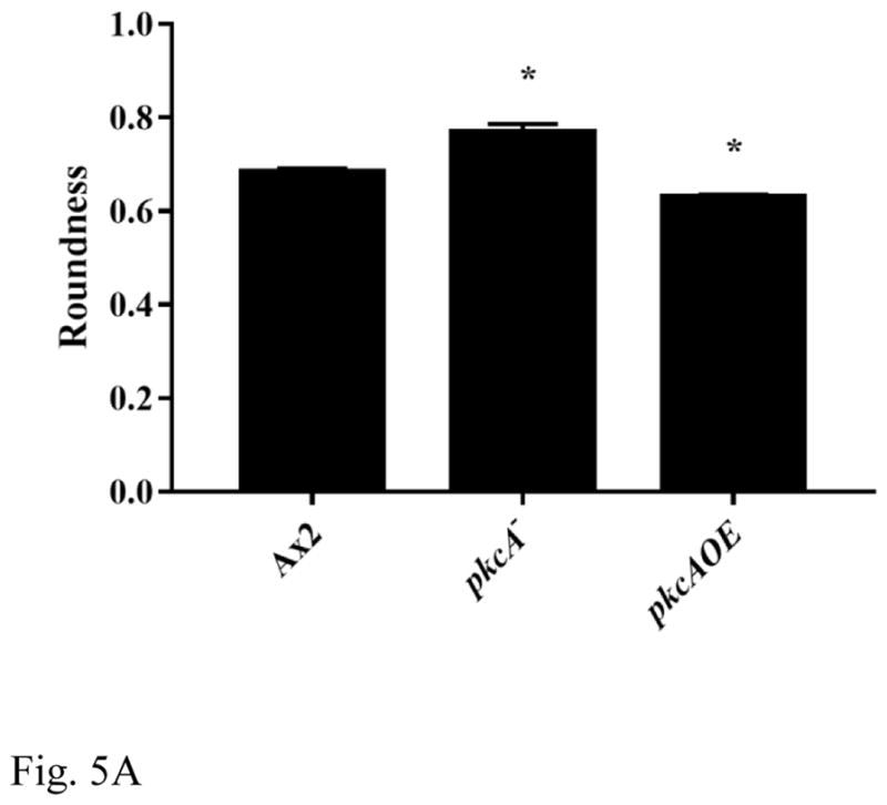
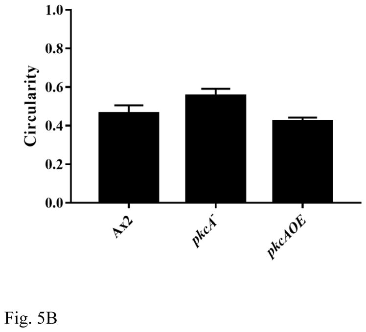
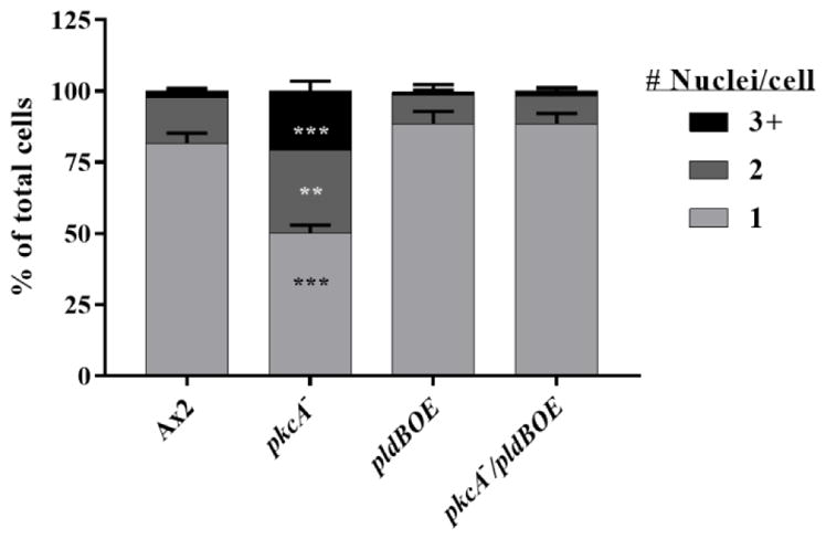
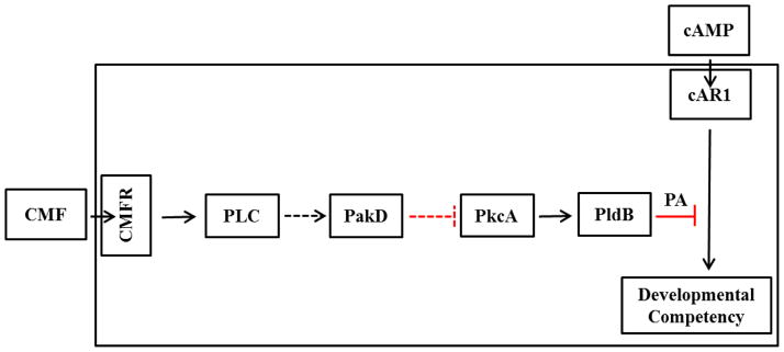
Multicellular development in Dictyostelium discoideum involves tightly regulated signaling events controlling the entry into development, initiation of aggregation and chemotaxis, and cellular differentiation. Here we show that PkcA, a Dictyostelium discoideum Protein Kinase C-orthologue, is involved in quorum sensing and the initiation of development, as well as cAMP sensing during chemotaxis. Additionally, by epistasis analysis we provide evidence that PkcA and PldB (a Phospholipase D-orthologue) functionally interact to regulate aggregation, differentiation, and cell-cell adhesion during development. Finally, we show that PkcA acts as a positive regulator of intracellular PLD-activity during development. Taken together, our results suggest that PkcA act through PldB, by regulating PLD-activity, in order to control events during development.

Keywords: Actin cytoskeleton; Development; Dictyostelium; PKC; PLD; Quorum sensing.


Figures

Figure 1
Figure 1. Fruiting bodies on agar
Wild-type Ax2 cells, pkcA- cells, pkcAOE cells, pldBOE cells, and pkcA/pldBOE double mutant cells were starved on agar and allowed to develop for 24 hours. Bar, 0.5mm.
Figure 2
Figure 2. pkcA regulates spore formation
Chimeras comprised of 10% Ax2, and 90% of the indicated mutant cell line were created and the total spores harvested. Total spore yield of chimera is expressed as a percentage of total spores collected from control fruiting bodies comprised of only Ax2 cells. Values are mean + SEM, from at least 3 independent experiments. Asterisk represents a significant difference from 10% wild-type spore yield, dashed red line (*, P < 0.05; ***, P < 0.005), as determined by the t-test.
Figure 3
Figure 3. pkcA regulates cell-cell cohesion and cell-substrate adhesion
(A) Cells were starved for 3 hours, aggregates were dissociated by vortexing, then allowed to re-adhere for 40 minutes. Single/duplex cells were counted on a hemocytometer and the percentage of cells bound in aggregates calculated. (B) Cells were allowed to adhere to a glass bottomed flask for 2 hours. After gentle agitation for 5 minutes, cells in the supernatant were counted on a hemocytometer and the percentage of adherent cells calculated. Values shown are mean + SEM, from at least 3 independent experiments. Asterisk represents a significant difference from wild-type (P < 0.005), as determined by the t-test.
Figure 4
Figure 4. pkcA regulates PLD activity during development
Cells were starved for 6 hours on filter pads, collected, lysed and the PLD activity measured. Values shown are mean + SEM, from at least 4 independent trials. Asterisk represents a significant difference from wild-type (*, P < 0.05; ***, P < 0.005), as determined by the t-test.
Figure 5
Figure 5. pkcA regulates cell polarity, does not regulate filopodia formation
Cells were starved for 6 hours on a coverslip, then fixed and stained to visualize the actin cytoskeleton. (A) Roundness (minor axis/major axis) or (B) Circularity (4π[area]/perimeter2) were calculated using ImageJ software. Values are means of at least 3 independent trials + SEM, in which at least 100 cells were evaluated per trial. Asterisk represents a significant difference from wild-type (P < 0.05), as determined by the t-test.
Figure 6
Figure 6. pkcA regulates nuclear segregation
Cells were allowed to settle on a coverslip for one hour, fixed and the nuclei stained. Cells were imaged by both brightfield and epifluorescence microscopy, and the number of nuclei/cell were calculated. Values shown are means of at least 3 independent trials + SEM, in which at least 150 cells were evaluated per trial. Asterisks represent a significant difference from wild-type (**, P < 0.01; ***, P < 0.005) as determined by the t-test.
Figure 7
Figure 7. Proposed model of CMF signal transduction
At the onset of starvation, cAMP signaling through cAR1 is blocked by PldB-mediated phosphatidic acid (PA) generation, which is regulated by PkcA. When sufficient cells begin to starve, CMF binding to its receptor induces the downstream activation of PLC. Activated PLC may activate PakD, which may serve to inactivate PkcA. The downregulation of PkcA results in a corresponding reduction of PldB-activity, and phosphatidic acid levels. This allows for cAR1-mediated cAMP signaling to occur, allowing for aggregation and development to proceed.

Similar articles

Cited by

References

    1. Gross JD. Dictyostelium discoideum: A developmental system. Elsevier; 1975. - DOI
    1. Kessin RH. Dictyostelium: evolution, cell biology, and the development of multicellularity. Cambridge University Press; 2001.
    1. Strmecki L, Greene DM, Pears CJ. Developmental decisions in Dictyostelium discoideum. Dev Biol. 2005;284:25–36. doi: 10.1016/j.ydbio.2005.05.011. - DOI - PubMed
    1. Loomis WF. Cell signaling during development of dictyostelium. Dev Biol. 2014;391:1–16. doi: 10.1016/j.ydbio.2014.04.001. - DOI - PMC - PubMed
    1. Gomer RH, Jang W, Brazill D. Cell density sensing and size determination. Dev Growth Differ. 2011;53:482–494. doi: 10.1111/j.1440-169X.2010.01248.x. - DOI - PMC - PubMed

Publication types

MeSH terms

LinkOut - more resources