Review
. 2017 Apr:45:38-46.
doi: 10.1016/j.ceb.2017.02.008. Epub 2017 Mar 1.

RNP transport in cell biology: the long and winding road

Affiliations
Review

RNP transport in cell biology: the long and winding road

Carolina Eliscovich et al. Curr Opin Cell Biol. 2017 Apr.

Abstract

Regulation of gene expression is key determinant to cell structure and function. RNA localization, where specific mRNAs are transported to subcellular regions and then translated, is highly conserved in eukaryotes ranging from yeast to extremely specialized and polarized cells such as neurons. Messenger RNA and associated proteins (mRNP) move from the site of transcription in the nucleus to their final destination in the cytoplasm both passively through diffusion and actively via directed transport. Dysfunction of RNA localization, transport and translation machinery can lead to pathology. Single-molecule live-cell imaging techniques have revealed unique features of this journey with unprecedented resolution. In this review, we highlight key recent findings that have been made using these approaches and possible implications for spatial control of gene function.


Figures

Figure 1
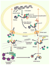
Figure 1. Messenger RNPs control the journey from birth to death
Dynamic association with RNA-binding proteins regulates the journey of an mRNA from transcription in the nucleus to translation and degradation in the cytoplasm. In the nucleus, pre-mRNAs are capped, spliced, cleaved and polyadenylated co-transcriptionally but then diffuse to nuclear pore complexes. Quality control mechanisms ensure that only properly processed mRNPs are remodeled and exported to the cytoplasm, where they undergo a series of diffusional and transported steps. Continuous mRNA remodeling allows proper RNA transport and localization to the subcellular sites of translation and degradation. Dotted orange arrows represent communication between the cytoplasm and the nucleus.
Figure 2
Figure 2. Single-molecule live-cell imaging techniques that monitor mRNP dynamic translation
(a) Nascent peptide imaging techniques (SINAPS, NTC and others) [74-78] visualize translation in real time. RNA reporter construct includes a labeling tag for the nascent peptide (SunTag or FLAG) in the coding sequence and MS2 or PP7 stem-loops in the 3′-UTR for labeling the mRNA. Co-expression of fluorescent antibodies that interact with SunTag and FLAG (green) allows the visualization of nascent peptide chains (blue). Fluorescent proteins (FPs) fused with the MCP or PCP (red) bind to the 3′-UTR and enables mRNA tracking. Actively translating mRNAs are visualized as yellow spots. (b) Translating RNA imaging by the coat protein knock-off (TRICK) method [73] discriminates between mRNAs that have undergone the first round of translation (red spots) and mRNAs that have never been translated (yellow spots) by tagging both the coding sequence and 3′-UTR with RNA stem-loops that bind distinct FPs (green and red). The FP bound to the tags in the coding region is knocked-off by the translating ribosome. (c) Fluorescence fluctuation spectroscopy (FFS) [19] allows spatiotemporal quantification of the association between fluorescent labeled ribosomes (red) and MS2-tagged mRNAs (green) as they pass through a femtoliter volume illuminated by a two-photon spot. (d) Co-tracking of fluorescent labeled ribosomes (red) and mRNAs using the MS2 system (green) shows translation “hot-spots” and dynamics of polysomes in living cells [55].

References

    1. Buxbaum AR, Haimovich G, Singer RH. In the right place at the right time: visualizing and understanding mRNA localization. Nat Rev Mol Cell Biol. 2015;16:95–109. - PMC - PubMed
    1. Eliscovich C, Buxbaum AR, Katz ZB, Singer RH. mRNA on the move: the road to its biological destiny. J Biol Chem. 2013;288:20361–20368. - PMC - PubMed
    1. Lecuyer E, Yoshida H, Parthasarathy N, Alm C, Babak T, Cerovina T, Hughes TR, Tomancak P, Krause HM. Global analysis of mRNA localization reveals a prominent role in organizing cellular architecture and function. Cell. 2007;131:174–187. - PubMed
    1. Katz ZB, Wells AL, Park HY, Wu B, Shenoy SM, Singer RH. beta-Actin mRNA compartmentalization enhances focal adhesion stability and directs cell migration. Genes Dev. 2012;26:1885–1890. - PMC - PubMed
    1. Lawrence JB, Singer RH. Intracellular localization of messenger RNAs for cytoskeletal proteins. Cell. 1986;45:407–415. - PubMed