. 2017 May;58(5):895-906.
doi: 10.1194/jlr.M074112. Epub 2017 Mar 3.

Oncostatin M receptor β deficiency attenuates atherogenesis by inhibiting JAK2/STAT3 signaling in macrophages

Affiliations

Oncostatin M receptor β deficiency attenuates atherogenesis by inhibiting JAK2/STAT3 signaling in macrophages

Xin Zhang et al. J Lipid Res. 2017 May.

Abstract

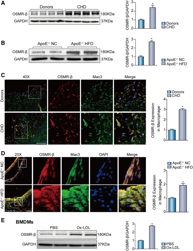
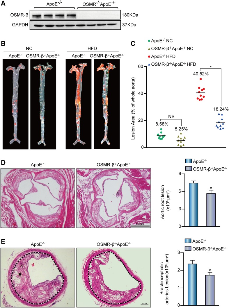
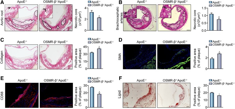
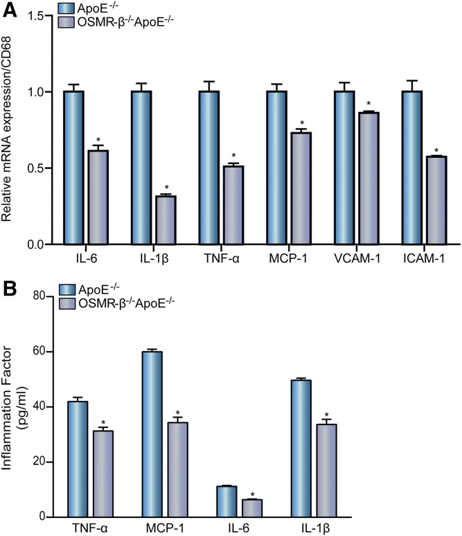
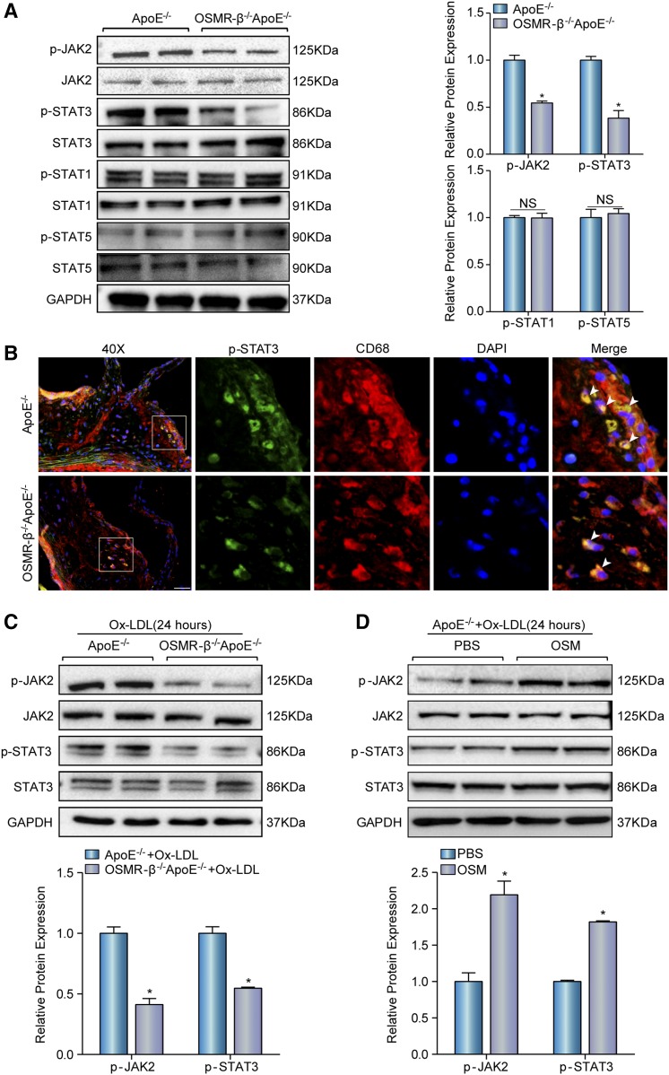
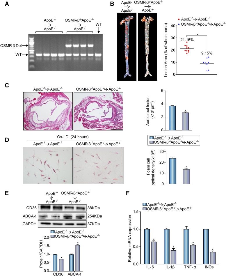
Oncostatin M (OSM) is a secreted cytokine mainly involved in chronic inflammatory and cardiovascular diseases through binding to OSM receptor β (OSMR-β). Recent studies demonstrated that the presence of OSM contributed to the destabilization of atherosclerotic plaque. To investigate whether OSMR-β deficiency affects atherosclerosis, male OSMR-β-/-ApoE-/- mice were generated and utilized. Here we observed that OSMR-β expression was remarkably upregulated in both human and mouse atherosclerotic lesions, which were mainly located in macrophages. We found that OSMR-β deficiency significantly ameliorated atherosclerotic burden in aorta and aortic root relative to ApoE-deficient littermates and enhanced the stability of atherosclerotic plaques by increasing collagen and smooth muscle cell content, while decreasing macrophage infiltration and lipid accumulation. Moreover, bone marrow transplantation of OSMR-β-/- hematopoietic cells to atherosclerosis-prone mice displayed a consistent phenotype. Additionally, we observed a relatively reduced level of JAK2 and signal transducer and activator of transcription (STAT)3 in vivo and under Ox-LDL stimulation in vitro. Our findings suggest that OSMR-β deficiency in macrophages improved high-fat diet-induced atherogenesis and plaque vulnerability. Mech-anistically, the protective effect of OSMR-β deficiency on atherosclerosis may be partially attributed to the inhibition of the JAK2/STAT3 activation in macrophages, whereas OSM stimulation can activate the signaling pathway.

Keywords: Janus kinase 2/signal transducer and activator of transcription 3; atherosclerosis; inflammation.


Figures

Fig. 1.
Fig. 1.
Enhanced OSMR-β expression in the atherosclerotic plaques of ApoE−/− mice and patients with CHD. A: The expression of OSMR-β in the coronary arteries of normal donors and patients with CHD (n = 4, *P < 0.05 versus donor). B: The expression of OSMR-β in aortas from ApoE−/− mice fed NC or a HFD (n = 4, *P < 0.05 versus control). C, D: Double immunofluorescence staining for OSMR-β (red) and Mac3 (macrophage, green) in the coronary arteries of normal donors and patients with CHD and in the aortic sinuses from ApoE−/− mice fed NC or a HFD (scale bar = 50 μm). The quantification was carried out by normalizing the fluorescence intensity of the OSMR-β-positive area with the Mac3-positive area in the atherosclerotic plaque. E: OSMR-β expression in BMDMs upon 15 μg/ml Ox-LDL stimulation for 24 h. *P < 0.05 versus control group.
Fig. 2.
Fig. 2.
Deletion of OSMR-β ameliorated the development of atherosclerosis. A: Genotyping of OSMR-β−/−ApoE−/− mice and ApoE−/− littermates. B, C: En face analysis of aortas from OSMR-β−/−ApoE−/− mice and ApoE−/− littermates fed NC or a HFD; aortas were stained with Oil Red O (n = 10). D, E: Left panel, representative images of the aortic sinus or brachiocephalic arteries from OSMR-β−/−ApoE−/− and ApoE−/− mice stained with H&E. Right panel, quantification of the atherosclerotic lesion area (n = 6). *P < 0.05 compared with ApoE−/−.
Fig. 3.
Fig. 3.
OSMR-β ablation reduces the area of necrotic core and enhances plaque stability. A, B: Left panel, representative images used to analyze the necrotic area in aortic roots and brachiocephalic arteries from OSMR-β−/−ApoE−/− and ApoE−/− mice. Right panel, quantitation of the necrotic area (n = 6). C–E: Histological analysis of plaque stability in the aortic sinus. C: PSR staining for the detection of collagen content. D: Immunofluorescence analysis of SMA for the detection of SMC content. E: Immunofluorescence analysis of CD68 for the detection of macrophage infiltration. F: Oil Red O staining for the detection of lipid accumulation. *P < 0.05 versus ApoE−/−.
Fig. 4.
Fig. 4.
OSMR-β deficiency decreases atherosclerosis-induced inflammation. A: Real-time PCR analysis of pro-inflammatory cytokine expression in the whole aortas of OSMR-β−/−ApoE−/− and ApoE−/− mice compared with CD68 expression (n = 3). B: Detection of MCP-1, TNF-α, IL-6, and IL-1β expression in the serum with ELISA (n = 6). *P < 0.05 compared with ApoE−/−.
Fig. 5.
Fig. 5.
OSMR-β deficiency inhibits the activation of JAK2/STAT3 signaling. A: Western blot analysis of the expression of phosphorylated and total JAK2, STAT3, STAT1, and STAT5 in the whole aortas of OSMR-β−/−ApoE−/− and ApoE−/− littermates. Quantitation of relative phosphorylated protein expression after normalization to each total protein expression, respectively (n = 3). B: Immunofluorescence costaining of phosphorylated STAT3 (green) and CD68 (red) in atherosclerotic plaques (scale bar = 50 μm). C: Western blot analysis of phosphorylated and total JAK2 and STAT3 in peritoneal macrophages from OSMR-β−/−ApoE−/− and ApoE−/− littermates upon 15 μg/ml Ox-LDL treatment for 24 h. The results present phosphorylated protein expression compared with the total protein expression, respectively (n = 3). D: The JAK2 and STAT3 expression in macrophages from ApoE−/− mice upon PBS or OSM treatment. *P < 0.05 compared with control group.
Fig. 6.
Fig. 6.
The absence of OSMR-β from bone marrow-derived cells attenuates atherogenesis. A: DNA was isolated from whole blood for genotyping to confirm genome reorganization with PCR. B: Left panel, Oil Red O staining for en face analysis of atherosclerotic plaques in aortas from OSMR-β−/−ApoE−/−→ApoE−/− (n = 8) and ApoE−/−→ApoE−/− (n = 10) mice. Right panel, analysis of the atherosclerotic lesion area (n = 10 or 8, each group). C: Left panel, H&E staining of the aortic root from OSMR-β−/−ApoE−/−→ApoE−/− mice and ApoE−/−→ApoE−/− mice. Right panel, quantification of the atherosclerotic lesion area (scale bar = 500 μm, n = 6). D: Oil Red O staining of peritoneal macrophages from OSMR-β−/−ApoE−/−→ApoE−/− mice and ApoE−/−→ApoE−/− mice treated with Ox-LDL. E: Western blot analysis of CD36 and ABCA-1 expression in OSMR-β−/−ApoE−/−→ApoE−/− and ApoE−/−→ApoE−/− mice. F: Real-time PCR analysis of pro-inflammatory cytokine expression in macrophages from OSMR-β−/−ApoE−/−→ApoE−/− mice and ApoE−/−→ApoE−/− mice treated with Ox-LDL (n = 3). *P < 0.05 compared with ApoE−/−→ApoE−/− group.

References

    1. Hansson G. K. 2005. Inflammation, atherosclerosis, and coronary artery disease. N. Engl. J. Med. 352: 1685–1695. - PubMed
    1. Yang Z., and Hall A. G.. 2008. The financial burden of overweight and obesity among elderly Americans: the dynamics of weight, longevity, and health care cost. Health Serv. Res. 43: 849–868. - PMC - PubMed
    1. Choudhury R. P., Fuster V., and Fayad Z. A.. 2004. Molecular, cellular and functional imaging of atherothrombosis. Nat. Rev. Drug Discov. 3: 913–925. - PubMed
    1. Mestas J., and Ley K.. 2008. Monocyte-endothelial cell interactions in the development of atherosclerosis. Trends Cardiovasc. Med. 18: 228–232. - PMC - PubMed
    1. Boyle J. J., Weissberg P. L., and Bennett M. R.. 2002. Human macrophage-induced vascular smooth muscle cell apoptosis requires NO enhancement of Fas/Fas-L interactions. Arterioscler. Thromb. Vasc. Biol. 22: 1624–1630. - PubMed

Publication types

MeSH terms

Substances