. 2017 May 1;312(5):L748-L759.
doi: 10.1152/ajplung.00393.2016. Epub 2017 Mar 3.

Effect of α7 nicotinic acetylcholine receptor activation on cardiac fibroblasts: a mechanism underlying RV fibrosis associated with cigarette smoke exposure

Affiliations

Effect of α7 nicotinic acetylcholine receptor activation on cardiac fibroblasts: a mechanism underlying RV fibrosis associated with cigarette smoke exposure

Alexander Vang et al. Am J Physiol Lung Cell Mol Physiol. .

Abstract

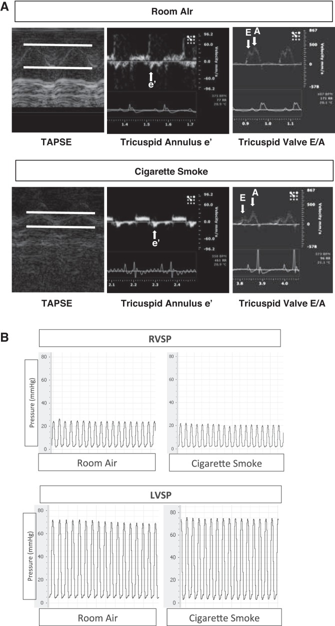
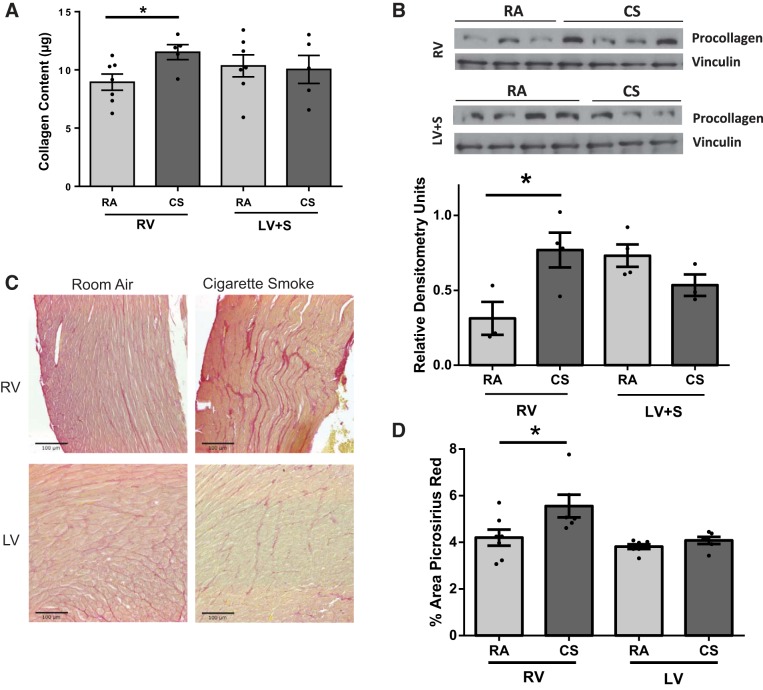
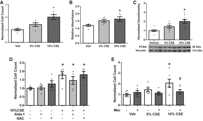
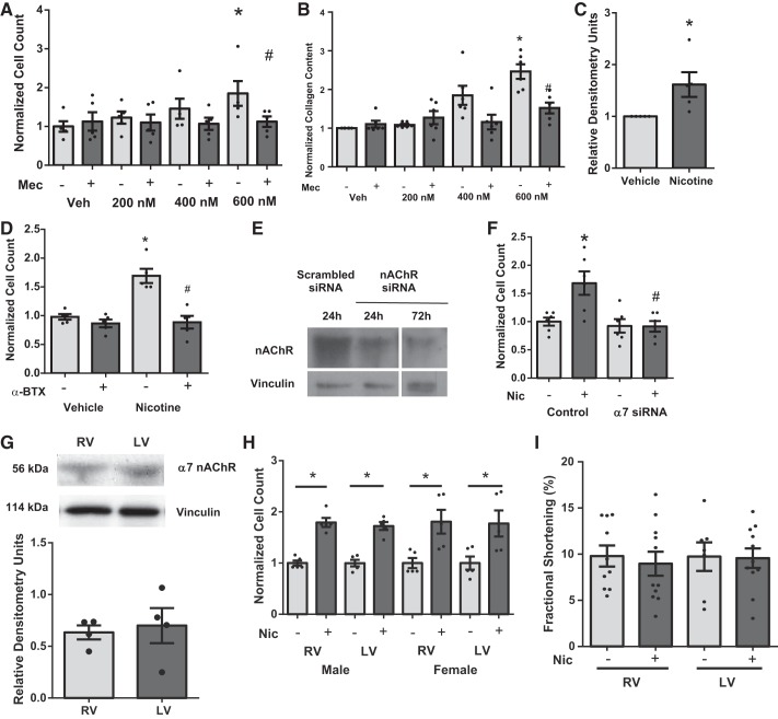
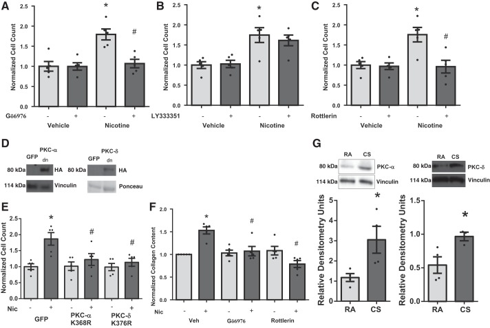
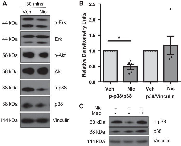
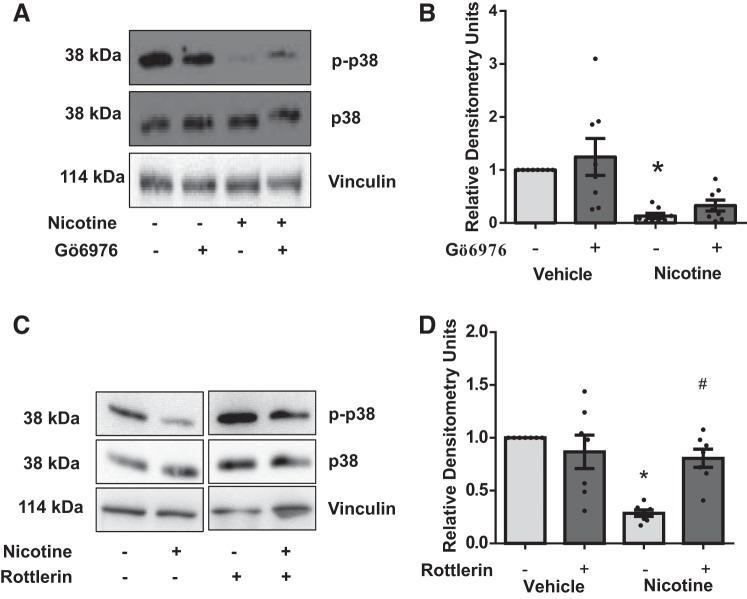
Right ventricular (RV) dysfunction is associated with numerous smoking-related illnesses, including chronic obstructive pulmonary disease (COPD), in which it is present even in the absence of pulmonary hypertension. It is unknown whether exposure to cigarette smoke (CS) has direct effects on RV function and cardiac fibroblast (CF) proliferation or collagen synthesis. In this study, we evaluated cardiac function and fibrosis in mice exposed to CS and determined mechanisms of smoke-induced changes in CF signaling and fibrosis. AKR mice were exposed to CS for 6 wk followed by echocardiography and evaluation of cardiac hypertrophy, collagen content, and pulmonary muscularization. Proliferation and collagen content were evaluated in primary isolated rat CFs exposed to CS extract (CSE) or nicotine. Markers of cell proliferation, fibrosis, and proliferative signaling were determined by immunoblot or Sircol collagen assay. Mice exposed to CS had significantly decreased RV function, as determined by tricuspid annular plane systolic excursion. There were no changes in left ventricular parameters. RV collagen content was significantly elevated, but there was no change in RV hypertrophy or pulmonary vascular muscularization. CSE directly increased CF proliferation and collagen content in CF. Nicotine alone reproduced these effects. CSE and nicotine-induced fibroblast proliferation and collagen content were mediated through α7 nicotinic acetylcholine receptors and were dependent on PKC-α, PKC-δ, and reduced p38-MAPK phosphorylation. CS and nicotine have direct effects on CFs to induce proliferation and fibrosis, which may negatively affect right heart function.

Keywords: chronic obstructive pulmonary disease; fibrosis; nicotine; nicotinic acetylcholine receptor; right ventricle.


Figures

Fig. 1.
Fig. 1.
Assessment of right ventricular (RV) and left ventricular (LV) function in room air (RA)- and cigarette smoke (CS)-exposed AKR mice. A: representative images using echocardiography demonstrating tricuspid annular plane systolic excursion (TAPSE), tricuspid annulus e′, and tricuspid valve E-to-A ratio, n = 6. B: representative hemodynamic tracings obtained with a Millar pressure catheter placed into the RV and the LV. RVSP, RV systolic pressure; LVSP, LV systolic pressure.
Fig. 2.
Fig. 2.
Analysis of cardiac fibrosis in RV and LV of RA- and CS-exposed mice. RV and LV + septum (S) tissue of mice exposed to CS exhibited increased collagen content (Sircol assay, per 100 μg of protein) (A) and procollagen expression (B). Sircol assay n = 5–7, procollagen expression n = 3 to 4. *P < 0.05 vs. RA, Student’s t-test. Similar increases in fibrosis were apparent with Picrosirius red-stained paraffin-embedded sections. C: representative images of RA- and CS-exposed RV and LV. D: significantly increased Picrosirius staining in CS-exposed RV but not LV, as determined by quantitation of images. *P < 0.05 vs. RA, Student’s t-test, n = 6 to 7. Scale on images = 100 μM.
Fig. 3.
Fig. 3.
Effects of CS extract (CSE) on fibroblast proliferation and role of nicotinic acetylcholine receptors (nAChRs). Cardiac fibroblasts isolated from rats were quiesced for 24 h, exposed to CSE for 24 h. CSE dose dependently increases cell count (n = 5) (A) and cell viability (B), as assessed by 3-(4, 5-dimethylthiazolyl-2)-2, 5-diphenyltetrazolium bromide (MTT) assay (n = 5). C: CSE increases expression of proliferation marker proliferating cell nuclear antigen (PCNA) (n = 9). Data were normalized to vinculin for loading control. D: CSE-induced cell proliferation in cardiac fibroblasts was not attenuated by activation of aldehyde dehydrogenase (Alda-1, 25 μM) or reactive oxygen species (ROS) scavenging (N-acetyl cysteine, NAC, 25 mM) (n = 5). E: mecamylamine (Mec, 20 μM) inhibited CSE-induced fibroblast proliferation (n = 5). Data are normalized to vehicle (Veh) and presented as means ± SE. *P < 0.05 compared with Veh; #P < 0.05 compared with CSE alone.
Fig. 4.
Fig. 4.
Effect of nicotine on isolated cardiac fibroblast proliferation and dependence of α7 nAChR. Nicotine increases cell counts (n = 5) (A) and collagen content (n = 5) (B) in cardiac fibroblasts through nAChR activation that was inhibited by Mec (20 μM). C: nicotine also increases procollagen expression in cardiac fibroblasts (n = 5). D: α-bungarotoxin (α-BTX; 100 nM) blocks nicotine-induced proliferation (n = 5). Knockdown of α7 nAChR with siRNA (E) blocks nicotine-induced proliferation (F) (n = 5). Quiescent cells were pretreated with either vehicle, Mec, α-BTX, or α7 nAChR siRNA, followed by vehicle or nicotine (600 nM) for 24 h. Data are normalized to vehicle treatment and presented as means ± SE. *P < 0.05 compared with Veh; #P < 0.05 compared with Mec/vehicle. G: there were no differences in expression of α7 nAChR in rat LV and RV tissues (n = 4). H: there were no changes in response to nicotine (Nic) in cardiac fibroblasts isolated from LV or RV or male or female rats. Nicotine significantly increased proliferation in all groups compared with respective untreated groups (*P < 0.05) (n = 3–4 animals/group performed in triplicate, 3-way ANOVA, Student-Newman-Keuls post hoc). I: there were no significant changes (t-test) in RV or LV cardiomyocyte contractile function with acute nicotine treatment.
Fig. 5.
Fig. 5.
Role of PKC signaling in nicotine-induced cardiac fibroblast proliferation. Small-molecule inhibitors of PKC-α (100 nM) (A) or -δ (3 μM) (C), but not PKC-β (50 nM) (B), block nicotine-induced cardiac fibroblast proliferation (n = 5). Expression of dominant-negative (Κ368Ρ) PKC-α (D) or dominant-negative (K376R) PKC-δ (E) also blocks nicotine-induced cardiac fibroblast proliferation (n = 5). GFP, green fluorescent protein; HA, hemagglutinin. Chemical inhibition of PKC-α and -δ blocks nicotine-induced increased collagen content (F). Cells were quiesced in serum-free medium for 24 h before nicotine stimulation. *P < 0.05 vs. Veh/Veh, and #P < 0.05 vs. Veh/Nic using 2-way ANOVA, Student-Newman-Keuls post hoc test. CS-exposed mice have increased expression of PKC-α and -δ in RV tissue. G, top: representative blot is shown. Bottom: quantitation of data in G (n = 4). *P < 0.05 vs. RA.
Fig. 6.
Fig. 6.
Effects of nicotine on phosphorylation of Akt, Erk, and p38-MAPK. A: nicotine treatment for 30 min does not change Akt or Erk phosphorylation but does acutely decrease phosphorylated p38-MAPK. Representative blots are shown. B: quantitation of the ratio of p-p38 to total p38 and total p38 to the loading control vinculin from A. Data are normalized to Veh. C: reduced p38-MAPK phosphorylation is blocked by the nAChR antagonist Mec. A representative blot is shown, n = 5. *P < 0.05 vs. vehicle, unpaired t-test.
Fig. 7.
Fig. 7.
Determination of upstream signals regulating nicotine-dependent reduced p38-MAPK phosphorylation. A: chemical inhibition of PKC-α does not reduce p38-MAPK phosphorylation. A representative blot is shown. B: quantitation of results in A, n = 8. C: nicotine-mediated reduction of p38-MAPK phosphorylation is blocked by rottlerin. D: quantitation of results in C, n = 8. *P < 0.05 vs. control/vehicle group. #P < 0.05 from both vehicle and nicotine control groups; 2-way ANOVA, Tukey’s test. Any samples in cropped images were run on the same gel with groups irrelevant to the study removed.

Similar articles

Cited by

References

    1. Albuquerque EX, Pereira EF, Alkondon M, Rogers SW. Mammalian nicotinic acetylcholine receptors: from structure to function. Physiol Rev 89: 73–120, 2009. doi:10.1152/physrev.00015.2008. - DOI - PMC - PubMed
    1. Bogaard HJ, Natarajan R, Henderson SC, Long CS, Kraskauskas D, Smithson L, Ockaili R, McCord JM, Voelkel NF. Chronic pulmonary artery pressure elevation is insufficient to explain right heart failure. Circulation 120: 1951–1960, 2009. doi:10.1161/CIRCULATIONAHA.109.883843. - DOI - PubMed
    1. Braz JC, Bueno OF, Liang Q, Wilkins BJ, Dai YS, Parsons S, Braunwart J, Glascock BJ, Klevitsky R, Kimball TF, Hewett TE, Molkentin JD. Targeted inhibition of p38 MAPK promotes hypertrophic cardiomyopathy through upregulation of calcineurin-NFAT signaling. J Clin Invest 111: 1475–1486, 2003. doi:10.1172/JCI200317295. - DOI - PMC - PubMed
    1. Casserly B, Mazer JM, Vang A, Harrington EO, Klinger JR, Rounds S, Choudhary G. C-type natriuretic peptide does not attenuate the development of pulmonary hypertension caused by hypoxia and VEGF receptor blockade. Life Sci 89: 460–466, 2011. doi:10.1016/j.lfs.2011.07.009. - DOI - PMC - PubMed
    1. Chichger H, Vang A, O’Connell KA, Zhang P, Mende U, Harrington EO, Choudhary G. PKC d and bII regulate angiotensin II-mediated fibrosis through p38: a mechanism of RV fibrosis in pulmonary hypertension. Am J Physiol Lung Cell Mol Physiol 308: L827–L836, 2015. doi:10.1152/ajplung.00184.2014. - DOI - PMC - PubMed

Publication types

MeSH terms

Substances

LinkOut - more resources