. 2017 Jun 1;312(6):L969-L982.
doi: 10.1152/ajplung.00530.2016. Epub 2017 Mar 3.

Newly divided eosinophils limit ozone-induced airway hyperreactivity in nonsensitized guinea pigs

Affiliations

Newly divided eosinophils limit ozone-induced airway hyperreactivity in nonsensitized guinea pigs

Sarah A Wicher et al. Am J Physiol Lung Cell Mol Physiol. .

Abstract

Ozone causes vagally mediated airway hyperreactivity and recruits inflammatory cells, including eosinophils, to lungs, where they mediate ozone-induced hyperreactivity 1 day after exposure but are paradoxically protective 3 days later. We aimed to test the role of newly divided eosinophils in ozone-induced airway hyperreactivity in sensitized and nonsensitized guinea pigs. Nonsensitized and sensitized guinea pigs were treated with 5-bromo-2-deoxyuridine (BrdU) to label newly divided cells and were exposed to air or ozone for 4 h. Later (1 or 3 days later), vagally induced bronchoconstriction was measured, and inflammatory cells were harvested from bone marrow, blood, and bronchoalveolar lavage. Ozone induced eosinophil hematopoiesis. One day after ozone, mature eosinophils dominate the inflammatory response and potentiate vagally induced bronchoconstriction. However, by 3 days, newly divided eosinophils have reached the lungs, where they inhibit ozone-induced airway hyperreactivity because depleting them with antibody to IL-5 or a TNF-α antagonist worsened vagally induced bronchoconstriction. In sensitized guinea pigs, both ozone-induced eosinophil hematopoiesis and subsequent recruitment of newly divided eosinophils to lungs 3 days later failed to occur. Thus mature eosinophils dominated the ozone-induced inflammatory response in sensitized guinea pigs. Depleting these mature eosinophils prevented ozone-induced airway hyperreactivity in sensitized animals. Ozone induces eosinophil hematopoiesis and recruitment to lungs, where 3 days later, newly divided eosinophils attenuate vagally mediated hyperreactivity. Ozone-induced hematopoiesis of beneficial eosinophils is blocked by a TNF-α antagonist or by prior sensitization. In these animals, mature eosinophils are associated with hyperreactivity. Thus interventions targeting eosinophils, although beneficial in atopic individuals, may delay resolution of airway hyperreactivity in nonatopic individuals.

Keywords: 5-bromo-2-deoxyuridine; airway hyperreactivity; eosinophil hematopoiesis; ozone; sensitization.


Figures

Fig. 1.
Fig. 1.
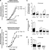
Ozone [O3, 2.0 parts per million (ppm), 4 h] significantly increased vagally mediated bronchoconstriction and lung inflammation measured 1 day later in nonsensitized guinea pigs. Electrical stimulation of both vagus nerves (1–25 Hz, 10 V, 0.2 ms, 5-s train at 1-min intervals) produced frequency-dependent bronchoconstriction that was significantly increased by ozone (●, A) compared with air (○, A). In contrast, intravenous methacholine (MCh)-induced bronchoconstriction was not similarly potentiated by ozone (B). In bronchoalveolar lavage (C), eosinophils (Eos), neutrophils (PMN), macrophages (Mac), and lymphocytes (Lymp) were all significantly increased by ozone. These increases were comprised of 5-bromo-2-deoxyuridine-positive (BrdU+) and BrdU cells although the increase in BrdU cells was only significant in macrophages and combined cells. Note that each column represents the total cell number and is subdivided to show BrdU (white shading) and BrdU+ (black shading) cells. The percentage of BrdU+ cells is included under each column. In bone marrow, ozone significantly potentiated the percentage of BrdU+ eosinophils (black portion, D, final column). Data are expressed as in C. Data are shown as means ± SE, n = 4–9, *P ≤ 0.05 in all cases. In C and D, statistical comparisons of total cells are shown as horizontal lines above bars. Statistical comparisons for BrdU+ and BrdU cells are shown within subdivisions.
Fig. 2.
Fig. 2.
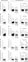
Ozone had no effect on total leukocytes, eosinophils, neutrophils, monocytes, and lymphocytes in blood 1 day (A–E) or 3 days (F–J) after exposure in nonsensitized (black/white shading) and sensitized (gray/white shading) guinea pigs. Note that, as in Fig. 1, each column represents the total and is subdivided to show BrdU (white shading) and BrdU+ (black or gray shading) cells. The percentage of BrdU+ cells is included under each column. Etanercept significantly increased circulating monocytes (I). Etanercept increased circulating monocytes (I) and lymphocytes (J) after ozone in both nonsensitized and sensitized animals. Data are means ± SE; n = 4–9. Statistics are shown as in Fig. 1, P ≤ 0.05, *significantly different from air, #significantly different from untreated ozone.
Fig. 3.
Fig. 3.
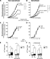
Ozone (O3, 2.0 ppm, 4 h) significantly increased vagally mediated bronchoconstriction and total inflammatory cells in nonsensitized and sensitized guinea pigs (S) measured 3 days later. Electrical stimulation of both vagus nerves (same parameters as Fig. 1) produced frequency-dependent bronchoconstriction that was significantly increased 3 days after ozone in nonsensitized (A) and sensitized (B) guinea pigs. Antibody to IL-5 (AbIL-5) significantly potentiated ozone-induced airway hyperreactivity in nonsensitized guinea pigs (A) but significantly inhibited hyperreactivity in sensitized animals (B). Etanercept (Et) significantly increased ozone-induced hyperreactivity in nonsensitized animals (A) but had no effect in sensitized animals (B). Bronchoconstriction induced by intravenous methacholine in vagotomized animals was not potentiated by ozone, AbIL-5, or etanercept in nonsensitized (C), or sensitized guinea pigs (D). Ozone significantly increased inflammatory cells in bronchoalveolar lavage in nonsensitized (black/white) and sensitized (gray/white) guinea pigs 3 days later (E). In nonsensitized animals, ozone significantly increased both BrdU+ (black shading) and BrdU (white shading) inflammatory cells, whereas, in sensitized guinea pigs, ozone increased only BrdU (white) inflammatory cells (E). Etanercept had no effect on total inflammatory cells in air- or ozone-exposed animals (F). Data are shown as means ± SE, n = 4–9, *P ≤ 0.05 in all cases. In E and F statistical comparisons of total cells are shown as horizontal lines above bars. Statistical comparisons for BrdU+ and BrdU cells are shown within subdivisions.
Fig. 4.
Fig. 4.
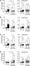
Ozone significantly increased eosinophils (A), neutrophils (B), and macrophages (C) but not lymphocytes (D) in bronchoalveolar lavage of nonsensitized (black/white) guinea pigs 3 days later. Note that, as in Fig. 1, each column represents the total and is subdivided to show BrdU (white shading) and BrdU+ (black or gray shading) cells. The percentage of BrdU+ cells is included under each column. In nonsensitized animals, the ozone-induced increase comprised BrdU+ cells. Only eosinophils were significantly potentiated by sensitization (A, compare 1st 2 sets of bars). In sensitized guinea pigs, ozone significantly increased eosinophils (A), neutrophils (B), macrophages (C), and lymphocytes (D) in bronchoalveolar lavage. However, in sensitized animals, ozone did not increase BrdU+ cells. Etanercept potentiated the ability of ozone to recruit eosinophils, macrophages, and lymphocytes in nonsensitized animals (3rd set of columns, black/white shading) and potentiated the ability of ozone to recruit neutrophils in sensitized animals (4th set of columns, gray/white shading). Statistics are shown as in Fig. 1, P ≤ 0.05, *significantly different from air, #significantly different from untreated ozone.
Fig. 5.
Fig. 5.
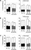
Ozone specifically and significantly increased eosinophil hematopoiesis in nonsensitized (black/white shading) guinea pigs 3 days later (A). Note that, as in Fig. 1, each column represents the total and is subdivided to show BrdU (white shading) and BrdU+ (black or gray shading) cells. The percentage of BrdU+ cells is included under each column. Ozone increased BrdU+ (black shading) eosinophils in bone marrow (1st set of columns, black shading, A) in nonsensitized guinea pigs; however, this increase failed to occur in nonsensitized (2nd set of columns, gray, A) or etanercept-treated nonsensitized (3rd set of columns, black shading, A) animals. Neutrophils (B) and monocytes (C) in were not changed by ozone, by sensitization, or by etanercept (B). Statistics are shown as in Fig. 1, P ≤ 0.05, *significantly different from air, #significantly different from untreated ozone.
Fig. 6.
Fig. 6.
Eosinophils in bronchoalveolar lavage were depleted by AbIL-5 in nonsensitized and sensitized guinea pigs 3 days after ozone. 3 days after ozone combined, eosinophils, neutrophils, and macrophages were significantly increased compared with air in both nonsensitized and sensitized guinea pigs. In nonsensitized animals, AbIL-5 significantly decreased eosinophils in ozone-exposed guinea pigs. In sensitized animals, total, eosinophils, and lymphocytes were decreased following AbIL-5 following ozone. Data are means ± SE, n = 5–9, P ≤ 0.05, *significantly different from nontreated animals.
Fig. 7.
Fig. 7.
Pretreatment with etanercept had no effect on ozone-induced airway hyperreactivity. Electrical stimulation of both vagus nerves (same parameters as Fig. 1) produced frequency-dependent bronchoconstriction that was significantly increased 1 day after ozone (○ compared with ● data regraphed from Fig. 1A). Etanercept had no effect on ozone-induced airway hyperreactivity (▲, A). Methacholine-induced bronchoconstriction was not changed by etanercept (B). Both BrdU+ and BrdU neutrophils in bronchoalveolar lavage were significantly increased by ozone in etanercept-treated animals (C). Etanercept had no effect on circulating inflammatory cells (D). In bone marrow, etanercept significantly decreased the percentage of BrdU+ eosinophils after ozone compared with nontreated animals in Fig. 1 (E). Data are means ± SE, n = 4–9. Statistics are shown as in Fig. 1, P ≤ 0.05, *significantly different from air, #significantly different from 1 day after ozone.
Fig. 8.
Fig. 8.
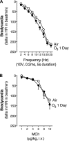
Vagally induced (A) and methacholine-induced (B) bradycardia was not changed by ozone at 1 day. Electrical stimulation of both vagus nerves (1–25 Hz, 10 V, 0.2 ms, 5-s train, at 1-min intervals) produced frequency-dependent bradycardia. Data are shown as means ± SE, n = 4–9. HR, heart rate.
Fig. 9.
Fig. 9.
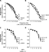
Vagally induced and methacholine-induced bradycardia was not changed by ozone sensitization, etanercept, or AbIL-5 3 days after ozone. Electrical stimulation of both vagus nerves (same parameters as Fig. 1) produced frequency-dependent bradycardia that was not affected by ozone (A) sensitization (B), etanercept (A and B), or AbIL-5 (A). Methacholine-induced bradycardia was not changed by ozone (C) or sensitization (D) or etanercept (C and D). Data are shown as means ± SE, n = 4–9.

Similar articles

Cited by

References

    1. Alexis NE, Lay JC, Haczku A, Gong H, Linn W, Hazucha MJ, Harris B, Tal-Singer R, Peden DB. Fluticasone propionate protects against ozone-induced airway inflammation and modified immune cell activation markers in healthy volunteers. Environ Health Perspect 116: 799–805, 2008. doi:10.1289/ehp.10981. - DOI - PMC - PubMed
    1. American Lung Association. State of the Air 2016. http://www.lung.org/local-content/california/our-initiatives/state-of-th.... Chicago, IL: ALA, 2016. Date accessed: 1 March 2017.
    1. Aris RM, Christian D, Hearne PQ, Kerr K, Finkbeiner WE, Balmes JR. Ozone-induced airway inflammation in human subjects as determined by airway lavage and biopsy. Am Rev Respir Dis 148: 1363–1372, 1993. doi:10.1164/ajrccm/148.5.1363. - DOI - PubMed
    1. Arjomandi M, Wong H, Donde A, Frelinger J, Dalton S, Ching W, Power K, Balmes JR. Exposure to medium and high ambient levels of ozone causes adverse systemic inflammatory and cardiac autonomic effects. Am J Physiol Heart Circ Physiol 308: H1499–H1509, 2015. doi:10.1152/ajpheart.00849.2014. - DOI - PMC - PubMed
    1. Bascom R, Naclerio RM, Fitzgerald TK, Kagey-Sobotka A, Proud D. Effect of ozone inhalation on the response to nasal challenge with antigen of allergic subjects. Am Rev Respir Dis 142: 594–601, 1990. doi:10.1164/ajrccm/142.3.594. - DOI - PubMed

Publication types

MeSH terms

LinkOut - more resources