. 2017 May 5;7(5):1417-1427.
doi: 10.1534/g3.117.040212.

Dynamic Notch Signaling Specifies Each Cell Fate in Drosophila Spermathecal Lineage

Affiliations

Dynamic Notch Signaling Specifies Each Cell Fate in Drosophila Spermathecal Lineage

Wei Shen et al. G3 (Bethesda). .

Abstract

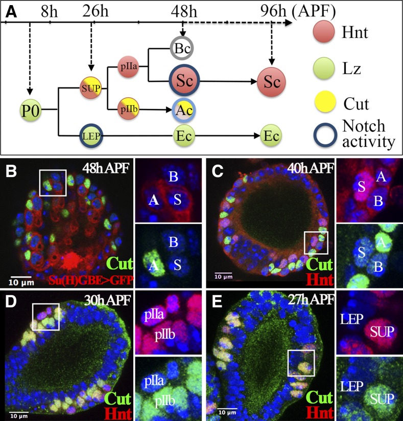
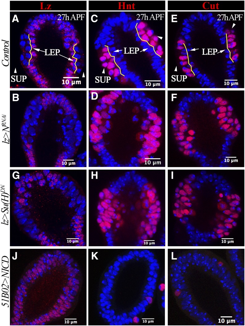
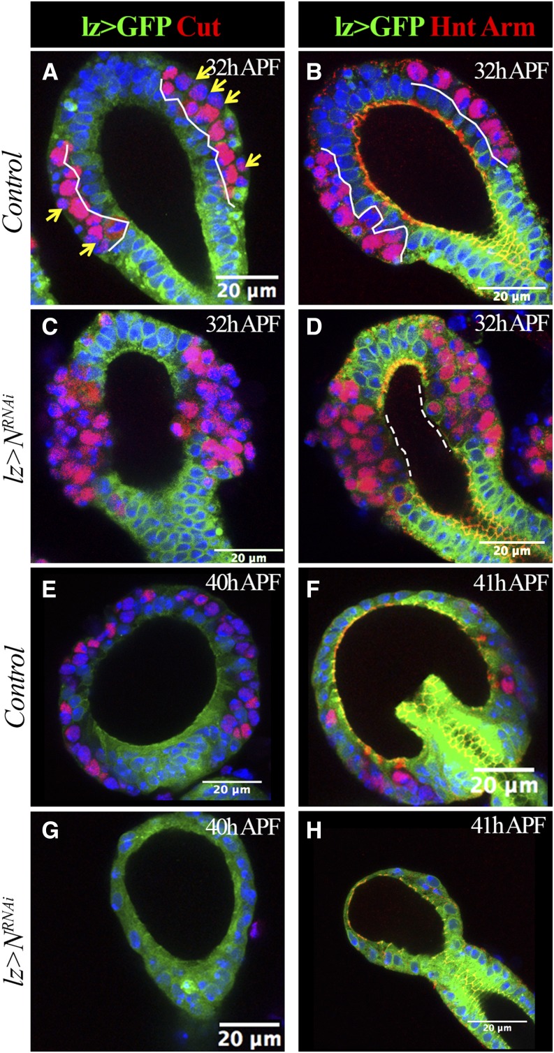
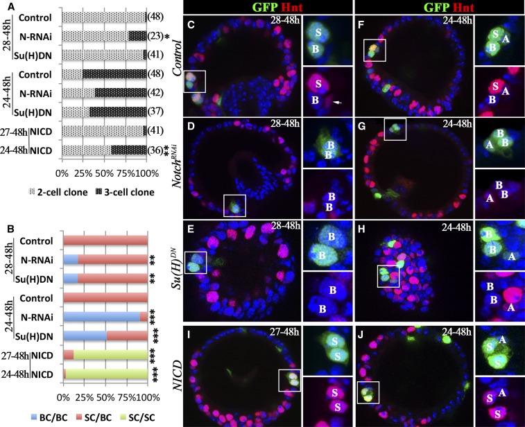
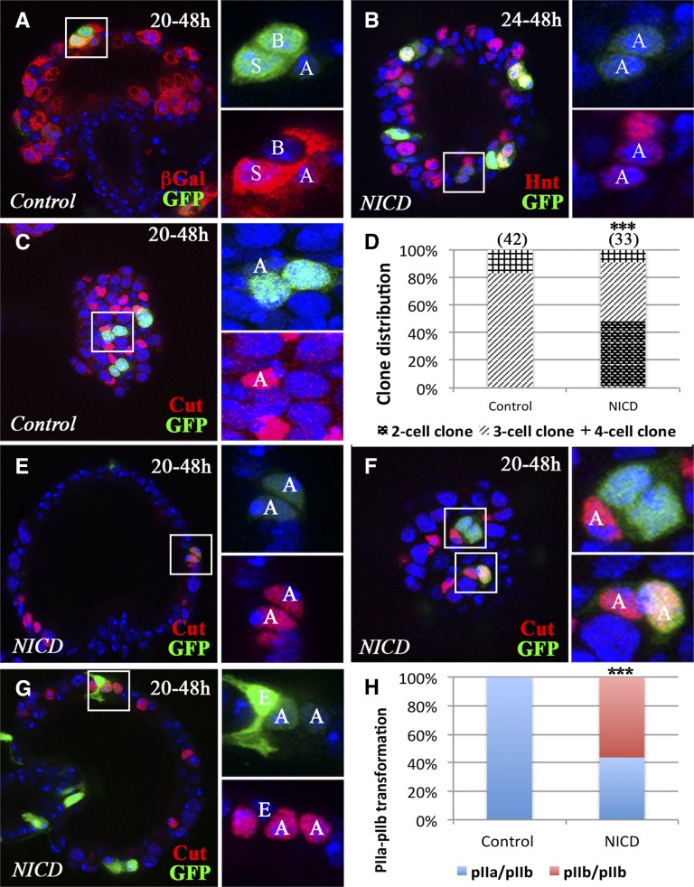
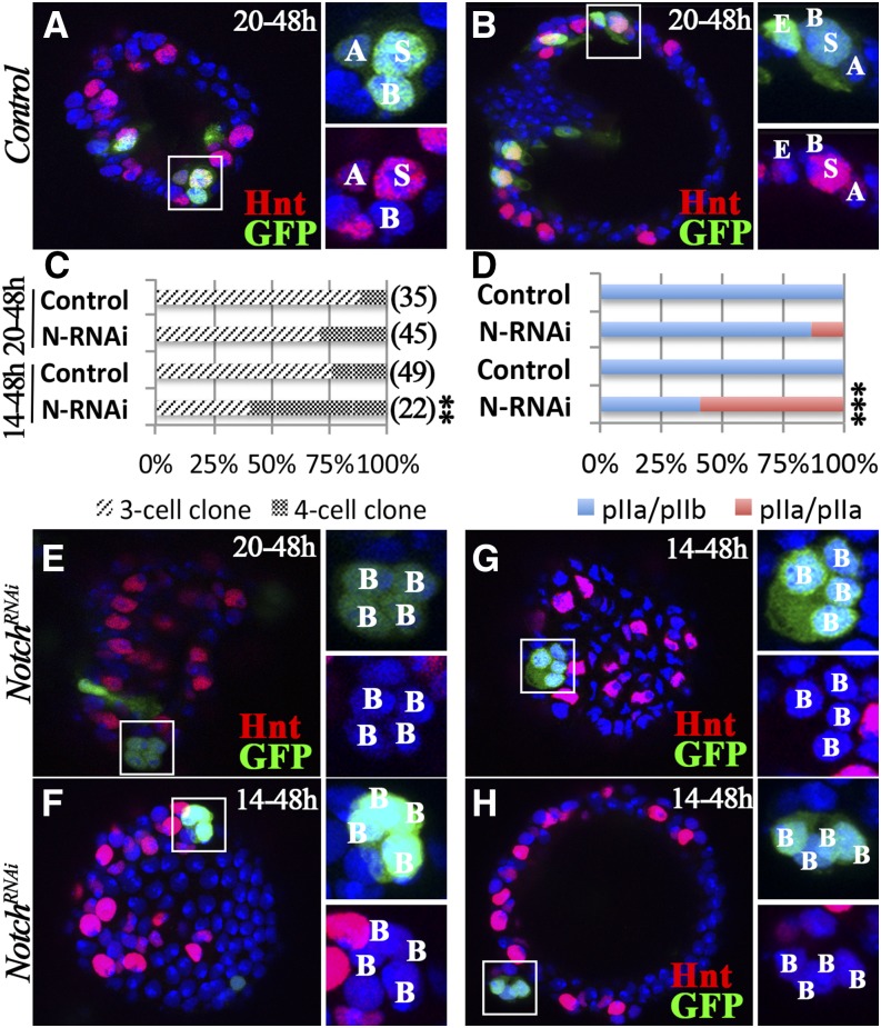
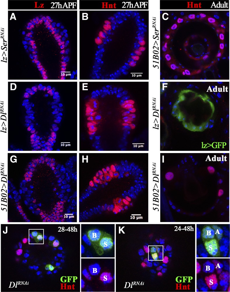
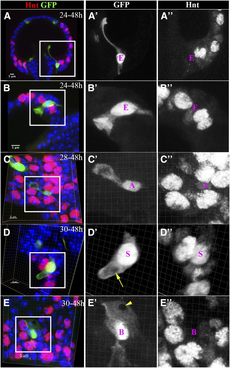
Spermathecae are glandular organs in the insect female reproductive tract that play essential roles in insect reproduction; however, the molecular mechanism involved in their development is largely unknown. Drosophila spermathecae consist of class-III secretory units, in which each secretory cell (SC) discharges its products to the central lumen through an end-apparatus and a canal. Secretory unit formation in Drosophila spermathecae utilizes a fixed cell lineage, in which each secretory unit precursor (SUP) divides to produce one pIIb cell and one pIIa cell. The former differentiates into an apical cell (AC), whereas the latter divides again to produce an SC and a basal cell (BC). It is unclear how each cell acquires its identity and contributes to secretory unit formation. Here, we demonstrate that Notch signaling is required and sufficient for the specification of lumen epithelial precursors (LEPs; vs. SUPs), pIIb (vs. pIIa), and SCs (vs. BCs) sequentially. To our surprise, Notch activation in LEPs and SCs apparently utilizes different ligand mechanisms. In addition, Notch signaling both suppresses and activates transcription factors Hindsight (Hnt) and Cut during spermathecal lineage specification, supporting the notion that Notch signaling can have opposite biological outcomes in different cellular environments. Furthermore, LEP-derived epithelial cells (ECs) and ACs show distinct cellular morphology and are essential for securing secretory units to the epithelial lumen. Our work demonstrates, for the first time, the dynamic role of Notch signaling in binary cell fate determination in Drosophila spermathecae and the role of ECs and ACs in secretory unit formation.

Keywords: Cut; Notch signaling; binary cell fate determination; class-III secretory gland; spermathecae.


Figures

Figure 1
Figure 1
Cut expression in the spermathecal lineage. (A) Diagram depicting the spermathecal lineage during Drosophila pupal development. Hnt, Lz, and Cut expression are labeled in red, green, and yellow, respectively. Notch activity is indicated by the blue circle. A light blue circle was used in the AC to reflect the lower expression of Notch activity reporter at 48 hr APF. (B) Cut expression (green) in spermathecae at 48 hr APF. Notch activity is indicated by Su(H)GBE-Gal4 driving expression of UAS-GFP (Su(H)GBE > GFP; shown in red). Enlarged pictures of the squared area with two channels are shown in the right two panels. The AC, BC, and SC are marked by A, B, and S, respectively. DNA staining with DAPI is shown in blue in all figures. (C–E) Expression of Cut (green) and Hnt (red) in spermathecae at 40 (C), 30 (D), and 27 hr (E) APF. All figures depict the spermathecal heads oriented with distal head (vault) pointed upwards and the duct pointed downward. AC, apical cell; APF, after puparium formation; BC, basal cell; DAPI, 4’,6-diamidino-2-phenylindole; Ec, epithelial cell; GFP, green fluorescent protein; Hnt, Hindsight; LEP, lumen epithelial precursors; Lz, Lozenge; SC, secretory cell; Su(H), Suppressor of Hairless; SUP, secretory unit precursors.
Figure 2
Figure 2
Notch is required and sufficient for LEP fate. All spermathecae are at 26–28 hr APF and at least 10 spermathecae are examined and show the same phenotype. (A–F) Expression of Lz (A and B), Hnt (C and D), and Cut (E and F) in spermathecal head. (A, C, and E) Spermathecae from the control group; (B, D, and F) depict spermathecae from N-knockdown pupae using lz-Gal4 (lz > NRNAi). Yellow lines demarcate the apical and basal layers; LEPs in apical layers are pointed with arrows, whereas SUPs in basal layers are pointed with arrowheads. (G–I) Expression of Lz (G), Hnt (H), and Cut (I) in spermathecae with overexpression of lz-Gal4 driving Su(H)DN. (J–L) Expression of Lz (J), Hnt (K), and Cut (L) in spermathecae with 51B02-Gal4 driving overexpression of NICD. APF, after puparium formation; DN, dominant negative; Hnt, Hindsight; LEP, lumen epithelial precursors; Lz, Lozenge; N, Notch; NICD, Notch intracellular domain; RNAi, RNA interference; Su(H), Suppressor of Hairless; SUP, secretory unit precursors.
Figure 3
Figure 3
Loss of LEP causes dissociation of secretory units from the central lumen. lz-Gal4 driving expression of UAS-mCD8:GFP (lz > GFP) is shown in green. Cut expression is shown in red in (A, C, E, and G). Hnt (nucleus red signal) and Arm (membrane red signal) expression are shown in (B, D, F, and H). Arm marks adherent junctions. At least 10 spermathecae are examined and shown the same pattern. (A and B) Control spermathecae at 32 hr APF. White lines demarcate the epithelial layer (GFP+ cells). Yellow arrows indicate pIIa cells with no or very low expression of Cut. (C and D) N-knockdown spermathecae at 32 hr APF. Notice the missing GFP+ epithelial cells in the middle region of the spermathecal head. Dashed lines demarcate the region without Arm staining. (E–H) Control (E and F) and N-knockdown (G and H) spermathecae at 40–41 hr APF. Only one or two Cut+ or Hnt+ cells were observed in N-knockdown spermathecae. APF, after puparium formation; GFP, green fluorescent protein; Hnt, Hindsight; LEP, lumen epithelial precursors; Lz, Lozenge; N, Notch; RNAi, RNA interference.
Figure 4
Figure 4
Notch signaling is required and sufficient for SC fate. Flip-out clones are marked by GFP expression (green in C–J), and Hnt expression is shown in red (C–J). (A and B) Quantification of SC-clone distribution according to clone size (A) or clone composition (B) when induced at multiple time points. The number of clones analyzed is shown in parentheses. The category in (B) only indicates the BC and SC, regardless of whether or not the clone contains the AC. Fisher’s exact test was used for assessing statistical significance (*P < 0.05, **P < 0.01, and ***P < 0.001). (C–E) Representative spermathecae with control (C), N-knockdown (D), or Su(H)DN-overexpressing (E) clones induced at 28 hr and observed at 48 hr APF (28–48 hr). The square areas are showed at higher magnification with only two channels in the right two subpanels. The clone cell identity is marked by A, B, or S for AC, BC, or SC, respectively. The arrow in (C) points to an AC with elongated nuclei and faint Hnt expression, which is not in the clone. (F–H) Representative spermathecae with control (F), N-knockdown (G), or Su(H)DN-overexpressing (H) clones induced at 24 hr and observed at 48 hr APF (24–48 hr). (I–J) Representative spermathecae with NICD-overexpressing clones induced at 27 (I) and 24 hr (J), and observed at 48 hr APF (27–48 and 24–48 hr, respectively). AC, apical cell; APF, after puparium formation; BC, basal cell; DN, dominant negative; GFP, green fluorescent protein; Hnt, Hindsight; N, Notch; NICD, Notch intracellular domain; RNAi, RNA interference; SC, secretory cell; Su(H), Suppressor of Hairless.
Figure 5
Figure 5
Notch signaling is sufficient for pIIb fate. Flip-out clones are marked by GFP expression (green in A–C and E–G). (A) A representative control clone induced at 20 hr APF shows weak Notch activity in the AC and strong Notch activity in the SC. βGal expression (red) from Su(H)GBE-LacZ reporter is used to mark Notch activity. (B) Representative NICD-overexpressing clones induced at 24 hr and examined at 48 hr APF. The square area is magnified in the right two panels and shows a clone with two AC-like cells with faint GFP (green) and Hnt (red) expression. (C) Representative control clones induced at 20 hr and examined at 48 hr APF. The three-cell clone in the square area shows the AC with Cut expression (red). The other two clones have the same composition. (D) Quantification of clone distribution according to clone size. Clones were induced at 20 hr APF and examined at 48 hr APF. (E–G) Representative NICD-overexpressing clones induced at 20 hr and examined at 48 hr APF. (E) A two-cell clone (faint GFP) is composed of two ACs (Cut+). (F) A three-cell clone (upper panel) contains one AC (Cut+); a two-cell clone (one with faint GFP and one with strong GFP; lower panel) is composed of two ACs (Cut+). (G) A three-cell clone is composed of one EC (strong GFP and distinct cellular morphology) and two ACs (Cut+). (H) Quantification of clone distribution according to clone composition. pIIa/pIIb: clones containing one pIIa and pIIb cell during division. pIIb/pIIb: clones containing two pIIb cells during division, which gives rise to two ACs at 48 hr APF. Fisher’s exact test was used (***P < 0.001). AC, apical cell; APF, after puparium formation; βGal, β-galactosidase; EC, epithelial cell; GFP, green fluorescent protein; Hnt, Hindsight; NICD, Notch intracellular domain; SC, secretory cell; Su(H), Suppressor of Hairless.
Figure 6
Figure 6
Notch signaling is required for pIIb fate. (A and B) Representative control clones induced at 20 hr and examined at 48 hr APF. A three-cell clone is shown in (A), and a four-cell clone is shown in (B) with clone composition labeled according to Hnt expression (red). (C and D) Quantification of clone distribution according to clone size (C) and composition (D). The Fisher’s exact test was used (**P < 0.01 and ***P < 0.001). (E) A representative N-knockdown clone induced at 20 hr APF shows four BC-like cells without Hnt expression (red) at 48 hr APF. (F–H) Representative N-knockdown clones induced at 14 hr and examined at 48 hr APF. (F) A four-cell clone with four BCs (same size). (G) A four-cell clone with four BCs (two small and two big). (H) A four-cell clone with four BCs detaching from the lumen. APF, after puparium formation; BC, basal cell; GFP, green fluorescent protein; Hnt, Hindsight; N, Notch; RNAi, RNA interference.
Figure 7
Figure 7
Dl, but not Ser, is required for LEP fate specification. (A and B) Ser knockdown with lz-Gal4 shows normal Lz (A) and Hnt (B) expression in spermatheca at 27 hr APF. (C) Adult spermathecae with Ser knockdown in 51B02-Gal4-expressing cells have normal secretory cells marked by Hnt expression. (D–I) Dl knockdown in lz-Gal4- (D–F) or 51B02-Gal4-expressing cells (G–I) leads to loss of Lz (D and G) and gain of Hnt (E and H) expression at the middle region of spermathecal head at 27 hr APF. Few secretory cells were formed in adult spermathecae (F–I). (J–K) Dl-knockdown clones induced at 28 (J) or 24 hr (K) and examined at 48 hr APF. These clones have normal cell composition. Hnt expression (red) is used to marked the SCs. APF, after puparium formation; Dl, Notch ligand δ; Hnt, Hindsight; LEP, lumen epithelial precursor; Lz, Lozenge; RNAi, RNA interference; SC, secretory cell; Ser, Serrate.
Figure 8
Figure 8
Cellular morphology of spermathecal lineage cells. (A and B) Single-EC clones induced at 24 hr and examined at 48 hr APF. (A) The EC localized at the junction of spermathecal lumen, and the duct has a long cytoplasmic protrusion in line with the introvert. (B) The EC localized in the middle region of the spermathecal head has an inverted umbrella-shaped apical membrane protruding into the lumen. The cytoplasm is marked by GFP [green in (A and B) and white in (A’–B’)]. (C) A single-AC clone shows the AC with an apical cytoplasmic bulge (C’). The AC is recognizable by the faint Hnt expression (white in C”). (D) A single-SC clone shows a finger-like apical protrusion with a hole in the middle (yellow arrow in D’). The SC is marked by strong Hnt expression (white in D”). (E) A single-BC clone shows a mesh-like apical membrane with a hole in the apical tip (arrowhead in E’). The BC is recognizable by the absence of Hnt expression (white in E”). The images in (B–E) are generated from three-dimensional volume rendering. AC, apical cell; BC, basal cell; EC, epithelial cell; GFP, green fluorescent protein; Hnt, Hindsight; SC, secretory cell.

Similar articles

Cited by

References

    1. Allen A. K., Spradling A. C., 2008. The Sf1-related nuclear hormone receptor Hr39 regulates Drosophila female reproductive tract development and function. Development 135: 311–321. - PubMed
    1. Anderson R. C., 1945. A study of the factors affecting fertility of lozenge females of Drosophila melanogaster. Genetics 30: 280–296. - PMC - PubMed
    1. Artavanis-Tsakonas S., Rand M. D., Lake R. J., 1999. Notch signaling: cell fate control and signal integration in development. Science 284: 770–776. - PubMed
    1. Bhat K. M., 2014. Notch signaling acts before cell division to promote asymmetric cleavage and cell fate of neural precursor cells. Sci. Signal. 7: ra101. - PubMed
    1. Billen J., 2011. Exocrine glands and their key function in the communication system of social insects. Formos. Entomol 31: 75–84.

Publication types

LinkOut - more resources