. 2017 Mar 22;37(12):3364-3377.
doi: 10.1523/JNEUROSCI.3010-16.2017. Epub 2017 Mar 3.

Inhibition of L-Type Ca2+ Channels by TRPC1-STIM1 Complex Is Essential for the Protection of Dopaminergic Neurons

Affiliations

Inhibition of L-Type Ca2+ Channels by TRPC1-STIM1 Complex Is Essential for the Protection of Dopaminergic Neurons

Yuyang Sun et al. J Neurosci. .

Abstract

Loss of dopaminergic (DA) neurons leads to Parkinson's disease; however, the mechanism(s) for the vulnerability of DA neurons is(are) not fully understood. We demonstrate that TRPC1 regulates the L-type Ca2+ channel that contributes to the rhythmic activity of adult DA neurons in the substantia nigra region. Store depletion that activates TRPC1, via STIM1, inhibits the frequency and amplitude of the rhythmic activity in DA neurons of wild-type, but not in TRPC1-/-, mice. Similarly, TRPC1-/- substantia nigra neurons showed increased L-type Ca2+ currents, decreased stimulation-dependent STIM1-Cav1.3 interaction, and decreased DA neurons. L-type Ca2+ currents and the open channel probability of Cav1.3 channels were also reduced upon TRPC1 activation, whereas increased Cav1.3 currents were observed upon STIM1 or TRPC1 silencing. Increased interaction between Cav1.3-TRPC1-STIM1 was observed upon store depletion and the loss of either TRPC1 or STIM1 led to DA cell death, which was prevented by inhibiting L-type Ca2+ channels. Neurotoxins that mimic Parkinson's disease increased Cav1.3 function, decreased TRPC1 expression, inhibited Tg-mediated STIM1-Cav1.3 interaction, and induced caspase activation. Importantly, restoration of TRPC1 expression not only inhibited Cav1.3 function but increased cell survival. Together, we provide evidence that TRPC1 suppresses Cav1.3 activity by providing an STIM1-based scaffold, which is essential for DA neuron survival.SIGNIFICANCE STATEMENT Ca2+ entry serves critical cellular functions in virtually every cell type, and appropriate regulation of Ca2+ in neurons is essential for proper function. In Parkinson's disease, DA neurons are specifically degenerated, but the mechanism is not known. Unlike other neurons, DA neurons depend on Cav1.3 channels for their rhythmic activity. Our studies show that, in normal conditions, the pacemaking activity in DA neurons is inhibited by the TRPC1-STIM1 complex. Neurotoxins that mimic Parkinson's disease target TRPC1 expression, which leads to an abnormal increase in Cav1.3 activity, thereby causing degeneration of DA neurons. These findings link TRPC1 to Cav1.3 regulation and provide important indications about how disrupting Ca2+ balance could have a direct implication in the treatment of Parkinson's patients.

Keywords: Cav1.3; Parkinson's disease; SOCE; TRPC1-STIM1; calcium.


Figures

Figure 1.
Figure 1.
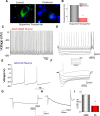
Functional characterization of DA and GABAergic neurons in the SN region. A, Paraffin-embedded sections of postmortem human SN region obtained from control and PD patients. Sections were immunostained using DAT. B, Quantification of the fluorescence obtained from 5 or 6 individual sections. Patterns of AP firing using tissue slices from SN area in a GABA neuron (C) and in adult DA neurons (E). Traces were recorded without current injection. D, F, Voltage traces were evoked by a series of 1000 ms current pulses (from −200 pA with increment −40 pA). G, H, Current traces evoked by −50 mV steps from holding potential −60 mV. I, Bar graph represents Ih ratio in GABA and DA neurons (n = 15). *p < 0.05.
Figure 2.
Figure 2.
Cav1.3 channels contribute to the rhythmic activity of DA neurons. In the whole-cell configuration, application of 0.5 μm TTX abolished spiking of GABA neurons in wild-type mice (A, B). C, Spontaneous APs and application of 0.5 μm TTX abolished spiking of DA neurons in juvenile mice (P14–P18 d) (D). E, Patterns of AP firing in adult (p > 28 d) DA neurons from the SN area. F, G, Application of 0.5 μm TTX decreased spiking in adult DA neurons. H, Application of 1 μm BayK8644 increased spiking. I, The addition of 1 μm nifedipine abolished spiking of DA neurons in adult mice. J, Application of 0.5 μm TTX along with removing external Ca2+ also abolished spiking of DA neurons in adult mice.
Figure 3.
Figure 3.
TRPC1 inhibits the rhythmic activity of DA neurons in the SN region. In the whole-cell configuration, patterns of AP firing of adult DA neurons (p > 28 d) in wild-type mice (A) and TRPC1−/− mice (E). B, F, AP firing upon application of 0.5 μm TTX in both wild-type and TRPC1−/− slices. C, G, Rhythmic activity in SN slices that were incubated with 1 μm Tg + TTX in wild-type and TRPC1−/− mice, receptively. D, H, Rhythmic activity in SN slices that were incubated with 1 μm Tg + TTX + 50 μm APB in wild-type and TRPC1−/− mice, receptively. I–L, Pooled time course of AP firing frequency under various conditions in WT and TRPC1−/− mice (n = 9). M, N, The frequencies and normalized amplitude under these conditions. *Significantly different values (p < 0.05) (n = 9).
Figure 4.
Figure 4.
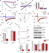
TRPC1 modulates Cav1.3 activity in SN area of mice. A, Representative trace showing store dependent (upon addition of 1 μm Tg Ca2+ entry at −70 mV holding potential) that induced a TRPC1-like current in DA neurons of WT mice but was abolished in the TRPC1−/− mice. Application 50 μm 2-APB also significantly inhibited the currents in wild-type mice. B, I–V curves under these conditions. C, Maximum current (at −70 mV holding potential) in wild-type with and without 2-APB and in TRPC1−/− mice (n = 11). D, Representative trace showed that calcium currents evoked in DA neurons from SN area using a step protocol from −60 to 10 mV in the whole-cell configuration in wild-type mice. E, Representative I–V relations under these conditions (n = 8). F, Representative trace showed that Ca2+ currents evoked in DA neurons from SN area in TRPC1−/− mice. G, The average (10–12 recordings) currents under various conditions (n = 10). *p < 0.05. H, A train of three APs induced Ca2+ currents in WT and TRPC1−/− mice (n = 5). *p < 0.05. I, Western blots of lysates from SN tissues from control and TRPC1−/− mice. Antibodies used are indicated in the figure. Densitometry for normalized Cav1.3 relative to β-actin bar graph is to show quantitation from at least three independent experiments. Data are mean ± SEM from three independent experiments. *p < 0.05.
Figure 5.
Figure 5.
TRPC1 provide STIM1 scaffold that protects TH-positive neurons in SN region. A, Western blots of coimmunoprecipitates of SN tissues using STIM1 antibody under control (CTRL) and stimulated (store depletion, +Tg) conditions. Bar graph represents densitometry for fold change in Cav1.3 pulldown to show quantitation from at least three independent experiments. B, Representative DAB images of brain sections showing full SN region stained with TH antibody from wild-type and TRPC1−/− mice. C, Bar graph represents stereology of TH staining from rostral to the caudal region. Data are mean ± SE. *Significantly different values (p < 0.05). n = 4. D, Immunostaining of TH cells in SN and VTA region obtained from wild-type and TRPC1−/− mice. E, Western blots of lysates from SN tissues from control and PD samples. Antibodies used are labeled in the figure. F, Densitometric quantitation for normalized TRPC1 or Cav1.3 relative to TH expression. Data are mean ± SEM from three independent experiments. *p < 0.05.
Figure 6.
Figure 6.
Cav1.3 channel activity in SH-SY5Y cells is regulated via TRPC1-STIM1. Representative Ca2+ traces in differentiated SH-SY5Y cells showed that silencing of STIM1 significantly inhibited Ca2+ influx that was induced upon store depletion (+ 1 μm Tg) (A). STIM1 or TRPC1 silencing increases L-type calcium currents (B and G, respectively). The average (8–10 recordings) I–V relations under these conditions are shown in C and H, respectively (n = 9). *p < 0.05. Mean steady-state inactivation curves were obtained for L-type current in control and siTRPC1 (D). n = 5. Silencing of Cav1.3 (E, F) abolished the effect of siTRPRC1 on the L-type calcium current. Insets, Western blots from lysates of differentiated SH-SY5Y cells showing decreased expression of TRPC1, Cav1.3, and STIM1 (as well as overexpression of STIM1 in siSTIM1 cells, rescue) using respective siRNAs. Overexpression of full-length STIM1 in siSTIM1-expressing cells (rescue) showed an increase in Ca2+ influx (I, J). K, Average histogram for current density in various conditions. *p < 0.05.
Figure 7.
Figure 7.
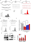
TRPC1 affect Cav1.3 channels in SH-SY5Y cells. Single-channel recording (from individual differentiated 10–12 SH-SY5Y cells in each condition) using step protocol from −40 mV to 10 mV in control (A), nifedipine (1 μm) (B), Tg-treated (1 μm) cells (C), and siCav1.3 cells (D, E). Variance histograms were generated with Gaussian fits and are shown at the bottom of the traces. F, Western blots of coimmunoprecipitates of SH-SY5Y cells using Cav1.3 or STIM1 antibodies in control (CTRL) or stimulated (+Tg) conditions. Antibodies used for Western blots are labeled in the figure. Quantification of the data from 3 to 5 independent experiments is shown. *Significantly different values (p < 0.05). n = 8. G, Cell viability under various conditions in SH-SY5Y cells is shown. *Significantly different from untreated cells (p < 0.05). n = 4.
Figure 8.
Figure 8.
TRPC1 regulates Cav1.3 function in neurotoxin PD model. A, Representative trace showed that calcium currents were evoked in differentiated SH-SY5Y cells with a step protocol from −60 to 10 mV under various conditions. Cells treated with MPP+ facilitated calcium current, but overexpression of TRPC1 abolished this effect. B, I–V curves. E, F, Western blots of lysates in control and MPP+-treated SH-SY5Y cells (500 μm, 24 h). C, D, Individual traces of L-type calcium currents in Cav1.3 silenced cells with and without MPP+ and/or Tg treatment. G, H, Coimmunoprecipitates of SH-SY5Y cells using Cav1.3 and STIM1 antibodies under various conditions. I, Western blots of lysates from differentiated SH-SY5Y cells using cleaved caspase3, Bak, and β-actin antibodies under various conditions. J, Western blots of lysates from mice SN region using cleaved caspase3, Bak, Bid, and β-actin antibodies under various conditions. For MPTP treatment, mice received MPTP-HCl 25 mg/kg (i.p. for 5 consecutive days at 24 h intervals). Densitometric quantitation for normalized relative to β-actin is shown below the blots. Values are mean ± SEM from three independent experiments. *p < 0.05. K, Cell viability under various conditions in SH-SY5Y cells. *Significantly different values from untreated cells (p < 0.05). n = 8.

Similar articles

Cited by

References

    1. Abou-Sleiman PM, Muqit MM, Wood NW (2006) Expanding insights of mitochondrial dysfunction in Parkinson's disease. Nat Rev Neurosci 7:207–219. 10.1038/nrn1868 - DOI - PubMed
    1. Berridge MJ, Lipp P, Bootman MD (2000) The versatility and universality of calcium signalling. Nat Rev Mol Cell Biol 1:11–21. 10.1038/35036035 - DOI - PubMed
    1. Björklund A, Dunnett SB (2007) Dopamine neuron systems in the brain: an update. Trends Neurosci 30:194–202. 10.1016/j.tins.2007.03.006 - DOI - PubMed
    1. Bollimuntha S, Singh BB, Shavali S, Sharma SK, Ebadi M (2005) TRPC1-mediated inhibition of 1-methyl-4-phenylpyridinium ion neurotoxicity in human SH-SY5Y neuroblastoma cells. J Biol Chem 280:2132–2140. 10.1074/jbc.M407384200 - DOI - PMC - PubMed
    1. Bollimuntha S, Ebadi M, Singh BB (2006) TRPC1 protects human SH-SY5Y cells against salsolinol-induced cytotoxicity by inhibiting apoptosis. Brain Res 1099:141–149. 10.1016/j.brainres.2006.04.104 - DOI - PMC - PubMed

Publication types

Substances

LinkOut - more resources