

Atg7 Deficiency Intensifies Inflammasome Activation and Pyroptosis in Pseudomonas Sepsis
- PMID: 28258192
- PMCID: PMC5382979
- DOI: 10.4049/jimmunol.1601196
Atg7 Deficiency Intensifies Inflammasome Activation and Pyroptosis in Pseudomonas Sepsis
Abstract
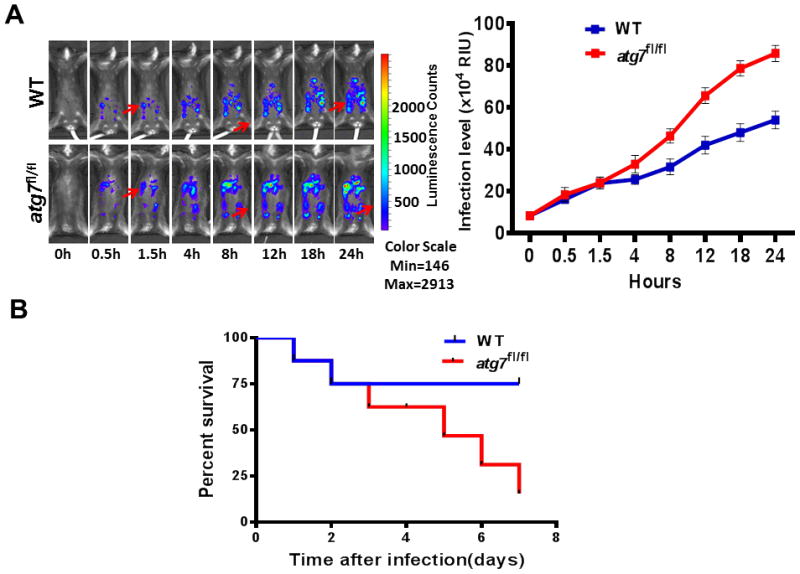
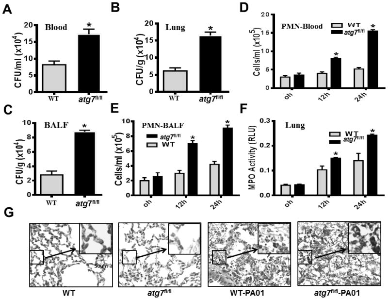
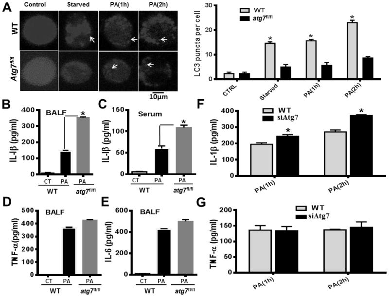
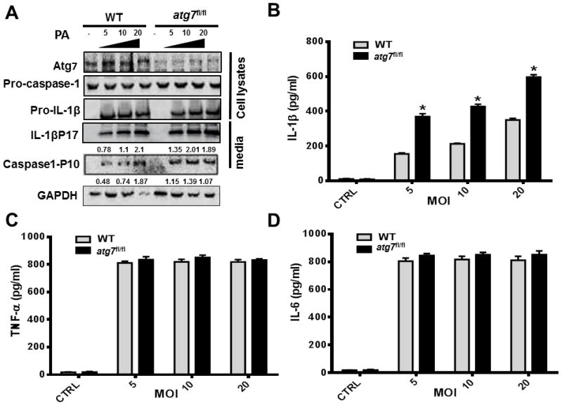
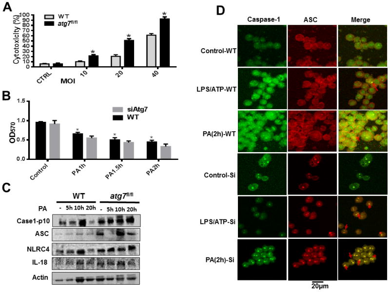
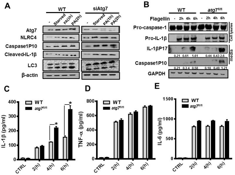
Sepsis is a severe and complicated syndrome that is characterized by dysregulation of host inflammatory responses and organ failure, with high morbidity and mortality. The literature implies that autophagy is a crucial regulator of inflammation in sepsis. In this article, we report that autophagy-related protein 7 (Atg7) is involved in inflammasome activation in Pseudomonas aeruginosa abdominal infection. Following i.p. challenge with P. aeruginosa, atg7fl/fl mice showed impaired pathogen clearance, decreased survival, and widespread dissemination of bacteria into the blood and lung tissue compared with wild-type mice. The septic atg7fl/fl mice also exhibited elevated neutrophil infiltration and severe lung injury. Loss of Atg7 resulted in increased production of IL-1β and pyroptosis, consistent with enhanced inflammasome activation. Furthermore, we demonstrated that P. aeruginosa flagellin is a chief trigger of inflammasome activation in the sepsis model. Collectively, our results provide insight into innate immunity and inflammasome activation in sepsis.
Copyright © 2017 by The American Association of Immunologists, Inc.
Conflict of interest statement
Figures
Similar articles
-
Pseudomonas aeruginosa type-3 secretion system dampens host defense by exploiting the NLRC4-coupled inflammasome.Am J Respir Crit Care Med. 2014 Apr 1;189(7):799-811. doi: 10.1164/rccm.201307-1358OC. Am J Respir Crit Care Med. 2014. PMID: 24555512
-
Triggering Receptors Expressed on Myeloid Cells 2 Promotes Corneal Resistance Against Pseudomonas aeruginosa by Inhibiting Caspase-1-Dependent Pyroptosis.Front Immunol. 2018 May 25;9:1121. doi: 10.3389/fimmu.2018.01121. eCollection 2018. Front Immunol. 2018. PMID: 29887864 Free PMC article.
-
Activation of inflammasome signaling mediates pathology of acute P. aeruginosa pneumonia.J Clin Invest. 2013 Apr;123(4):1630-7. doi: 10.1172/JCI66142. Epub 2013 Mar 8. J Clin Invest. 2013. PMID: 23478406 Free PMC article.
-
Inflammation: A Double-Edged Sword in the Response to Pseudomonas aeruginosa Infection.J Innate Immun. 2017;9(3):250-261. doi: 10.1159/000455857. Epub 2017 Feb 22. J Innate Immun. 2017. PMID: 28222444 Free PMC article. Review.
-
Neutrophil pyroptosis: new perspectives on sepsis.Cell Mol Life Sci. 2019 Jun;76(11):2031-2042. doi: 10.1007/s00018-019-03060-1. Epub 2019 Mar 14. Cell Mol Life Sci. 2019. PMID: 30877336 Free PMC article. Review.
Cited by
-
Targeting the miR-665-3p-ATG4B-autophagy axis relieves inflammation and apoptosis in intestinal ischemia/reperfusion.Cell Death Dis. 2018 May 1;9(5):483. doi: 10.1038/s41419-018-0518-9. Cell Death Dis. 2018. PMID: 29706629 Free PMC article.
-
Mechanisms and Targeted Therapeutic Strategies in Sepsis-Induced Myocardial Dysfunction: The Role of NLRP3 Inflammasome-Mediated Inflammation.J Inflamm Res. 2025 Jul 5;18:8875-8897. doi: 10.2147/JIR.S521655. eCollection 2025. J Inflamm Res. 2025. PMID: 40635763 Free PMC article. Review.
-
Autophagy, cell death, and cytokines in K. pneumoniae infection: therapeutic perspectives.Emerg Microbes Infect. 2023 Dec;12(1):2140607. doi: 10.1080/22221751.2022.2140607. Emerg Microbes Infect. 2023. PMID: 36287114 Free PMC article. Review.
-
ATG7/GAPLINC/IRF3 axis plays a critical role in regulating pathogenesis of influenza A virus.PLoS Pathog. 2024 Jan 16;20(1):e1011958. doi: 10.1371/journal.ppat.1011958. eCollection 2024 Jan. PLoS Pathog. 2024. PMID: 38227600 Free PMC article.
-
Regulation of eosinophil functions by autophagy.Semin Immunopathol. 2021 Jun;43(3):347-362. doi: 10.1007/s00281-021-00860-1. Epub 2021 May 21. Semin Immunopathol. 2021. PMID: 34019141 Free PMC article. Review.
References
-
- Bone RC, Sibbald WJ, Sprung CL. The ACCP-SCCM consensus conference on sepsis and organ failure. Chest. 1992;101:1481–1483. - PubMed
-
- Angus DC, Linde-Zwirble WT, Lidicker J, Clermont G, Carcillo J, Pinsky MR. Epidemiology of severe sepsis in the United States: analysis of incidence, outcome, and associated costs of care. Crit Care Med. 2001;29:1303–1310. - PubMed
-
- Martin GS, Mannino DM, Eaton S, Moss M. The epidemiology of sepsis in the United States from 1979 through 2000. N Engl J Med. 2003;348:1546–1554. - PubMed
-
- Stubljar D, Skvarc M. Effective Strategies for Diagnosis of Systemic Inflammatory Response Syndrome (SIRS) due to Bacterial Infection in Surgical Patients. Infect Disord Drug Targets. 2015;15:53–56. - PubMed
-
- Medzhitov R. Origin and physiological roles of inflammation. Nature. 2008;454:428–435. - PubMed
MeSH terms
Substances
Grants and funding
LinkOut - more resources
Full Text Sources
Other Literature Sources
Medical
Molecular Biology Databases
