. 2017 Apr 3;149(4):465-481.
doi: 10.1085/jgp.201611668. Epub 2017 Mar 3.

Mechanism of sodium channel block by local anesthetics, antiarrhythmics, and anticonvulsants

Affiliations

Mechanism of sodium channel block by local anesthetics, antiarrhythmics, and anticonvulsants

Denis B Tikhonov et al. J Gen Physiol. .

Abstract

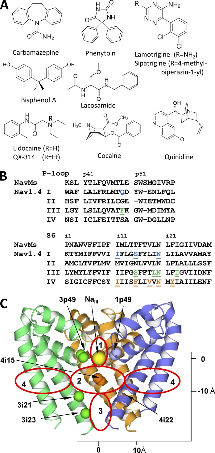
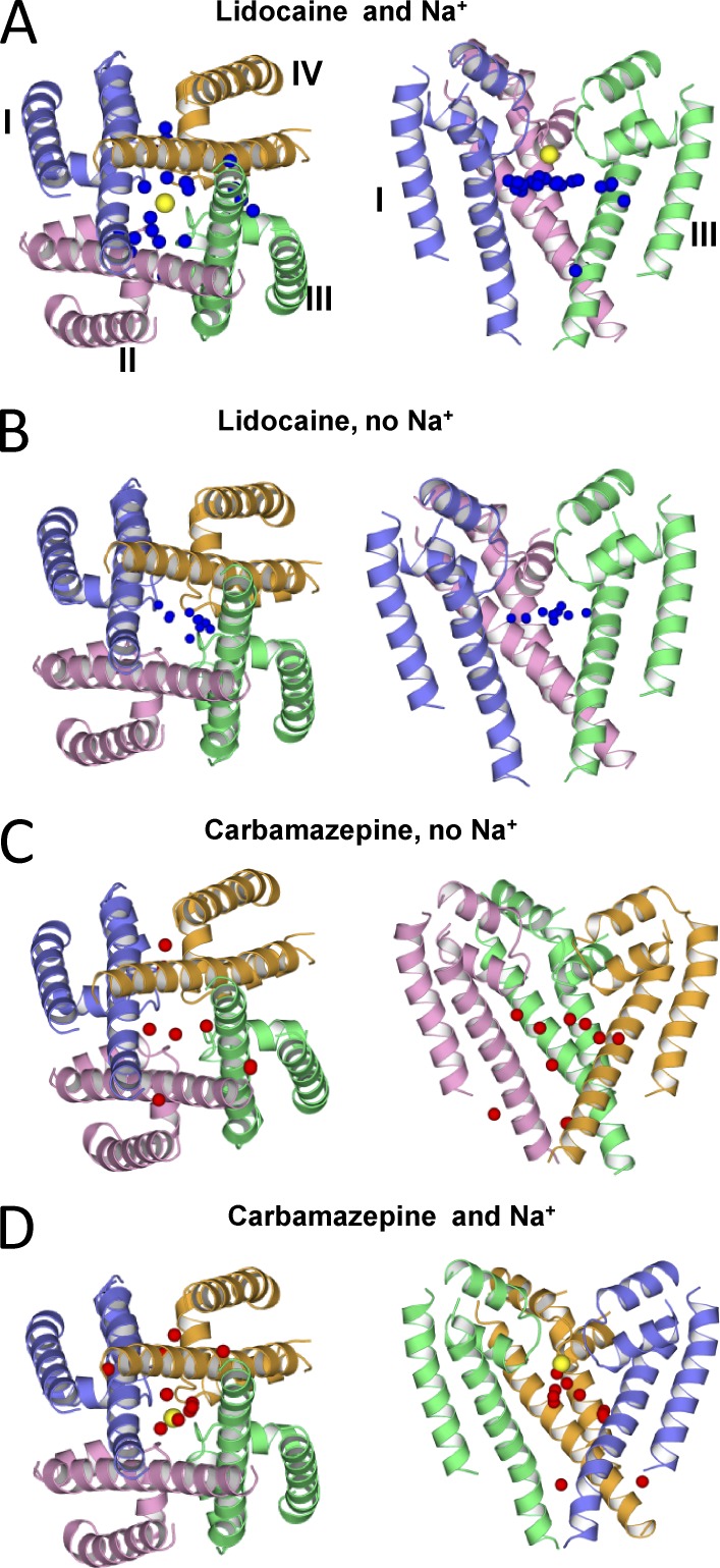
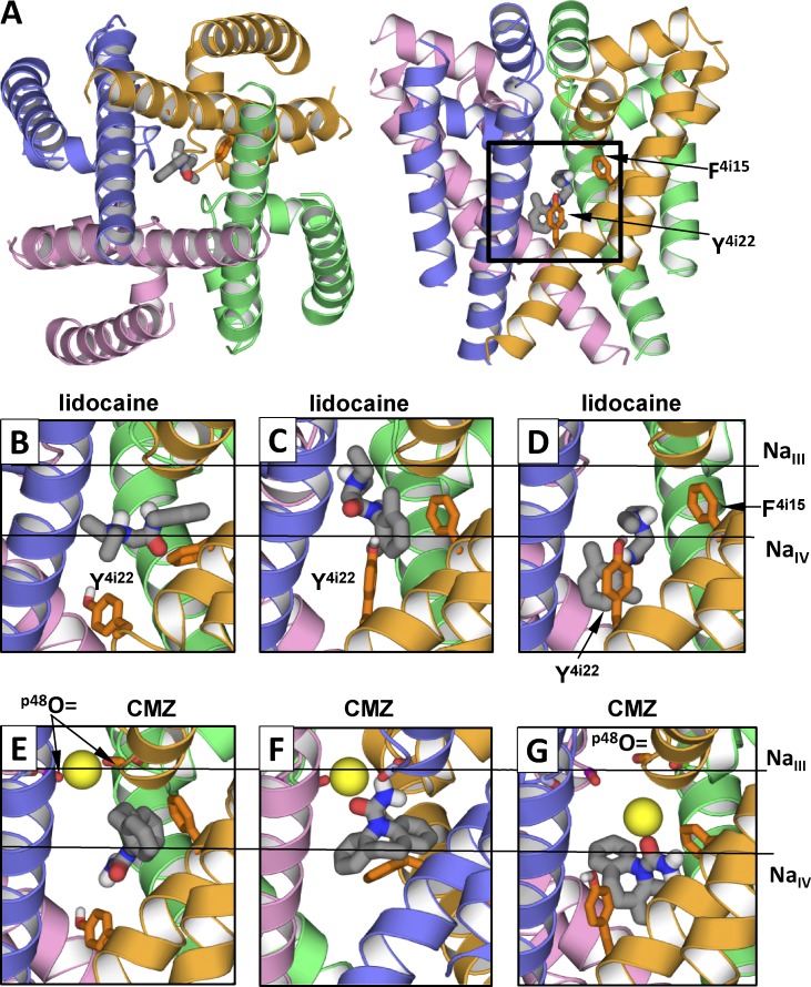
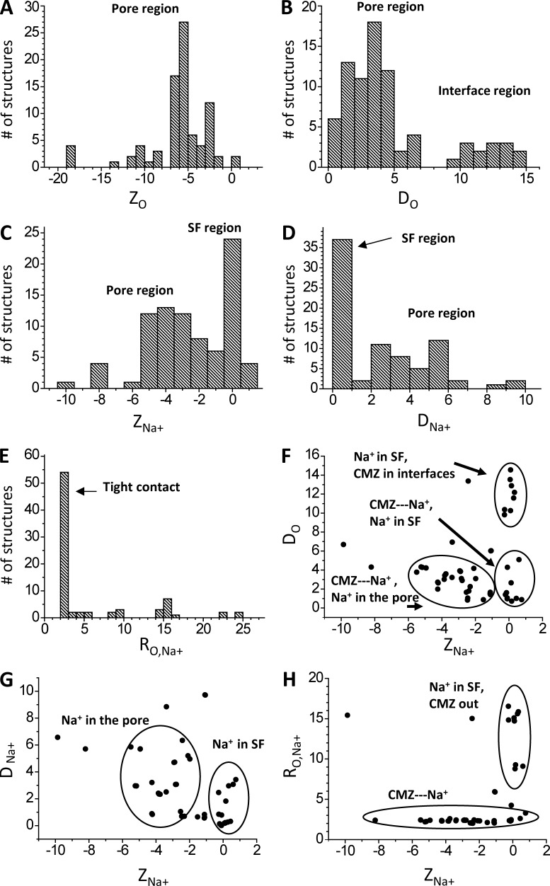
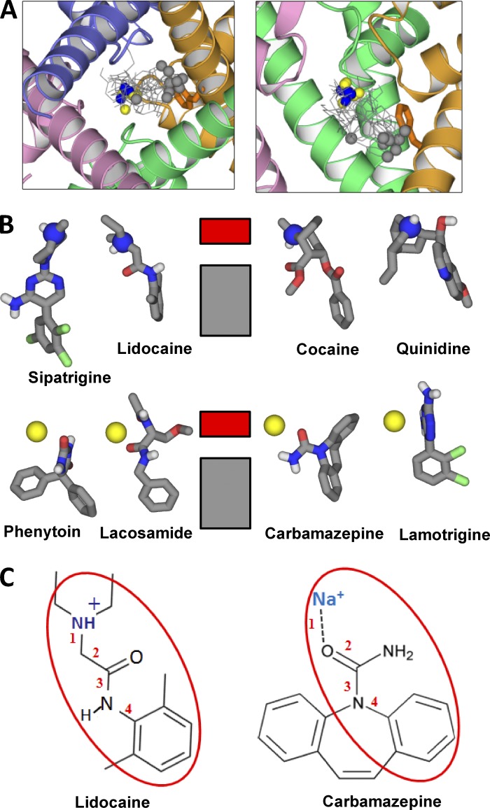
Local anesthetics, antiarrhythmics, and anticonvulsants include both charged and electroneutral compounds that block voltage-gated sodium channels. Prior studies have revealed a common drug-binding region within the pore, but details about the binding sites and mechanism of block remain unclear. Here, we use the x-ray structure of a prokaryotic sodium channel, NavMs, to model a eukaryotic channel and dock representative ligands. These include lidocaine, QX-314, cocaine, quinidine, lamotrigine, carbamazepine (CMZ), phenytoin, lacosamide, sipatrigine, and bisphenol A. Preliminary calculations demonstrated that a sodium ion near the selectivity filter attracts electroneutral CMZ but repels cationic lidocaine. Therefore, we further docked electroneutral and cationic drugs with and without a sodium ion, respectively. In our models, all the drugs interact with a phenylalanine in helix IVS6. Electroneutral drugs trap a sodium ion in the proximity of the selectivity filter, and this same site attracts the charged group of cationic ligands. At this position, even small drugs can block the permeation pathway by an electrostatic or steric mechanism. Our study proposes a common pharmacophore for these diverse drugs. It includes a cationic moiety and an aromatic moiety, which are usually linked by four bonds.


Figures

Figure 1.
Figure 1.
Ligand-sensing residues in the inner-pore region of Nav1.x channels. (A) Ligands considered in this study. (B) Sequences alignment of NavMs and Nav1.4. Key positions where mutations affect action of inner pore blockers (Mike and Lukacs, 2010) are highlighted. (C) The NavMs structure. Shown are Cα-Cβ bonds in key ligand-sensing residues and NaIII. Repeats I, III, and IV are blue, green, and orange, respectively. Repeat II is not depicted for clarity. Red ovals mark approximate borders of the selectivity filter region (1), the inner pore region (2), the activation gate region (3), and two of the four repeat interfaces (4).
Figure 2.
Figure 2.
Hands-free docking of lidocaine and CMZ in the Nav1.4 model with and without NaIII (yellow spheres). The oxygen atom of CMZ and the protonated nitrogen of lidocaine are shown as red and blue spheres, respectively. Distribution of ligands, as indicated by their key atoms, strongly depends on the presence of NaIII. In the presence of NaIII, low-energy positions of lidocaine and CMZ are scattered and clustered, respectively, and in the absence or presence of NaIII, the opposite distribution is observed. These results suggest that the NaIII site is a hotspot for ligands.
Figure 3.
Figure 3.
Statistics of lidocaine-binding modes in the channel model without NaIII. Each binding mode is characterized by the Z-coordinates of the ammonium nitrogen, ZN+, and the carbon atom in para-position of the aromatic ring, ZCp, and deviations of these atoms from the Z-axis, DN+ and DCp. (A–D) Distributions of the characteristics of the ensemble of low-energy binding modes. (E–H) Correlation fields between the characteristics. Dots in E, which are close to the dashed line (ZCp = ZN), correspond to horizontal binding modes of lidocaine. All coordinates and distances are given if angstroms.
Figure 4.
Figure 4.
Examples of low-energy binding modes of lidocaine and CMZ. Side chains of F4i15 and Y4i22 are shown as sticks. Horizontal lines in B–G show levels of site NaIII and putative site NaIV. (A) Location of ligand-binding region. (B–G) Representative structures from the ensembles of low-energy binding modes. (B) Horizontal binding mode of lidocaine. The aromatic group protrudes into the III/IV interface. (C and D) Vertical binding modes with the ammonium group at the levels of NaIII site and NaIV site, respectively. (E) CMZ without tight contacts with NaIII. (F and G) CMZ bound to NaIII. The sodium ion is bound at its innate site seen in the NavMs structure (F) or shifted toward putative site NaIV (G). Note that interactions of lidocaine and CMZ with F4i15 and Y4i22 depend on the ligand-binding modes.
Figure 5.
Figure 5.
Statistics of CMZ and Na+ binding in the channel model. Each binding mode is characterized by the Z-coordinates of carbonyl oxygen, ZO, and sodium ion, ZNa, as well as deviations of these atoms from the Z-axis, DO and DNa. (A–E) Distributions of the characteristics of the ensemble of low-energy binding modes. (F–H) Correlation fields between the characteristics. All coordinates and distances are given in angstroms.
Figure 6.
Figure 6.
Cationic and sodium-bound electroneutral ligands in the channel. (A) The triethylammonium group of QX-314 is close to the NaIII site, the aromatic ring binds between F4i15 and Y4i22, and the ligand also interacts with Q1p49 and F3p49 in agreement with mutational data (Yamagishi et al., 2009). (B) The bulky moiety of quinidine fits the hotspot at the NaIII site, whereas the hydroxyl group donates an H bond to 3p48O=C. (C and D) Two binding modes of cocaine. In one binding mode (C), the cocaine amino group binds at the NaIII site. In another binding mode (D), the amino group binds close to the putative NaIV site and forms cation-π contacts with F4i15, whereas the opposite end of cocaine reaches I3i23. (E and F) The long sipatrigine molecule can bind in the horizontal (E) and vertical (F) modes, forming many contacts with the channel. In the vertical mode, sipatrigine extends from the NaIII site to the hydrophobic region at levels i22-i23. (G and H) Side and extracellular views of lamotrigine. In the side view (G), repeat II is removed for clarity. In the extracellular view (H), lamotrigine and backbone carbonyls p48O=C are space filled. Lamotrigine binds to NaIII, which is coordinated by 3p48O=C and 4p48O=C, donates an H bond to 2p48O=C, interacts with 1p48O=C, π-stacks with F4i15, and approaches Y4i22. (I and J) Side and extracellular views at two similar binding modes of bisphenol A. Both aromatic rings are involved in π-cation interactions with NaIII, two hydroxyls donate H bonds to carbonyls p48O=C, and methyl groups bind between F4i15 and Y4i22. (K and L) Side views of lacosamide and phenytoin. Both ligands interact with NaIII and form H bonds with Q1p49, whereas their aromatic rings bind between F4i15 and Y4i22.
Figure 7.
Figure 7.
Similarity of binding modes of charged and neutral blockers. (A) Cytoplasmic and side views at superposition of the channel-bound ligands. The charged nitrogens and ligand-bound sodium ions are blue and yellow, respectively. The carbon atoms at the opposite end of the ligands are gray. Note the clustering of blue/yellow atoms at the NaIII site and gray atoms at F4i15. The gray outlier belongs to bisphenol A, which lacks an aromatic ring protruding into the pore. (B) Scheme of ligand–channel interactions. The charged nitrogen or ligand-bound sodium ion interacts with the ring of carbonyls p48O=C (NaIII site) indicated by the red rectangle. The aromatic groups interact with hydrophobic residues in the inner pore (gray rectangle), among which F4i15 plays the major role. (C) Lidocaine and sodium-bound CMZ have a common pharmacophore.

Comment in

Similar articles

Cited by

References

    1. Ahern C.A., Eastwood A.L., Dougherty D.A., and Horn R.. 2008. Electrostatic contributions of aromatic residues in the local anesthetic receptor of voltage-gated sodium channels. Circ. Res. 102:86–94. 10.1161/CIRCRESAHA.107.160663 - DOI - PubMed
    1. Anger T., Madge D.J., Mulla M., and Riddall D.. 2001. Medicinal chemistry of neuronal voltage-gated sodium channel blockers. J. Med. Chem. 44:115–137. 10.1021/jm000155h - DOI - PubMed
    1. Bagnéris C., DeCaen P.G., Naylor C.E., Pryde D.C., Nobeli I., Clapham D.E., and Wallace B.A.. 2014. Prokaryotic NavMs channel as a structural and functional model for eukaryotic sodium channel antagonism. Proc. Natl. Acad. Sci. USA. 111:8428–8433. 10.1073/pnas.1406855111 - DOI - PMC - PubMed
    1. Bernèche S., and Roux B.. 2001. Energetics of ion conduction through the K+ channel. Nature. 414:73–77. 10.1038/35102067 - DOI - PubMed
    1. Boiteux C., Vorobyov I., French R.J., French C., Yarov-Yarovoy V., and Allen T.W.. 2014. Local anesthetic and antiepileptic drug access and binding to a bacterial voltage-gated sodium channel. Proc. Natl. Acad. Sci. USA. 111:13057–13062. 10.1073/pnas.1408710111 - DOI - PMC - PubMed

MeSH terms

Associated data