. 2017 Apr;173(4):2399-2410.
doi: 10.1104/pp.16.01512. Epub 2017 Mar 3.

Antiviral Resistance Protein Tm-22 Functions on the Plasma Membrane

Affiliations

Antiviral Resistance Protein Tm-22 Functions on the Plasma Membrane

Tianyuan Chen et al. Plant Physiol. 2017 Apr.

Abstract

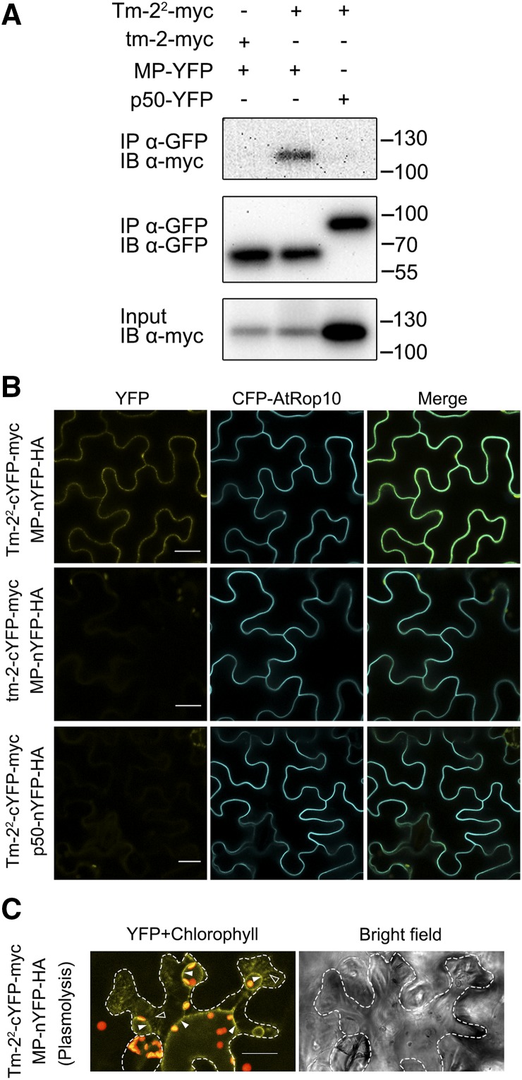
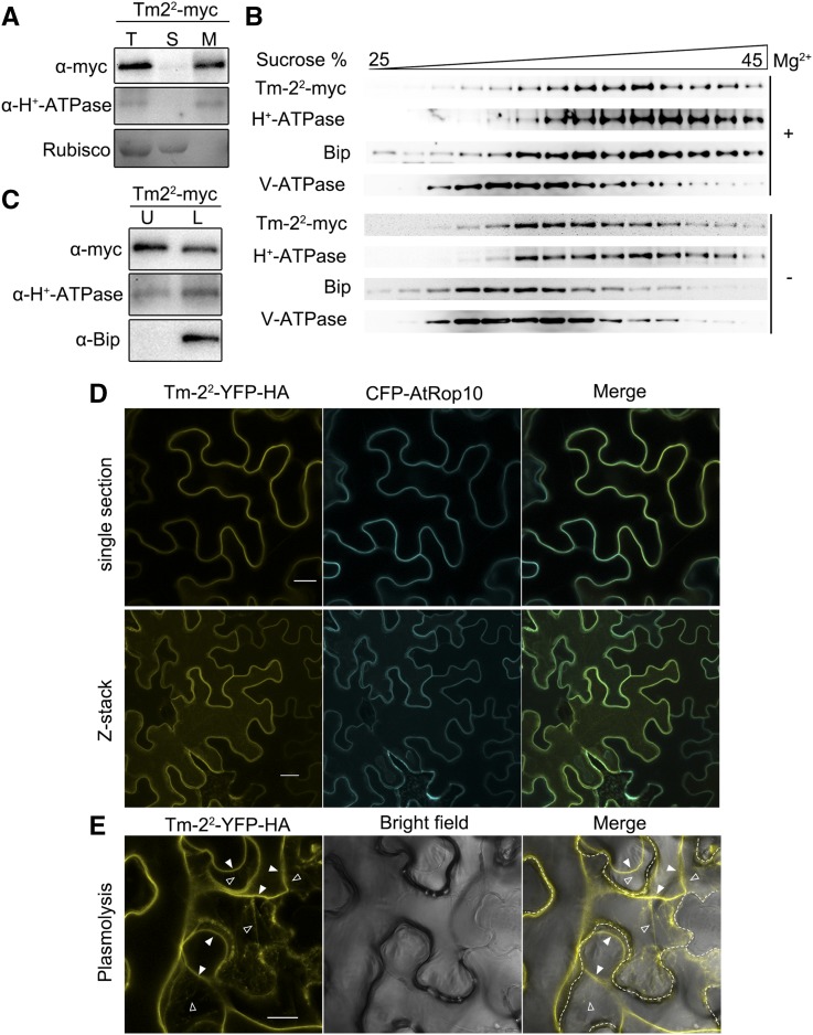
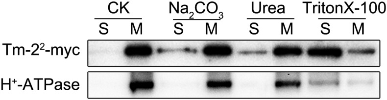
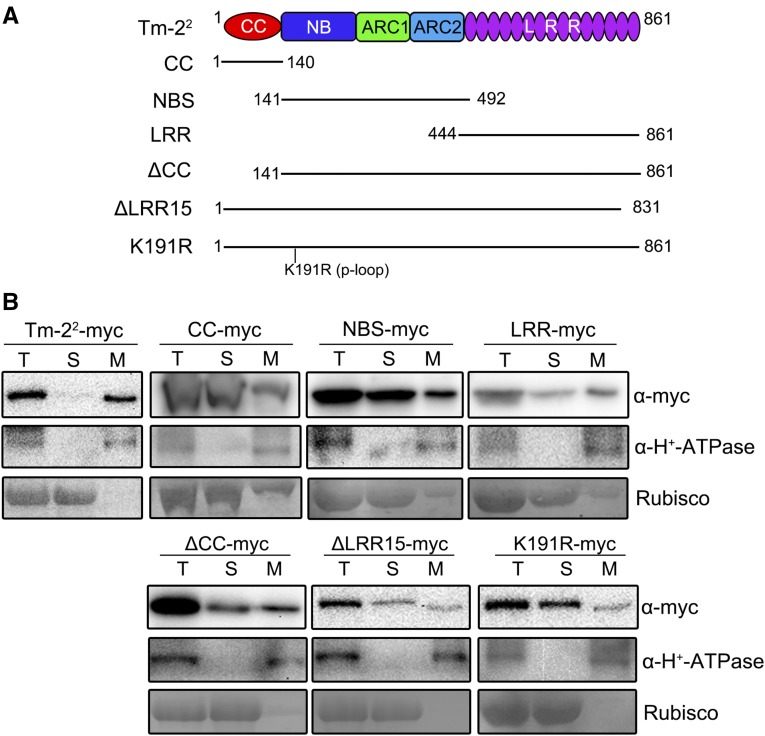
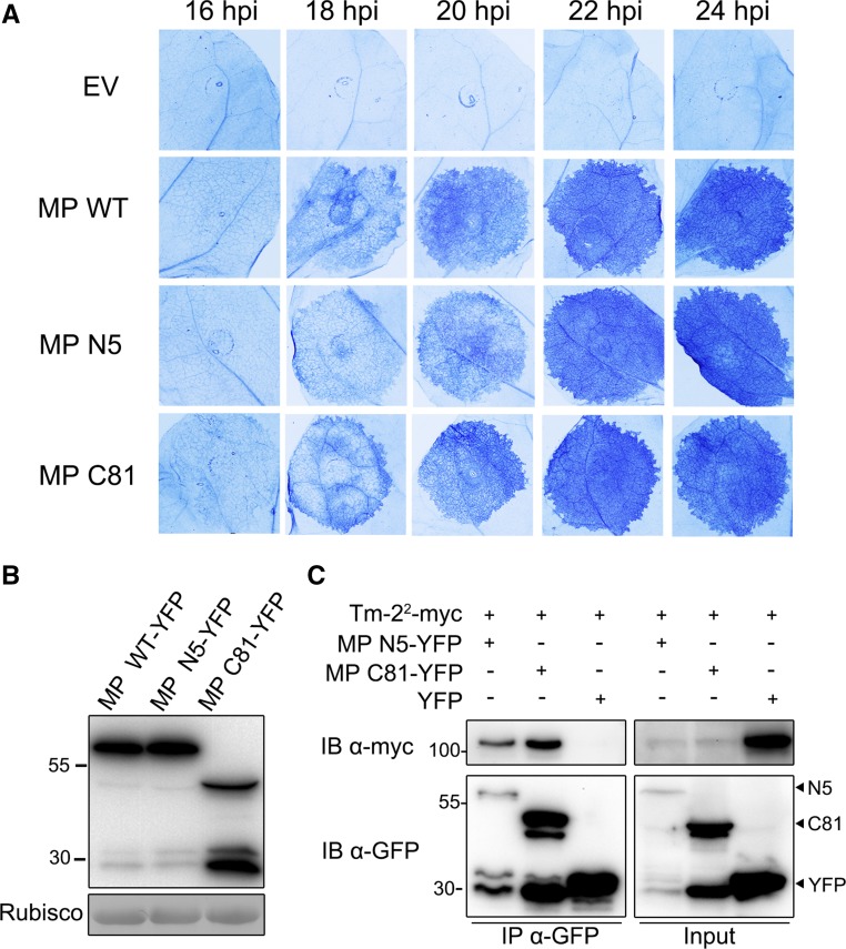
The tomato Tobacco mosaic virus resistance-22 (Tm-22 ) gene encodes a coiled-coil-nucleotide binding site-Leu-rich repeat protein lacking a conventional plasma membrane (PM) localization motif. Tm-22 confers plant extreme resistance against tobamoviruses including Tobacco mosaic virus (TMV) by recognizing the avirulence (Avr) viral movement protein (MP). However, the subcellular compartment where Tm-22 functions is unclear. Here, we demonstrate that Tm-22 interacts with TMV MP to form a protein complex at the PM We show that both inactive and active Tm-22 proteins are localized to the PM When restricted to PM by fusing Tm-22 to the S-acylated PM association motif, the Tm-22 fusion protein can still induce a hypersensitive response cell death, consistent with its activation at the PM Through analyses of viral MP mutants, we find that the plasmodesmata (PD) localization of the Avr protein MP is not required for Tm-22 function. These results suggest that Tm-22-mediated resistance takes place on PM without requirement of its Avr protein to be located to PD.


Figures

Figure 1.
Figure 1.
Tm-22 forms a complex with TMV MP in N. benthamiana. A, Tm-22 coimmunoprecipitates with MP. Tm-22-myc was transiently expressed with MP-YFP or p50-YFP in N. benthamiana leaves, and tm-2-myc was also coexpressed with MP-YFP. Agroinfiltration is followed by LaCl3 treatment at 16 hpi. Protein extracts from the infiltrated leaves at 36 hpi were subjected to anti-GFP immunoprecipitation (IP) followed by immunoblotting (IB) with the indicated antibodies. The size of protein molecular weight markers (kD) is on the right. B, The BiFC assays show the interaction of Tm-22 with MP. Tm-22-cYFP-myc was coexpressed with MP-nYFP-HA or p50-nYFP-HA, and tm-2-cYFP-myc was coexpressed with MP-nYFP-HA. All the combinations were coexpressed with the PM fluorescent marker CFP-AtROP10. C, YFP fluorescence from the interaction of Tm-22-cYFP-myc with MP-nYFP-HA was observed after cell plasmolysis. Cell plasmolysis was performed by treatment of 5% NaCl for 5 min. Red color indicates the chloroplast autofluorescence. Hechtian strands, typical connections of PM-cell wall, are indicated by outlined triangles, and the retracted PM is indicated by filled triangles. The cell wall is highlighted by dotted lines. Bars = 20 μm.
Figure 2.
Figure 2.
Tm-22 is localized to the PM. A, Tm-22-myc is a membrane-associated protein. The total protein (T) extracted from leaves expressing Tm-22-myc was fractionated into soluble (S) and membrane (M) fractions by ultracentrifugation at 100,000g. B, Tm-22 cofractionated with the PM marker through Suc gradients centrifugation. C, Tm-22-myc is a PM-located protein. Upper phase (U) and lower phase (L) were obtained by aqueous two-phase partitioning. Fractions in A to C were analyzed by western blot using antibodies against myc epitope, H+-ATPase (PM marker), BiP (ER marker), and V-ATPase (tonoplast marker). Rubisco (soluble protein marker) was stained by Ponceau S. D, Confocal images illustrated that Tm-22-YFP-HA colocalized with CFP-AtRop10 at the PM. Upper, single image intersecting the epidermal cells; lower, projection from Z-stack images. E, Tm-22-YFP-HA was also detected in the Hechtian strands after plasmolysis. Hechtian strands are indicated by outlined triangles, and the retracted PM is indicated by filled triangles. The cell wall is highlighted by dotted lines. Bars = 20 μm.
Figure 3.
Figure 3.
Tm-22 is a peripheral membrane protein. Microsomal membranes purified from leaves expressing Tm-22-myc were treated to release peripheral membrane proteins as indicated. The remaining membranes (M) and the newly soluble proteins (S) were analyzed by immunoblot with indicated antibodies. CK, The extraction buffer.
Figure 4.
Figure 4.
The activated Tm-22 resides at the PM. A, HR cell death is induced by Tm-22 in the presence of TMV MP and autoactive MHD mutant D481V, but not by wild-type Tm-22 alone in N. benthamiana. Cell death was visualized by trypan blue staining (lower panel) at 48 hpi. Solid line circles indicate cell death; dashed line circles indicate no obvious cell death. B, Cell fractionation assays show that activated Tm-22 is associated with the membrane. Soluble (S) and microsomal membrane (M) fractions were separated by ultracentrifugation. C, Aqueous two-phase partitioning assays show that the activated Tm-22 is partitioned in the PM phase. Upper phase (U) and lower phase (L) were obtained by aqueous two-phase partitioning of microsomal membrane fractions from B.
Figure 5.
Figure 5.
Plasma membrane-tethered Tm-22 retains effector-mediated HR function. A, Schematic representations of Rop tag and mRop tag. B, Confocal images show the localization of YFP-Rop or YFP-mRop in normal condition or after plasmolysis. C, Confocal images show the localization of Tm-22-YFP-Rop or Tm-22-YFP-mRop in normal condition or after plasmolysis. Hechtian strands are indicated by outlined triangles, and the retracted PM is indicated by filled triangles. The cell wall is highlighted by dotted lines. D, Both Tm-22-YFP-Rop and Tm-22-YFP-mRop induced cell death when coexpressed with MP. Cell death was visualized by trypan blue staining (right).
Figure 6.
Figure 6.
Tm-22 requires CC, NBS, and LRR domains for the PM localization. A, Schematic diagram of Tm-22 mutants used for cell fractionation. B, Cell fractionation analysis of Tm-22 or its mutants. The total proteins extracted from N. benthamiana leaves expressing myc-tagged Tm-22 mutants were fractionated by ultracentrifugation at 100,000g. The fractions were detected by immunoblotting with anti-myc and anti-H+-ATPase antibodies. Rubisco was stained by Ponceau S.
Figure 7.
Figure 7.
TMV MP mutants defective in targeting plasmodesmata still trigger Tm-22-mediated cell death. A, The extent of HR cell death induced by MP and its mutants was analyzed in transgenic Tm-22 plants. MP (MP WT) and its PD-targeting defective mutants (MP N5 and MP C81) tagged with C-terminal YFP were agroinfiltrated in Tm-22 transgenic N. benthamiana, and leaves from 16 to 24 hpi were stained by trypan blue. B, Total protein from wild-type N. benthamiana leaves expressing MP or its mutants at 24 hpi was extracted and detected with anti-GFP antibody. C, Tm-22-myc can coimmunoprecipitate with MP mutant N5 or C81. YFP was employed as a negative control. Proteins were immunoprecipitated with anti-GFP beads, and the immunoblotting was performed with indicated antibodies. The sizes of protein molecular weight markers (kD) are indicated.

References

    1. Aarts N, Metz M, Holub E, Staskawicz BJ, Daniels MJ, Parker JE (1998) Different requirements for EDS1 and NDR1 by disease resistance genes define at least two R gene-mediated signaling pathways in Arabidopsis. Proc Natl Acad Sci USA 95: 10306–10311 - PMC - PubMed
    1. Ade J, DeYoung BJ, Golstein C, Innes RW (2007) Indirect activation of a plant nucleotide binding site-leucine-rich repeat protein by a bacterial protease. Proc Natl Acad Sci USA 104: 2531–2536 - PMC - PubMed
    1. Amari K, Di Donato M, Dolja VV, Heinlein M (2014) Myosins VIII and XI play distinct roles in reproduction and transport of tobacco mosaic virus. PLoS Pathog 10: e1004448. - PMC - PubMed
    1. Axtell MJ, Staskawicz BJ (2003) Initiation of RPS2-specified disease resistance in Arabidopsis is coupled to the AvrRpt2-directed elimination of RIN4. Cell 112: 369–377 - PubMed
    1. Bai S, Liu J, Chang C, Zhang L, Maekawa T, Wang Q, Xiao W, Liu Y, Chai J, Takken FL, Schulze-Lefert P, Shen QH (2012) Structure-function analysis of barley NLR immune receptor MLA10 reveals its cell compartment specific activity in cell death and disease resistance. PLoS Pathog 8: e1002752. - PMC - PubMed

Publication types

MeSH terms

Substances