. 2017 Apr 21;292(16):6728-6743.
doi: 10.1074/jbc.M117.778308. Epub 2017 Mar 3.

Structural analyses of Candida albicans sterol 14α-demethylase complexed with azole drugs address the molecular basis of azole-mediated inhibition of fungal sterol biosynthesis

Affiliations

Structural analyses of Candida albicans sterol 14α-demethylase complexed with azole drugs address the molecular basis of azole-mediated inhibition of fungal sterol biosynthesis

Tatiana Y Hargrove et al. J Biol Chem. .

Abstract

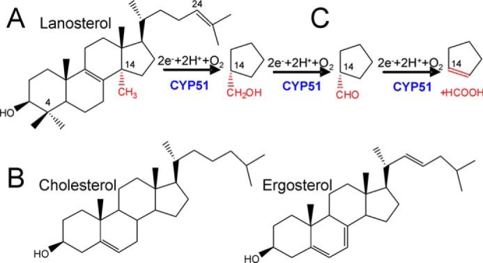
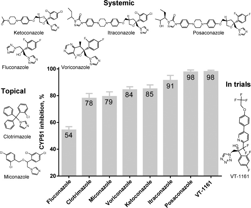
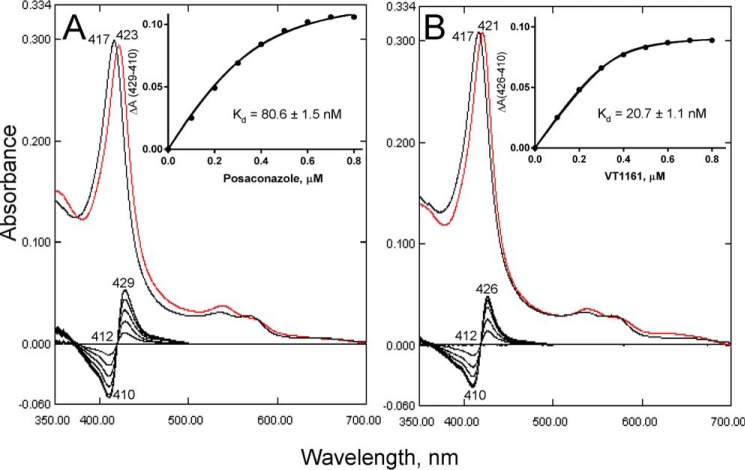
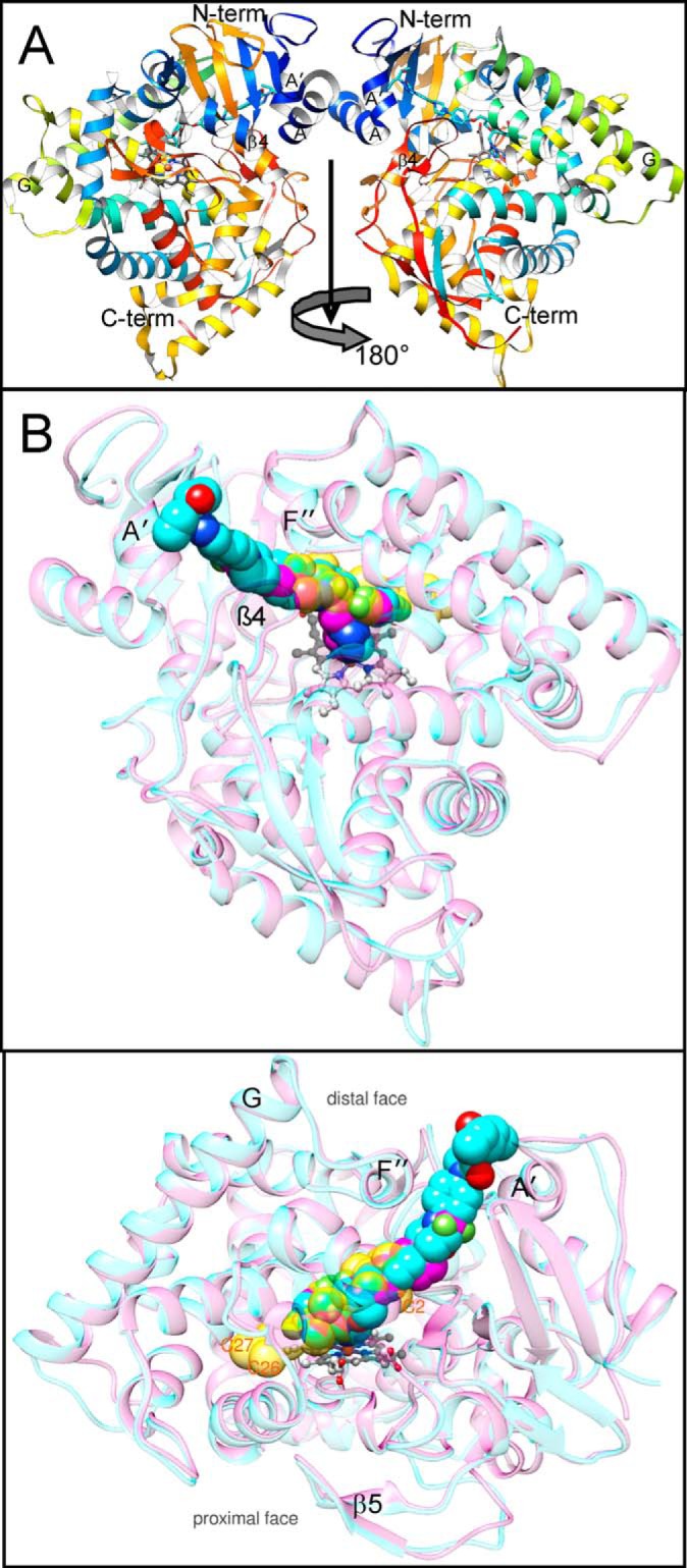
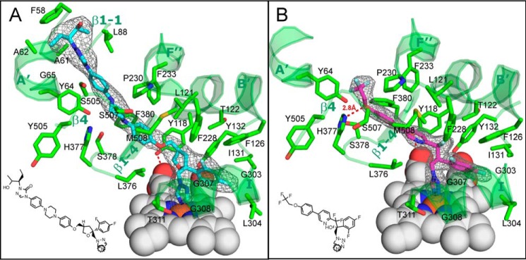
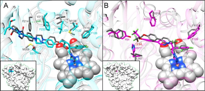
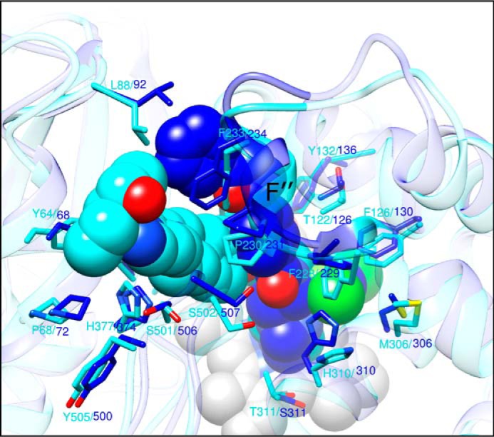
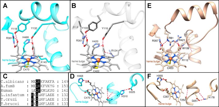
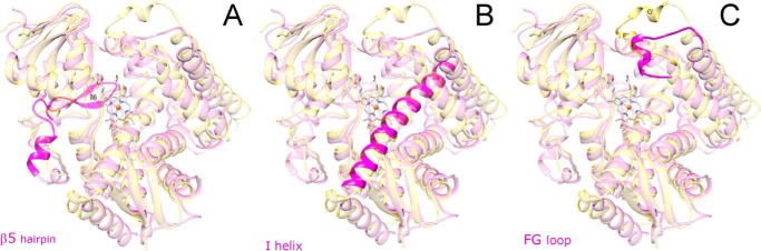
With some advances in modern medicine (such as cancer chemotherapy, broad exposure to antibiotics, and immunosuppression), the incidence of opportunistic fungal pathogens such as Candida albicans has increased. Cases of drug resistance among these pathogens have become more frequent, requiring the development of new drugs and a better understanding of the targeted enzymes. Sterol 14α-demethylase (CYP51) is a cytochrome P450 enzyme required for biosynthesis of sterols in eukaryotic cells and is the major target of clinical drugs for managing fungal pathogens, but some of the CYP51 key features important for rational drug design have remained obscure. We report the catalytic properties, ligand-binding profiles, and inhibition of enzymatic activity of C. albicans CYP51 by clinical antifungal drugs that are used systemically (fluconazole, voriconazole, ketoconazole, itraconazole, and posaconazole) and topically (miconazole and clotrimazole) and by a tetrazole-based drug candidate, VT-1161 (oteseconazole: (R)-2-(2,4-difluorophenyl)-1,1-difluoro-3-(1H-tetrazol-1-yl)-1-(5-(4-(2,2,2-trifluoroethoxy)phenyl)pyridin-2-yl)propan-2-ol). Among the compounds tested, the first-line drug fluconazole was the weakest inhibitor, whereas posaconazole and VT-1161 were the strongest CYP51 inhibitors. We determined the X-ray structures of C. albicans CYP51 complexes with posaconazole and VT-1161, providing a molecular mechanism for the potencies of these drugs, including the activity of VT-1161 against Candida krusei and Candida glabrata, pathogens that are intrinsically resistant to fluconazole. Our comparative structural analysis outlines phylum-specific CYP51 features that could direct future rational development of more efficient broad-spectrum antifungals.

Keywords: Candida albicans; X-ray crystallography; antifungal drugs; cytochrome P450; enzyme inhibitor; enzyme kinetics; sterol 14 alpha-demethylase (CYP51); sterol biosynthesis.


Conflict of interest statement

The authors declare that they have no conflicts of interest with the contents of this article

Figures

Figure 1.
Figure 1.
CYP51 in sterol biosynthesis. A, C. albicans CYP51 substrate lanosterol. B, products of the pathway in animals (cholesterol) and in fungi (ergosterol). C, three-step reaction of sterol 14α-demethylation. Each step involves one cycle of monooxygenation. 14α-Demethylation of lanosterol produces 4,4′-dimethylcholesta-8,14,24-triene-3β-ol (14α-desmethyllanosterol in Fig. 5).
Figure 2.
Figure 2.
Amino acid sequence alignment of six structurally characterized eukaryotic sterol 14α-demethylases. The secondary structural elements of C. albicans (3tz1) and T. brucei (3g1q) enzymes are indicated above and below the sequences, respectively. The 34 residues invariant across the whole CYP51 family are marked with blue triangles. CYP51 family signature motifs (signature 1 and signature 2) are underscored (blue line). Black rectangles mark two phyla-specific segments: αG (protozoa) and β5 hairpin (fungi). Sequence alignment was generated in ClustalW, and secondary structural information was added in ESPript.
Figure 3.
Figure 3.
Absorbance spectra of C. albicans CYP51. Absolute (black) and difference CO-binding (red) spectra, P450 concentration 3.3 μm, optical path length 1 cm.
Figure 4.
Figure 4.
Spectral response of C. albicans CYP51 to the addition of the sterol substrates. Type 1 shift (red line) in the absorbance spectra of C. albicans CYP51 (1.9 μm) upon titration with the four CYP51 sterol substrates is shown. Absolute (top) and difference (bottom) absorbance spectra are shown. A, titration under standard substrate titration conditions (50 mm potassium phosphate buffer (pH 7.4), containing 200 mm NaCl and 0.1 mm EDTA). B, in the presence of 0.1% Triton X-100 (v/v). The titration range was 0.5–7 μm; the titration step was 0.5 μm, and the optical path length was 1 cm. C, titration curves of A; D, titration curves from B. The high-spin form content (the portion of the substrate-bound CYP51 molecules) and the apparent Kd values were calculated as described under “Experimental procedures.”
Figure 5.
Figure 5.
Enzymatic activity of C. albicans CYP51. A, time course of lanosterol conversion (37 °C, 0.5 μm P450, 1 μm CPR, 25 μm lanosterol). Inset, HPLC profile of sterols extracted after a 1.5-min reaction. B, steady-state kinetics (1-min reaction). The experiments were performed in triplicate, and the results are presented as means ± S.E.
Figure 6.
Figure 6.
Comparative inhibitory effects of clinical antifungal drugs on the activity of C. albicans CYP51. The molar ratio of enzyme/inhibitor/substrate was 1:2:50, with the P450 concentration at 0.5 μm (37 °C, 60-min reaction). The experiments were performed in triplicate, and the results are presented as means ± S.D.
Figure 7.
Figure 7.
Spectral responses of C. albicans CYP51 to the addition of the triazole posaconazole and tetrazole VT-1161. Type 2 shifts are shown in the Soret band maximum in the absolute (top, red line) and difference (bottom) absorption spectra. The P450 concentration was ∼0.5 μm; the titration range was 0.1–0.8 μm; the titration step was 0.1 μm; and the optical path length was 5 cm. The titration was conducted in 50 mm potassium phosphate buffer (pH 7.4) containing 200 mm NaCl, 0.1 mm EDTA, and 0.1% Triton X-100 (v/v). Apparent Kd values were calculated as described under “Experimental procedures.” A, posaconazole. B, VT-1161. Insets, titration curves.
Figure 8.
Figure 8.
Complexes of C. albicans CYP51 with posaconazole and VT-1161, overall view of the structures. A, asymmetric unit, a view along the rotation axis that runs from top to bottom and relates two monomers. The protein main chain is shown in ribbon representation colored by secondary structure succession from the N (blue) to C (red) termini. Helices A, A′, G, the β4 hairpin, the N terminus, and the C terminus are marked. B, superimposed complexes with posaconazole (cyan) and VT-1161 (magenta). The protein main chains are shown as semitransparent ribbons of the corresponding color. The heme (gray) is depicted in a ball-and-stick representation. The inhibitors and the molecule of the sterol substrate (semitransparent gold, modeled from the costructure with T. brucei CYP51 (PDB code 3P99) are presented as spheres. One atom of the nucleus (C2) and two side chain atoms (C26 and C27) of the sterol molecule are marked as references. Distal (top) and 180°-rotated upper (bottom) view.
Figure 9.
Figure 9.
Posaconazole and VT-1161 bound in the C. albicans CYP51 active site. The 2FoFc omit electron density maps (gray mesh) of posaconazole (cyan) and VT-1161 (magenta) are contoured at 1.2 and 1.5 σ, respectively. A, posaconazole; B, VT-1161. The inhibitor contacting residues (within 4.5 Å, stick representation) and the corresponding secondary structural elements (ribbon representation) of C. albicans CYP51 are depicted in green and marked (see also Table 2). The heme is depicted as gray spheres. H-bonds are shown as red dashes. The heme-coordinating nitrogen atoms in the structural formulas of the inhibitors are circled.
Figure 10.
Figure 10.
Superimposition of C. albicans and T. cruzi (gray) CYP51 complexes with posaconazole (PDB code 3K1O) and VT-1161 (PDB code 5AJR). A, posaconazole; B, VT-1161. Some phyla-specific residues that line the enzyme substrate-binding cavity, altering the conformation of the inhibitors, are shown as examples. A complete list of the corresponding ligand-contacting residues aligned in the fungal and protozoan CYP51 structures is shown in Table 2. Insets, C. albicans CYP51 in a surface representation (same view). The C-atoms of posaconazole and VT-1161 are colored in cyan and magenta, respectively.
Figure 11.
Figure 11.
Superimposition of C. albicans CYP51 and A. fumigatus CYP51B (PDB code 4UYL) complexes with posaconazole (cyan) and VNI (blue), respectively. The heme (gray), posaconazole, and VNI are depicted as spheres; the binding cavity-forming residues, conserved in both proteins, are shown in stick representation and labeled.
Figure 12.
Figure 12.
Heme support and proton delivery route in CYP51. Six protein residues located within 3 Å of the heme are noted. A, C. albicans; B, T. cruzi CYP51. The H-bonds with the heme propionates are displayed as red dashes, and the iron-coordinated cysteine is seen at the bottom. C, fragment of CYP51 sequence alignment showing the porphyrin ring D supporting lysine in fungi/animals (Lys-143 in C. albicans) versus located one turn upstream of the C-helix arginine (Arg-124) in protozoa. D, surface-exposed Asp-225 in C. albicans CYP51, via the CYP51 signature His-310 and “conserved P450 threonine” (Thr-311), supplies protons to the oxygenated heme iron. E, heme support in human P450scc (CYP11A1 (PDB code 3N9Y)) is provided for comparison. F, proton delivery route in P450cam (CYP101A1 (PDB code 1DZ4)) (56) presented as a comparison. Overall, the heme support in CYP11A1 is probably stronger, because, opposite to CYP51, all the H-bonds here are formed between the N and O atoms (salt bridges). The charges in the conserved salt bridge pair involved into the proton delivery route in CYP101 (as well as in most other CYP families) are reversed.
Figure 13.
Figure 13.
Three phylum-specific segments (magenta) mapped on the structure of C. albicans CYP51 (semitransparent magenta). A, β5 hairpin is unique for fungal CYP51 and forms the proximal surface of the P450 molecule. B, similar to protozoan and opposite to human CYP51, the I-helix in the C. albicans enzyme does not have a loop-like region in the middle portion. C, FG-loop in C. albicans CYP51 is shorter and has only one α-helical region (F″), and the G′-helix is unique for the protozoan enzymes. Superimposition with the structure of T. brucei CYP51 (PDB code 3G1Q) is shown in semi-transparent gold. View is from distal face of the P450 molecule.
Figure 14.
Figure 14.
Amino acid substitutions in CYP51 from fluconazole-resistant clinical isolates of C. albicans that have not been found in fluconazole-sensitive strains, mapped on the C. albicans CYP51 structure. A, mutations within the substrate-binding cavity, distal view. B, mutations on the proximal P450 surface, proximal view. The side chains of the wild-type residues are shown as green lines, and the side chains of the mutant residues are presented as tan sticks. Fluconazole (red sticks) was modeled from the complex with Leishmania infantum CYP51 (PDB code 3L4D).

References

    1. Noble S. M., Gianetti B. A., and Witchley J. N. (2017) Candida albicans cell-type switching and functional plasticity in the mammalian host. Nat. Rev. Microbiol. 15, 96–108 - PMC - PubMed
    1. Kullberg B. J., and Arendrup M. C. (2015) Invasive candidiasis. N. Engl. J. Med. 373, 1445–1456 - PubMed
    1. Jampilek J. (2016) How can we bolster the antifungal drug discovery pipeline? Future Med. Chem. 8, 1393–1397 - PubMed
    1. da Silva Dantas A., Lee K. K., Raziunaite I., Schaefer K., Wagener J., Yadav B., and Gow N. A. (2016) Cell biology of Candida albicans–host interactions. Curr. Opin. Microbiol. 34, 111–118 - PMC - PubMed
    1. Ene I. V., Adya A. K., Wehmeier S., Brand A. C., MacCallum D. M., Gow N. A., and Brown A. J. (2012) Host carbon sources modulate cell wall architecture, drug resistance and virulence in a fungal pathogen. Cell. Microbiol. 14, 1319–1335 - PMC - PubMed

LinkOut - more resources