Review
. 2017 Mar;276(1):165-177.
doi: 10.1111/imr.12522.

TAM receptor tyrosine kinases as emerging targets of innate immune checkpoint blockade for cancer therapy

Affiliations
Review

TAM receptor tyrosine kinases as emerging targets of innate immune checkpoint blockade for cancer therapy

Yemsratch T Akalu et al. Immunol Rev. 2017 Mar.

Abstract

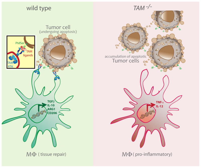
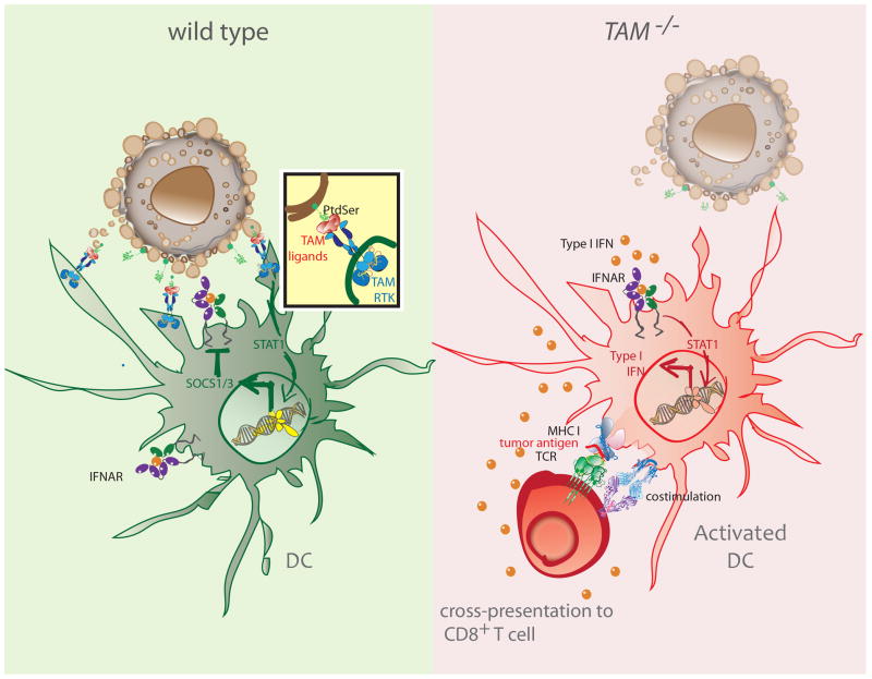
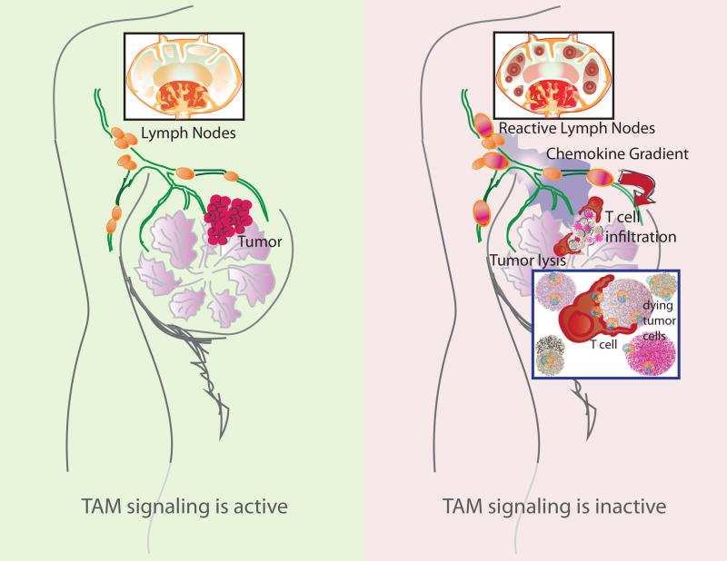
Cancer immunotherapy utilizing T-cell checkpoint inhibitors has shown tremendous clinical success. Yet, this mode of treatment is effective in only a subset of patients. Unresponsive patients tend to have non-T-cell-inflamed tumors that lack markers associated with the activation of adaptive anti-tumor immune responses. Notably, elimination of cancer cells by T cells is critically dependent on the optimal activity of innate immune cells. Therefore, identifying new targets that regulate innate immune cell function and promote the engagement of adaptive tumoricidal responses is likely to lead to the development of improved therapies against cancer. Here, we review the TAM receptor tyrosine kinases-TYRO3, AXL, and MERTK-as an emerging class of innate immune checkpoints that participate in key steps of anti-tumoral immunity. Namely, TAM-mediated efferocytosis, negative regulation of dendritic cell activity, and dysregulated production of chemokines collectively favor the escape of malignant cells. Hence, disabling TAM signaling may promote engagement of adaptive immunity and complement T-cell checkpoint blockade.

Keywords: AXL; MERTK; TYRO3; innate immune checkpoints.


Conflict of interest statement

Conflict of Interest:

The authors disclose that there is a potential conflict of interest as C.V.R. is a shareholder of Kolltan Pharmaceuticals and a member of the science advisory board at Surface Oncology.

Figures

Figure 1
Figure 1. TAM RTK-mediated efferocytosis promotes a pro-tumorigenic program in MΦs
Apoptotic tumor cells expose PtdSer on their surfaces. TAM agonists bind to PtdSer and trigger activation of TAM RTKs on the surface of phagocytes, such as MΦs. Efferocytosis by MΦs can lead to the expression of factors like ARG1, TGF-β and IL-10 (left). In the absence of TAM RTK signaling, MΦs in the TME maintain their production of pro-inflammatory factors that could contribute to the killing of tumor cells (right).
Figure 2
Figure 2. TAM signaling inhibits activation of intratumoral DCs
Type I IFN signaling leads to an IFNAR/STAT1 dependent induction of the TAM receptor AXL. Once engaged, the TAM receptor signaling pathway co-opts IFNAR and STAT1 to induce expression of SOCS1 and SOCS3, which suppress the type I IFN cascade and foster tolerogenic function of DCs (left). In the absence of TAM signaling, the induction of SOCS proteins is impaired. Enhanced production of type I IFNs is expected to promote cross-presentation of tumor antigens and upregulation of costimulatory molecules (right).
Figure 3
Figure 3. Loss of TAM RTK signaling favors cytolytic killing of tumor cells
TAM signaling on TIDCs is thought to limit the generation of chemokines that are needed for the recruitment of T cells into the TME (left). In the absence of TAM signaling, TIDCs are expected to produce increased amounts of T cell-attractant chemokines. This results in the enhanced recruitment of T cells from tissue draining LNs. Concomitantly, the accumulation of apoptotic tumor cells (Fig. 1), production of pro-inflammatory factors (Fig. 1), and increased activation of DCs (Fig. 2) are hypothesized to favor the engagement of TILs. These, together with an increased recruitment of TILs to the TME promote the effective killing of tumor cells (right).

Similar articles

Cited by

References

    1. Wiemann B, Starnes CO. Coley’s toxins, tumor necrosis factor and cancer research: a historical perspective. Pharmacol Ther. 1994;64(3):529–64. - PubMed
    1. Ansell SM, Lesokhin AM, Borrello I, et al. PD-1 Blockade with Nivolumab in Relapsed or Refractory Hodgkin’s Lymphoma. New Engl J Med. 2015;372(4):311–9. - PMC - PubMed
    1. Brahmer JR, Tykodi SS, Chow LQM, et al. Safety and Activity of Anti-PD-L1 Antibody in Patients with Advanced Cancer. New Engl J Med. 2012;366(26):2455–65. - PMC - PubMed
    1. Topalian SL, Hodi FS, Brahmer JR, et al. Safety, Activity, and Immune Correlates of Anti-PD-1 Antibody in Cancer. New Engl J Med. 2012;366(26):2443–54. - PMC - PubMed
    1. Robert C, Long GV, Brady B, et al. Nivolumab in previously untreated melanoma without BRAF mutation. N Engl J Med. 2015;372(4):320–30. - PubMed

Publication types

MeSH terms

Substances