

Review
doi: 10.1186/s40644-017-0110-z.
Imaging of hepatocellular carcinoma and image guided therapies - how we do it
Affiliations
- PMID: 28259177
- PMCID: PMC5336669
- DOI: 10.1186/s40644-017-0110-z
Item in Clipboard
Review
Imaging of hepatocellular carcinoma and image guided therapies - how we do it
Cancer Imaging.
.
Abstract
Treatment options for hepatocellular carcinoma have evolved over recent years. Interventional radiologists and surgeons can offer curative treatments for early stage tumours, and locoregional therapies can be provided resulting in longer survival times. Early diagnosis with screening ultrasound is the key. CT and MRI are used to characterize lesions and determine the extent of tumour burden. Imaging techniques are discussed in this article as the correct imaging protocols are essential to optimise successful detection and characterisation. After treatment it is important to establish regular imaging follow up with CT or MRI as local residual disease can be easily treated, and recurrence elsewhere in the liver is common.
Figures
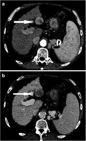
54 year old male with hepatitis C cirrhosis. CT shows an arterial enhancing nodule a with washout of contrast in the delayed phase b consistent with hepatocellular carcinoma
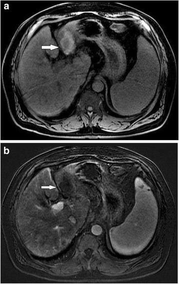
67 year old male with alchohol liver disease and cirrhosis. Venous phase MRI with gadolinium demonstrates an HCC nodule at the dome of the liver with capsular enhancement
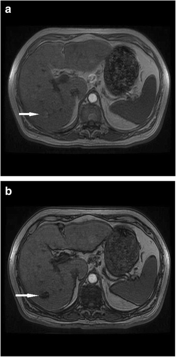
71 year old male with hepatitis C cirrhosis. Signal drop out on opposed phase imaging (b) in comparison with in phase imaging (a). The findings represent intracellular lipid in an HCC tumour
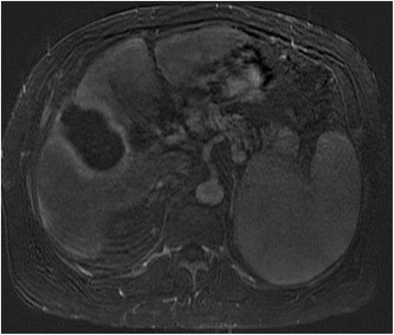
66 year old female with hepatitis C cirrhosis post microwave ablation of HCC Precontrast image post microwave ablation (a) show a cavity with intrinsic high signal on T1 weighted imaging. A subtraction image (b) removes the high signal resulting in no evidence of enhancement
63 year old male with cirrhosis and HCC treated with microwave ablation. A thin rim of enhancement post ablation, consistent with hyperemia adjacent to the ablation zone, is a normal finding and does not represent recurrent tumour
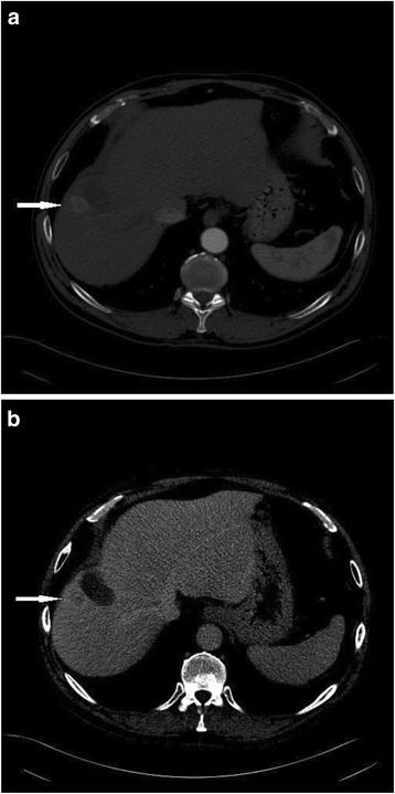
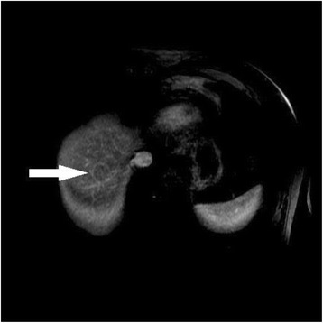
57 year old female with cirrhosis and HCC treated with RFA. CT in arterial (a) and venous (b) phases shows enhancement and washout of a nodule adjacent to an RFA ablation zone
References
Publication types
MeSH terms
LinkOut - more resources
Full Text Sources
Other Literature Sources
Medical
