. 2017 May 5;429(9):1305-1320.
doi: 10.1016/j.jmb.2017.02.014. Epub 2017 Mar 1.

Architecture of the Flagellar Switch Complex of Escherichia coli: Conformational Plasticity of FliG and Implications for Adaptive Remodeling

Affiliations

Architecture of the Flagellar Switch Complex of Escherichia coli: Conformational Plasticity of FliG and Implications for Adaptive Remodeling

Eun A Kim et al. J Mol Biol. .

Abstract

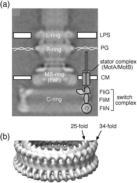
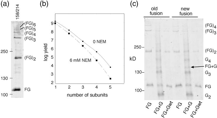
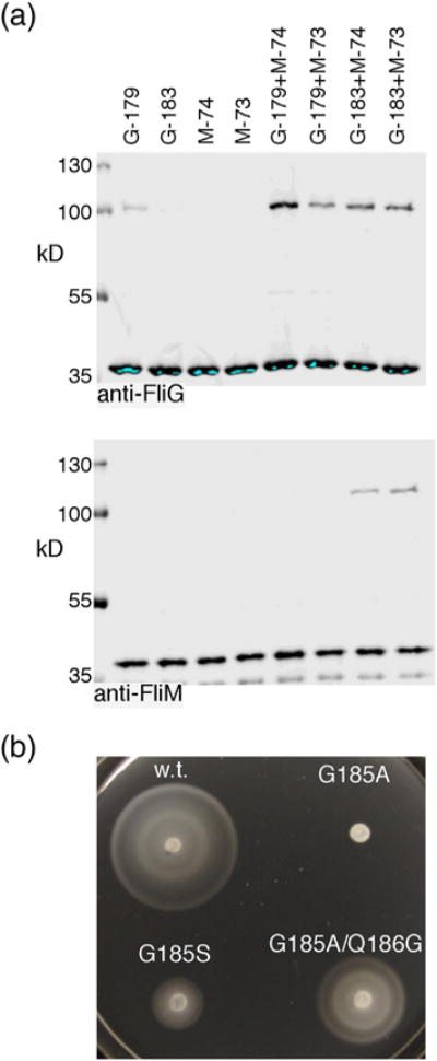
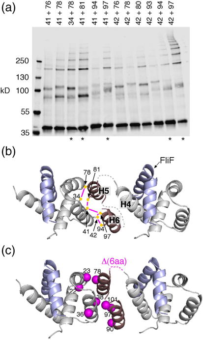
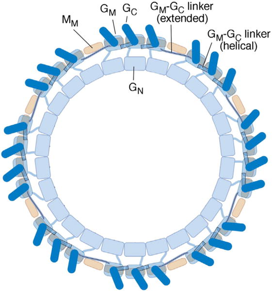
Structural models of the complex that regulates the direction of flagellar rotation assume either ~34 or ~25 copies of the protein FliG. Support for ~34 came from crosslinking experiments identifying an intersubunit contact most consistent with that number; support for ~25 came from the observation that flagella can assemble and rotate when FliG is genetically fused to FliF, for which the accepted number is ~25. Here, we have undertaken crosslinking and other experiments to address more fully the question of FliG number. The results indicate a copy number of ~25 for FliG. An interaction between the C-terminal and middle domains, which has been taken to support a model with ~34 copies, is also supported. To reconcile the interaction with a FliG number of ~25, we hypothesize conformational plasticity in an interdomain segment of FliG that allows some subunits to bridge gaps created by the number mismatch. This proposal is supported by mutant phenotypes and other results indicating that the normally helical segment adopts a more extended conformation in some subunits. The FliG amino-terminal domain is organized in a regular array with dimensions matching a ring in the upper part of the complex. The model predicts that FliG copy number should be tied to that of FliF, whereas FliM copy number can increase or decrease according to the number of FliG subunits that adopt the extended conformation. This has implications for the phenomenon of adaptive switch remodeling, in which the FliM copy number varies to adjust the bias of the switch.

Keywords: chemotaxis; molecular motors; motility; protein structure; self-assembly.


Figures

Fig. 1
Fig. 1
Morphology of the flagellar basal body of Salmonella. (a) EM reconstruction of the flagellar basal body from Thomas et al. [28]. The structure consists of a central rod and a set of rings. The membrane/supra-membrane (MS) ring is in and just above the cytoplasmic membrane (CM). The outer membrane (LPS) and peptidoglycan (PG) are also indicated. One stator complex is shown; motors can contain up to about a dozen independently functioning stator complexes, each attached to peptidoglycan. Approximate positions of the three switch complex proteins are indicated. (b) A tilted view of the switch complex from a higher-resolution EM reconstruction [6]. Density has been rotationally averaged to improve signal to noise. Most of the switch complex has ~34-fold symmetry, but the inner ring in the upper (membrane-proximal) part displays ~25-fold symmetry.
Fig. 2
Fig. 2
Properties of the original FliF–FliG fusion strain. The strain has a 7-nt deletion near the end of fliF that brings it into frame with fliG while replacing five C-terminal FliF residues with Ile (see Fig. S1 for details). (a) Chemotaxis defect of the strain in a soft-agar migration assay. A tryptone soft-agar plate was spotted with 3 μl of overnight cultures and incubated for 6 h at 32 °C. Under the microscope, cells displayed vigorous tumbling. (b) Anti-FliG immunoblots of cell fractions, and whole cells, showing that native-sized FliG is not present at a significant level. w.c., whole cell; mem, membrane; cyto, cytosol; w.c. + FliG, whole-cell proteins from the fusion strain with native-sized FliG expressed from a plasmid (pDB97, with no induction). (c) Improved motility of the original FliF–FliG fusion strain when FliG or certain FliG variants are expressed from plasmids. The control (cont) is the original fusion strain with nothing else expressed. Plasmids were derivatives of pKP619, induced with 200 μM IPTG. Plates were spotted with 3 μl of overnight cultures and incubated for 12 h at 32 °C. The strain expressing wild-type FliG from the plasmid migrates at ~20% of the wild-type rate. FliGN, FliG amino-terminal domain; FliGMC, FliG middle and C-terminal domains. The right-hand plate shows the effects of expressing point-mutant variants of FliG that have been shown previously to abolish the function of the full-length protein [34,35]. Rescue of the fusion strain by extra FliG is diminished by a mutation in the N-terminal domain (L13D) but not by mutations in the middle or C-terminal domains. (d) Examples of suppressors of the chemotaxis defect in the fusion strain. Strains retained the FliF–FliG fusion and had acquired the indicated mutations in FliG or FliM. The original, unsuppressed FliF–FliG fusion is also shown (labeled “cont”). Plates were incubated for 12 h at 32 °C. Suppressor mutations are listed in Table 1.
Fig. 3
Fig. 3
Properties of a different fusion strain that has a linker between FliF and FliG. (a) Nucleotide and amino-acid sequences at the FliF–FliG junction. (b) Motility of the linker-fusion strain in soft agar. A tryptone soft-agar plate was inoculated with 3 μl of overnight cultures and incubated for 8 h at 32 °C. Following a short delay in the onset of motility, the fusion-linker strain migrates at >80% of the wild-type rate. (c) Anti-FliG immunoblots of whole-cell samples and cell fractions showing that native-sized FliG is not present at a significant level in the fusion-linker strain. w.c., whole cell; mem, membrane; cyto, cytosol; w.c. + FliG, whole cells with native-sized FliG expressed from a plasmid (pDB97, with no induction). (d) Rotation speeds of tethered cells of the FliF–FliG fusion-linker strain and of the wild type. Cells were tethered to coverslips by anti-flagellin antibody, and rotation speeds were determined by slow-speed video playback.
Fig. 4
Fig. 4
Crosslinking between positions in the middle and C-terminal domains of FliG. (a) Examples of the crosslinking of double-Cys mutant proteins, using either plasmid-based expression (derivatives of pKP619 induced with 40 μM IPTG) or expression from the normal chromosomal locus. (b) Left: ladder of products observed with the chromosomally expressed 158/214 pair, resolved on a gel using less than the usual amount of bis-acrylamide (acrylamide:bis-acrylamide ratio 80:1). Right: Yield versus product size for the ladders produced by the 158/214 Cys pair. Bands were quantified by densitometry using Image-J [53]. (c) Blocking of the 158–214 crosslinking by pretreatment with NEM. Blocking was for 3 min prior to treatment with iodine for all except for the rightmost sample, which was blocked for 30 min. Proteins were resolved on a 4–20% gradient gel.
Fig. 5
Fig. 5
(a) Iodine-induced disulfide crosslinking of positions 158 and 214 in the contest of the new (i.e., linker-containing) FliF–FliG fusion protein, expressed from the chromosome. (b) Product yield versus size (number of crosslinked subunits), and effect of pretreatment with NEM (6 mM for 3 min). (c) Crosslinking of native-sized, plasmid-expressed FliG to the FliF–FliG fusion proteins. Results with both the old and new fusion constructs are shown. The FliF–FliG fusions contained the 158C/214C replacements. The plasmid-expressed FliG either carried the same Cys replacements or was wild type (with no Cys residues), as indicated; plasmids were derivatives of pKP619, induced with 40 μM IPTG.
Fig. 6
Fig. 6
(a) Crosslinking of position 183 in the FliGM–FliGC connecting segment to positions 73 and 74 at the top of FliMM. Mutant FliG and FliM proteins were expressed from the chromosome. Crosslinking was induced with 50 mM H2O2 in all lanes. (b) Effects of mutations in the FliGM–FliGC connecting helix.
Fig. 7
Fig. 7
Organization of the FliG N-terminal domain. (a) I2-induced crosslinking through pairs of Cys residues in FliGN. Mutant FliG proteins were expressed from the chromosome. Asterisks at the bottom indicate the five highest-yield pairs. (b) Model for the arrangement of FliGN domains based on the crosslinking. The five highest-yield Cys pairs are shown, connected by magenta lines. Dashed lines indicate segments with predicted non-regular secondary structure, 4–5 residues in length, whose conformation is uncertain. Numbers are for the protein of E. coli. (c) Mapping the suppressors of the original FliF–FliG fusion (Fig. 2d and Table 1) onto the model. Mutations cluster at the FliGN–FliGN interface. One of the suppressors is a 6-aa deletion that corresponds with the loop connecting H4 and H5.
Fig. 8
Fig. 8
Model for the organization of FliG subunits in the flagellar switch complex. The view is from the top (the membrane-proximal side). The three domains of FliG, and the middle domain of FliM, are shown. Copy numbers of FliG and FliM vary somewhat from specimen to specimen [46]; the complex pictured has 26 FliG and 34 FliM subunits. Each FliGM domain (present in 26 copies) rests on a FliMM domain (present in a 34-fold array). To accommodate the copy-number mismatch while allowing every FliGC to stack onto the FliGM domain of an adjacent subunit, the segment linking FliGM to FliGC takes on an extended rather than helical conformation at the gap positions.

References

    1. Berg HC, Anderson RA. Bacteria swim by rotating their flagellar filaments. Nature. 1973;245:380–382. - PubMed
    1. Larsen SH, Adler J, Gargus JJ, Hogg RW. Chemomechanical coupling without ATP: the source of energy for motility and chemotaxis in bacteria, Proc. Natl Acad Sci U S A. 1974;71:1239–1243. - PMC - PubMed
    1. Hirota N, Kitada M, Imae Y. Flagellar motors of alkalophilic Bacillus are powered by an electrochemical potential gradient of Na+ FEBS Lett. 1981;132:278–280.
    1. Larsen SH, Reader RW, Kort EN, Tso WW, Adler J. Change in direction of flagellar rotation is the basis of the chemotactic response in E. coli. Nature. 1974;249:74–77. - PubMed
    1. DePamphilis ML, Adler J. Fine structure and isolation of the hook-basal body complex of flagella from Escherichia coli and Bacillus subtilis. J Bacteriol. 1971;105:384–395. - PMC - PubMed

Publication types

MeSH terms

LinkOut - more resources