

Consensus classification of posterior cortical atrophy
- PMID: 28259709
- PMCID: PMC5788455
- DOI: 10.1016/j.jalz.2017.01.014
Consensus classification of posterior cortical atrophy
Abstract
Introduction: A classification framework for posterior cortical atrophy (PCA) is proposed to improve the uniformity of definition of the syndrome in a variety of research settings.
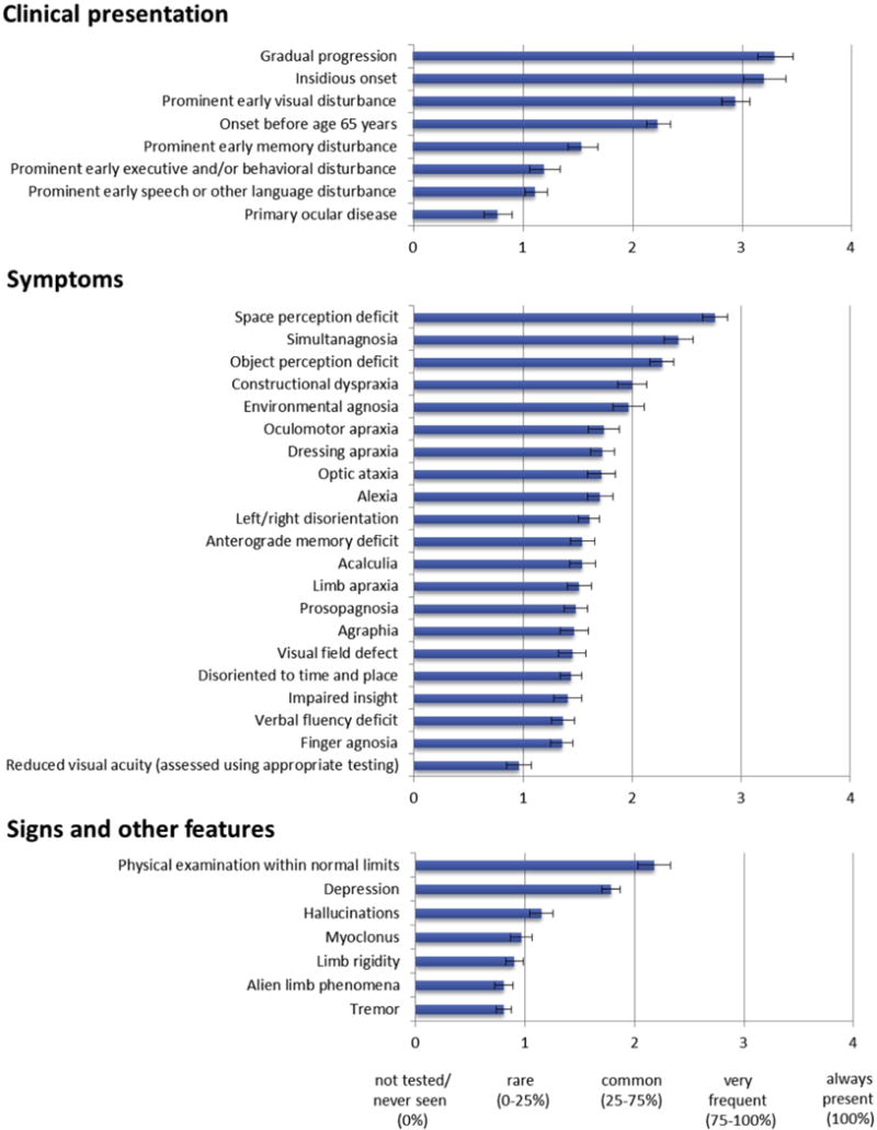
Methods: Consensus statements about PCA were developed through a detailed literature review, the formation of an international multidisciplinary working party which convened on four occasions, and a Web-based quantitative survey regarding symptom frequency and the conceptualization of PCA.
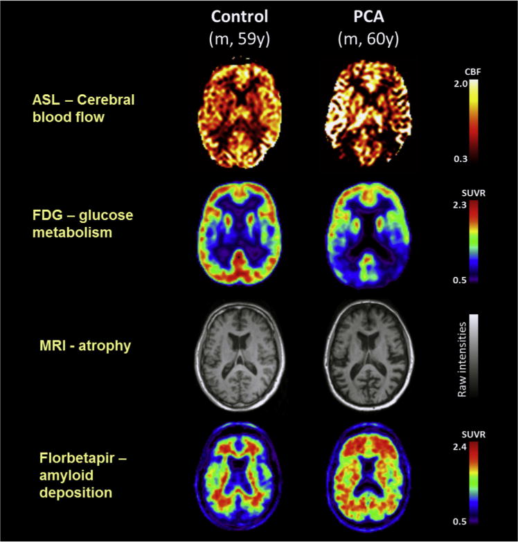
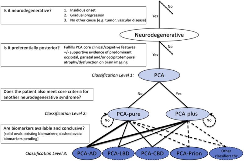
Results: A three-level classification framework for PCA is described comprising both syndrome- and disease-level descriptions. Classification level 1 (PCA) defines the core clinical, cognitive, and neuroimaging features and exclusion criteria of the clinico-radiological syndrome. Classification level 2 (PCA-pure, PCA-plus) establishes whether, in addition to the core PCA syndrome, the core features of any other neurodegenerative syndromes are present. Classification level 3 (PCA attributable to AD [PCA-AD], Lewy body disease [PCA-LBD], corticobasal degeneration [PCA-CBD], prion disease [PCA-prion]) provides a more formal determination of the underlying cause of the PCA syndrome, based on available pathophysiological biomarker evidence. The issue of additional syndrome-level descriptors is discussed in relation to the challenges of defining stages of syndrome severity and characterizing phenotypic heterogeneity within the PCA spectrum.
Discussion: There was strong agreement regarding the definition of the core clinico-radiological syndrome, meaning that the current consensus statement should be regarded as a refinement, development, and extension of previous single-center PCA criteria rather than any wholesale alteration or redescription of the syndrome. The framework and terminology may facilitate the interpretation of research data across studies, be applicable across a broad range of research scenarios (e.g., behavioral interventions, pharmacological trials), and provide a foundation for future collaborative work.
Keywords: Alzheimer's disease; Biomarker; Clinico-radiological syndrome; Pathophysiology; Posterior cortical atrophy.
Copyright © 2017 The Authors. Published by Elsevier Inc. All rights reserved.
Figures
References
-
- Benson DF, Davis RJ, Snyder BD. Posterior cortical atrophy. Arch Neurol. 1988;45:789–93. - PubMed
-
- Grünthal E. Zur hirnpathologischen Analyse der Alzheimerschen Krankheit. Psychiatrische und Neurologische Wochenschr. 1928;36:401–7.
-
- Morel F. Les aires striée, parastriée et péristriée dans les troubles de la fonction visuelle au cours de la maladie d’Alzheimer. Confinia Neurol. 1944;6:238–42. - PubMed
-
- Critchley M. The Parietal Lobes. New York: Hafner; 1953. pp. 182–3.
Publication types
MeSH terms
Grants and funding
LinkOut - more resources
Full Text Sources
Other Literature Sources
Medical
