. 2017 Apr;15(4):1613-1623.
doi: 10.3892/mmr.2017.6166. Epub 2017 Feb 3.

Ethyl acetate extract from Asparagus cochinchinensis exerts anti‑inflammatory effects in LPS‑stimulated RAW264.7 macrophage cells by regulating COX‑2/iNOS, inflammatory cytokine expression, MAP kinase pathways, the cell cycle and anti-oxidant activity

Affiliations

Ethyl acetate extract from Asparagus cochinchinensis exerts anti‑inflammatory effects in LPS‑stimulated RAW264.7 macrophage cells by regulating COX‑2/iNOS, inflammatory cytokine expression, MAP kinase pathways, the cell cycle and anti-oxidant activity

Hyun Ah Lee et al. Mol Med Rep. 2017 Apr.

Abstract

Asparagus cochinchinesis (A. cochinchinesis) is a medicine traditionally used to treat fever, cough, kidney disease, breast cancer, inflammatory disease and brain disease in northeast Asian countries. Although numerous studies of the anti‑inflammatory effects of A. cochinchinesis have been conducted, the underlying mechanisms of such effects in macrophages remain to be demonstrated. To investigate the mechanism of suppressive effects on the inflammatory response in macrophages, alterations of the nitric oxide (NO) level, the cell viability, inducible nitric oxide synthase (iNOS) and cyclooxygenase‑2 (COX‑2) expression levels, inflammatory cytokine expression, the mitogen-activated protein kinase (MAPK) signaling pathway, cell cycle arrest and reactive oxygen species (ROS) levels were measured in lipopolysaccharide (LPS)-activated RAW264.7 cells following treatment with ethyl acetate extract from A. cochinchinesis root (EaEAC). RAW264.7 cells pretreated two different concentrations of EaEAC prior to LPS treatment exhibited no significant toxicity. The concentration of NO was significantly decreased in the EaEAC + LPS treated group compared with the vehicle + LPS treated group. A similar decrease in mRNA transcript level of COX‑2, iNOS, pro-inflammatory cytokines [tumor necrosis factor‑α and interleukin (IL)‑1β] and anti‑inflammatory cytokines (IL‑6 and IL‑10) was detected in the EaEAC + LPS treated group compared with the vehicle + LPS treated group, although the decrease rate varied. Enhancement of the phosphorylation of MAPK family members following LPS treatment was partially rescued in the EaEAC pretreated group, and the cell cycle was arrested at the G2/M phase. Furthermore, the EaEAC pretreated group exhibited a reduced level of ROS generation compared with the vehicle + LPS treated group. Taken together, these results suggest that EaEAC suppresses inflammatory responses through inhibition of NO production, COX‑2 expression and ROS production, as well as differential regulation of inflammatory cytokines and cell cycle in RAW264.7 cells. In addition, these results provide strong evidence to suggest that EaEAC may be considered as an important candidate for the treatment of particular inflammatory diseases.


Figures

Figure 1.
Figure 1.
Free radical scavenging activity of EaEAC. DPPH radical scavenging activity was assayed in a mixture containing 0.1 mM DPPH and a range of concentrations of EaEAC (250–2,000 µg/ml). DPPH, 2,2-diphenyl-1-picrylhydrazyl radical; IC50, half maximal inhibitory concentration; EaEAC, ethyl acetate extract from Asparagus cochinchinesis root. Values are presented as the mean ± standard deviation of three replicates.
Figure 2.
Figure 2.
Toxicity of EaEAC. (A) Cell morphologies were observed under an inverted light microscope at ×400 magnification following incubation with LPS and 0 (vehicle), 100 or 200 µg/ml EaEAC in dimethyl sulfoxide for 24 h. (B) Cell viability of RAW264.7 cells treated with EaEAC + LPS was determined by 3-[4,5-dimethylthiazol-2-yl]-2,5-diphenyltetrazolium bromide (MTT) assay. Values are presented as the mean ± standard deviation of three replicates. NC, untreated control; EaEAC, ethyl acetate extract from Asparagus cochinchinesis root; LPS, lipopolysaccharide.
Figure 3.
Figure 3.
Determination of NO concentration, COX-2 and iNOS expression. (A) NO concentration was determined using supernatant collected from LPS-activated RAW264.7 cells treated with 0 (vehicle), 100 or 200 µg/ml EaEAC. (B) COX-2 and iNOS mRNA transcript levels in the NC, vehicle + LPS and EaEAC + LPS treated groups were examined by reverse transcription-quantitative polymerase chain reaction (RT-qPCR) using transcript-specific primers. Values are presented as the mean ± standard deviation of three replicates. *P<0.05 vs. NC; #P<0.05 vs. vehicle + LPS-treated group; ΔP<0.05 vs. 100 µg/ml EaEAC + LPS-treated group. NO, nitric oxide; NC, untreated control; EaEAC, ethyl acetate extract from Asparagus cochinchinesis root; LPS, lipopolysaccharide; COX-2, cyclooxygenase-2; iNOS, inducible nitric oxide synthase.
Figure 4.
Figure 4.
Analysis of pro- and anti-inflammatory cytokine expression. Following treatment of RAW264.7 cells with LPS and 0 (vehicle), 100 or 200 µg/ml of EaEAC, (A) IL-1β, TNF-α, IL-6 and IL-10 mRNA levels were assessed by semi-quantitative reverse transcription polymerase chain reaction using transcript-specific primers, and (B) IL-6 concentration was detected using an enzyme-linked immunosorbent assay kit with a minimum detection threshold of 9.3 pg/ml. Values are presented as the mean ± standard deviation of three replicates. *P<0.05 vs. NC; #P<0.05 vs. vehicle + LPS-treated group; ΔP<0.05 vs. 100 µg/ml EaEAC + LPS-treated group. LPS, lipopolysaccharide; NC, untreated control; EaEAC, ethyl acetate extract from A. cochinchinesis root; TNF-α, tumor necrosis factor-α; IL, interleukin.
Figure 5.
Figure 5.
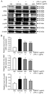
Activation of the mitogen-activated protein kinase signaling pathway proteins ERK, JNK, and p38. (A) p-ERK, ERK, p-JNK, JNK, p38, p-p38 and β-actin protein expression was assessed by western blot following stimulation of RAW264.7 cells with LPS and 0 (vehicle), 100 or 200 µg/ml of EaEAC. (B) Band intensity was determined using an imaging densitometer and expression levels calculated relative to the intensity of β-actin. Values are presented as the mean ± standard deviation of three replicates. *P<0.05 vs. NC; #P<0.05 vs. vehicle + LPS-treated group; ΔP<0.05 vs. 100 µg/ml EaEAC + LPS-treated group. LPS, lipopolysaccharide; NC, untreated control; EaEAC, ethyl acetate extract from Asparagus cochinchinesis root; p-, phosphorylated; ERK, extracellular signal-regulated kinase; JNK, c-Jun N-terminal kinase.
Figure 6.
Figure 6.
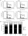
Cell cycle analysis. The cell cycle distribution was determined by flow cytometric analysis of the DNA content of nuclei of RAW264.7 cells following staining with PI. After treatment with EaEAC + LPS, the number of cells in the G0/G1, S and G2/M stage was determined. (A) Fluorescence-activated cell sorting analysis and (B) the relative percentage of cells in each cell cycle phase (G0/G1, S and G2/M). *P<0.05 vs. NC. #P<0.05 vs. Vehicle + LPS treated group. PI, propidium iodide; EaEAC, ethyl acetate extract from Asparagus cochinchinesis root; LPS, lipopolysaccharide; NC, untreated control.
Figure 7.
Figure 7.
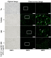
Determination of intracellular reactive oxygen species production. Cells were pretreated with LPS and 0 (vehicle), 100 or 200 µg/ml of EaEAC in dimethyl sulfoxide, then treated with DCFH-DA. Optical images (left column) were observed by light microscopy, and green fluorescence in cells was observed by fluorescence microscopy. Boxed cells in each ×200 magnification image (center column) were further examined under ×400 magnification (right column). Arrows indicate cells stained with DCFH-DA. DCFH-DA, 2′,7′-dichlorodihydrofluorescein diacetate; NC, untreated control; EaEAC, ethyl acetate extract from Asparagus cochinchinesis root; LPS, lipopolysaccharide.

Similar articles

Cited by

References

    1. Xiong DS, Yu LX, Yan X, Guo C, Xiong Y. Effects of root and stem extracts of Asparagus cochinchinensis on biochemical indicators related to aging in the brain and liver of mice. Am J Chinese Med. 2011;39:719–726. doi: 10.1142/S0192415X11009159. - DOI - PubMed
    1. Xiao PG. Modern Chinese material medica. Chemical Industry Press; Beijing: 2002. p. 150.
    1. Liu YZ, Qu FY, Zhang PX. Effect of chloroform extract of Tiandong on the brain antioxidation of D-galatose-induced senile mice. Heilongjiang Med Pharm. 2001;24:7–8.
    1. Ni JM, Zhao R, Wang R. Comparison on amino acid content in prepared and unprepared Asparagus cochinchinensis. Chin Tradit Herb Drugs. 1992;23:182–183.
    1. Tenji K, Junzo S. Studies on the constituents of Asparagi Radix. I. On the structures of furostanol oligosides of Asparagus cochinchinensis (LOUREIO) MERRILL. Chem Pharm Bull. 1979;27:3086–3094. doi: 10.1248/cpb.27.3086. - DOI

MeSH terms