

Baicalin ameliorates renal fibrosis via inhibition of transforming growth factor β1 production and downstream signal transduction
- PMID: 28260014
- PMCID: PMC5364985
- DOI: 10.3892/mmr.2017.6208
Baicalin ameliorates renal fibrosis via inhibition of transforming growth factor β1 production and downstream signal transduction
Abstract
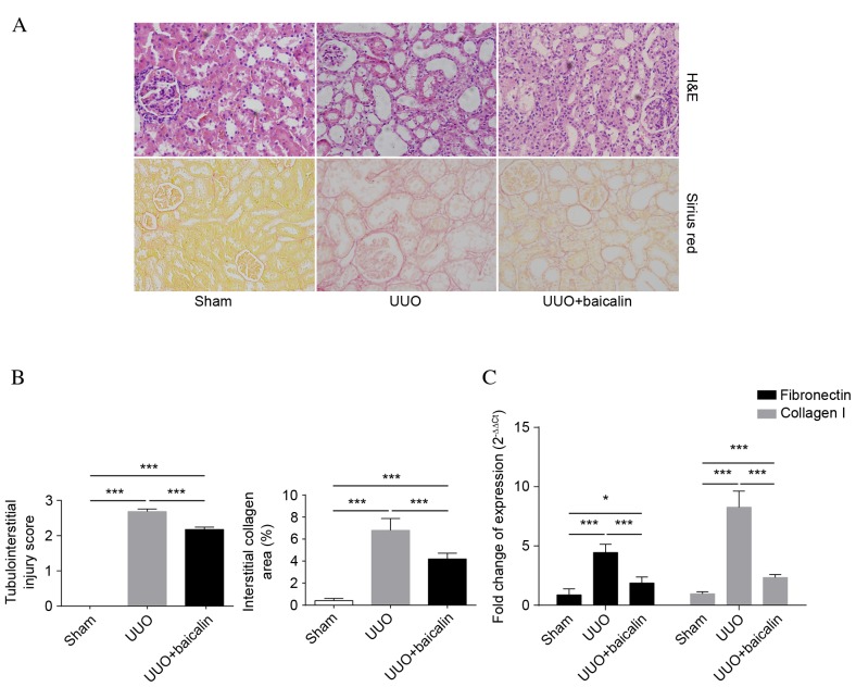
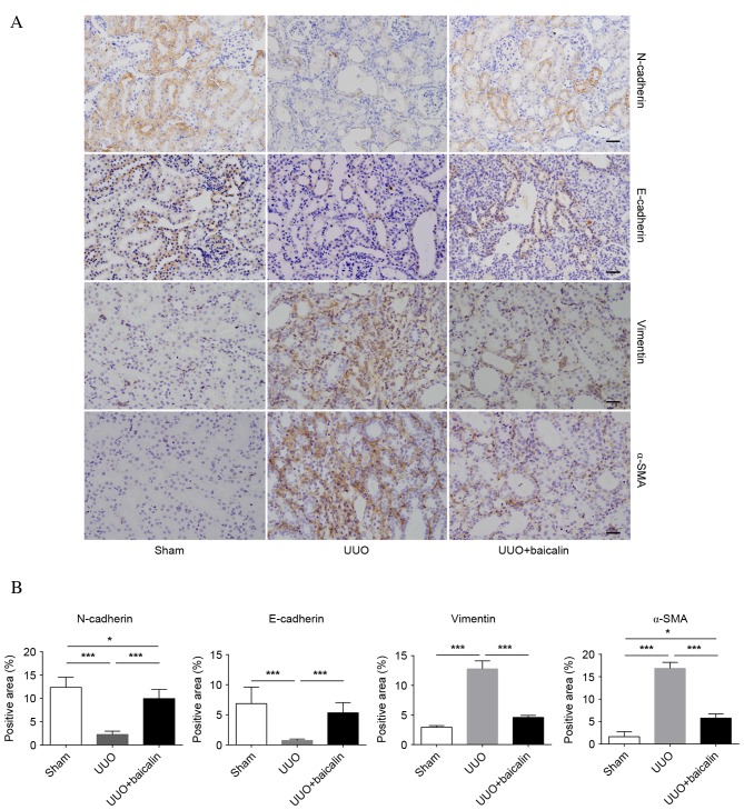
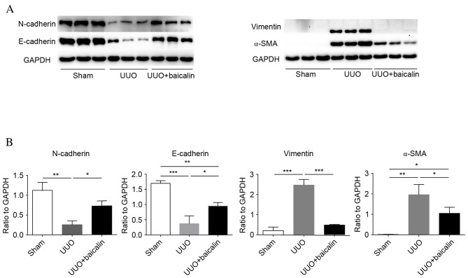
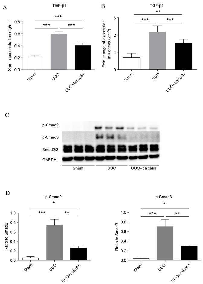
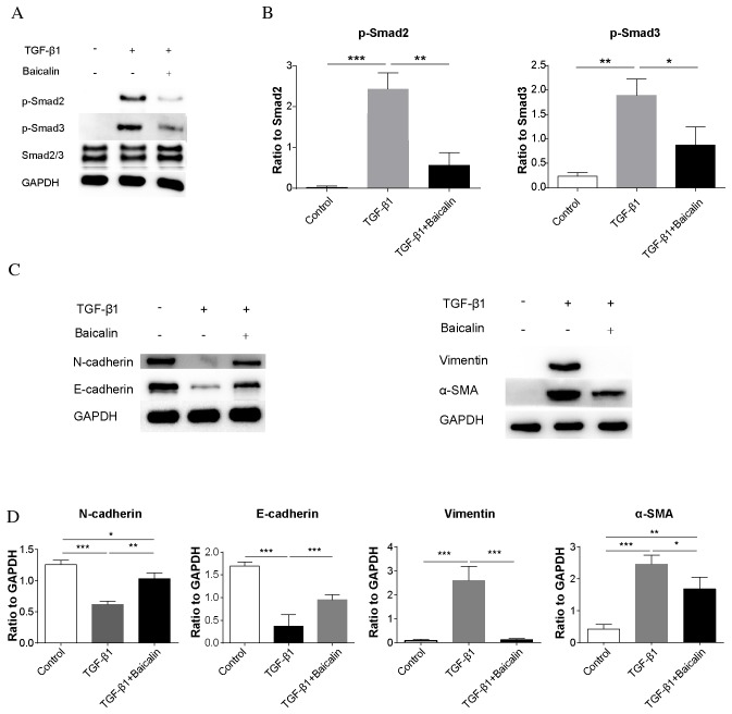
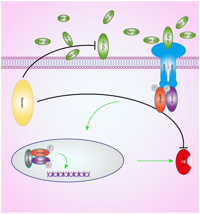
Previous studies have demonstrated the potential antifibrotic effects of baicalin in vitro, via examination of 21 compounds isolated from plants. However, its biological activity and underlying mechanisms of action in vivo remain to be elucidated. The present study aimed to evaluate the effect of baicalin on renal fibrosis in vivo, and the potential signaling pathways involved. A unilateral ureteral obstruction (UUO)‑induced renal fibrosis model was established using Sprague‑Dawley rats. Baicalin was administrated intraperitoneally every 2 days for 10 days. The degree of renal damage and fibrosis was investigated by histological assessment, and detection of fibronectin and collagen I mRNA expression levels. Epithelial‑mesenchymal transition (EMT) markers, transforming growth factor-β1 (TGF-β1) levels and downstream phosphorylation of mothers against decapentaplegic 2/3 (Smad2/3) were examined in vivo and in an NRK‑52E rat renal tubular cell line in vitro. Baicalin was demonstrated to markedly ameliorate renal fibrosis and suppress EMT, as evidenced by reduced interstitial collagen accumulation, decreased fibronectin and collagen I mRNA expression levels, upregulation of N‑ and E‑cadherin expression levels, and downregulation of α‑smooth muscle actin and vimentin expression. Furthermore, baicalin decreased TGF‑β1 expression levels in serum and kidney tissue following UUO, and suppressed Smad2/3 phosphorylation in rat kidney tissue. In vitro studies identified that baicalin may inhibit the phosphorylation of Smad2/3 under the same TGF‑β1 concentration. In conclusion, baicalin may protect against renal fibrosis, potentially via inhibition of TGF‑β1 production and its downstream signal transduction.
Figures
Similar articles
-
Inhibitory effects of fasudil on renal interstitial fibrosis induced by unilateral ureteral obstruction.Mol Med Rep. 2015 Dec;12(6):8010-20. doi: 10.3892/mmr.2015.4467. Epub 2015 Oct 21. Mol Med Rep. 2015. PMID: 26498136 Free PMC article.
-
Adenovirus-mediated P311 ameliorates renal fibrosis through inhibition of epithelial-mesenchymal transition via TGF-β1-Smad-ILK pathway in unilateral ureteral obstruction rats.Int J Mol Med. 2018 May;41(5):3015-3023. doi: 10.3892/ijmm.2018.3485. Epub 2018 Feb 12. Int J Mol Med. 2018. PMID: 29436600
-
Anti-renal fibrosis effect of asperulosidic acid via TGF-β1/smad2/smad3 and NF-κB signaling pathways in a rat model of unilateral ureteral obstruction.Phytomedicine. 2019 Feb;53:274-285. doi: 10.1016/j.phymed.2018.09.009. Epub 2018 Sep 5. Phytomedicine. 2019. PMID: 30668407
-
TGF-β1 → SMAD/p53/USF2 → PAI-1 transcriptional axis in ureteral obstruction-induced renal fibrosis.Cell Tissue Res. 2012 Jan;347(1):117-28. doi: 10.1007/s00441-011-1181-y. Epub 2011 Jun 4. Cell Tissue Res. 2012. PMID: 21638209 Free PMC article. Review.
-
Unilateral Ureteral Obstruction as a Model to Investigate Fibrosis-Attenuating Treatments.Biomolecules. 2019 Apr 8;9(4):141. doi: 10.3390/biom9040141. Biomolecules. 2019. PMID: 30965656 Free PMC article. Review.
Cited by
-
Early Progression of Xanthogranulomatous Pyelonephritis in Children Might Be Dependent on Vimentin Expression.Am J Case Rep. 2017 Oct 5;18:1066-1072. doi: 10.12659/ajcr.904376. Am J Case Rep. 2017. PMID: 28978905 Free PMC article.
-
Flavonoids in Kidney Health and Disease.Front Physiol. 2018 Apr 24;9:394. doi: 10.3389/fphys.2018.00394. eCollection 2018. Front Physiol. 2018. PMID: 29740333 Free PMC article. Review.
-
Baicalin: a potential therapeutic agent for acute kidney injury and renal fibrosis.Front Pharmacol. 2025 Jan 22;16:1511083. doi: 10.3389/fphar.2025.1511083. eCollection 2025. Front Pharmacol. 2025. PMID: 39911847 Free PMC article. Review.
-
Baicalin attenuates liver hypoxia/reoxygenation injury by inducing autophagy.Exp Ther Med. 2018 Aug;16(2):657-664. doi: 10.3892/etm.2018.6284. Epub 2018 Jun 8. Exp Ther Med. 2018. PMID: 30116320 Free PMC article.
-
TGF-β/Smad Signaling Pathway in Tubulointerstitial Fibrosis.Front Pharmacol. 2022 Mar 24;13:860588. doi: 10.3389/fphar.2022.860588. eCollection 2022. Front Pharmacol. 2022. PMID: 35401211 Free PMC article.
References
MeSH terms
Substances
LinkOut - more resources
Full Text Sources
Other Literature Sources
Medical
